腰椎终板Modic改变与椎间盘退变的相关性的定量MRI研究
龚静山,梅东东,朱进,周阳泱
腰椎终板Modic改变与椎间盘退变的相关性的定量MRI研究
龚静山*,梅东东,朱进,周阳泱
目的探讨腰椎终板Modic改变与相邻椎间盘退变的相关。材料与方法于66例腰椎结构和定量MR图像上,采用Pfirrmann分级半定量和测量椎间盘T2和T2*值定量评价椎间盘退变程度,比较腰椎终板Modic改变与椎间盘Pfirrmann评分、T2和T2*值的相关性。结果66例患者中,14例患者(21.2%)终板有19个椎间盘相邻的终板有Modic改变,包括2个椎间盘相邻的终板为Modic 1型改变,17个椎间盘相邻的终板为Modic 2型改变。正常终板、Modic 1型终板和Modic 2型终板相邻椎间盘的Pfirrmann评分分别为2.40±0.96、4.00±1.41和4.12±1.27,T2值分别为(95.38±51.88)、(70.50±36.06)和(58.65±38.47) ms,T2*值分别为(34.43±19.16)、(24.00±1.41)和(28.65±23.39) ms,差异均有统计学意义(P值分别为0.000、0.000和0.023)。Modic 2型终板相邻的椎体伴有严重的椎间盘退变(Pfirrmann评分升高,T2和T2*值降低),与正常终板相邻的椎间盘比较,差异具有统计学意义(P值分别为0.000、0.000和0.006)。结论腰椎终板Modic改变与相邻的椎间盘退变具有相关性,终板改变和椎间盘退变应作为腰椎退变的整体改变。
椎间盘退化;腰椎;磁共振成像
下腰痛是十分常见影响劳动生产力的疾患,约2/3的成年人一生中经历过下腰痛[1]。随着MRI的普及,腰椎MRI检查是临床医生确定下腰痛病因最常用的检查方法。椎间盘退变是常见腰椎MRI检查的异常征象,也是引起下腰痛的常见原因之一[2]。有严重椎间盘退变的成人发生下腰痛的风险是椎间盘无结构异常的两倍[3],但1/3无症状女性MRI检查可以检出腰椎椎间盘退变[4]。近年研究表明,椎间盘、终板和脊旁肌在脊柱退变中应作为一个“整体-器官”,退变的椎间盘邻近的终板经常伴有MRI信号改变,即Modic改变[5-6]。笔者假设腰椎终板Modic改变是腰椎退变的一部分,与椎间盘退变有相关性。为验证这一假设,特设计本研究探索腰椎终板Modic改变与椎间盘退变的关系。
1 材料与方法
1.1 一般资料
2015年3月至2015年6月间,75例因下腰痛患者在我院采用3.0 T MR扫描仪行结构和定量MR成像。2例患者MRI检出腰椎肿瘤并累及椎间盘,4例患者检查过程中未能很好配合,T2和T2*mapping序列图像伪影较多,1例患者有多个椎体融合改变,1例患者不能耐受额外T2和T2*mapping序列扫描,1例患者检出腰椎结核,共9例患者排除本研究,最终66例患者入选本研究。入选的66例患者中,男26例,女40例,年龄范围22~79岁(平均48.7岁)。全部患者检查前均签署书面知情同意书,医院伦理委员会同意本研究。
1.2 结构和定量MR成像
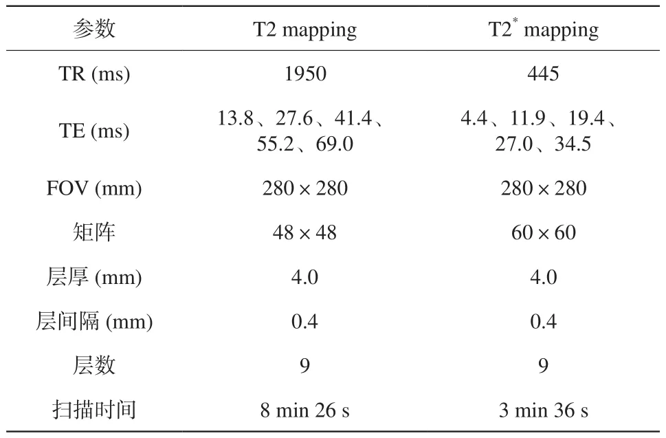
全部患者MRI检查采用32通道脊柱线圈,在Siemens Magnetom Skyro 3.0 T MR成像系统(Siemens Healthcare,Germany)进行。扫描方案见文献[7],扫描参数见表1。T2和T2*mapping 由在线商业软件MapIt (Siemens Healthcare,Germany)自动生成。
1.3 图像分析
由1名有多年MRI读片经验的放射诊断医师在正中矢状面T2 map和T2*map上并参照正中矢状面T2WI手动绘制尽可能包含髓核和内层纤维环的感兴趣区(region of interest,ROI)测量椎间盘的T2和T2*值。由2名有多年MRI读片经验的放射诊断医师在不知道椎间盘的T2和T2*值的情况下,基于矢状面T2加权图像对腰椎椎间盘进行Pfirrmann分级。Pfirrmann分级标准为:Ⅰ级,椎间盘为均匀高信号;Ⅱ级,椎间盘信号不均匀伴或不伴水平低信号带;Ⅲ级,椎间盘呈不均匀灰信号,高度正常或轻度降低,纤维环和髓核分界不清;Ⅳ级,椎间盘为不均匀灰信号,高度正常或中度降低,髓核和纤维环界限消失;Ⅴ级,椎间盘为黑信号并且塌陷[8]。根据矢状面T1WI和T2WI终板信号改变分为4型:Modic 0型(即正常终板),T1WI和T2WI无异常信号;Modic 1型,T1WI低信号,T2WI高信号;Modic 2型,T1WI和T2WI均为高信号;Modic 3型,T1WI和T2WI均为低信号[9]。
1.4 统计学处理
采用Kruskal-Wallis H检验比较腰椎终板改变间相邻椎间盘Pfirrmann评分差异。如差异有统计学意义,采用Mann-Whitney U检验行两两比较。统计软件采用IBM SPSS Statistics 24,P<0.05认为有统计学意义。
2 结果
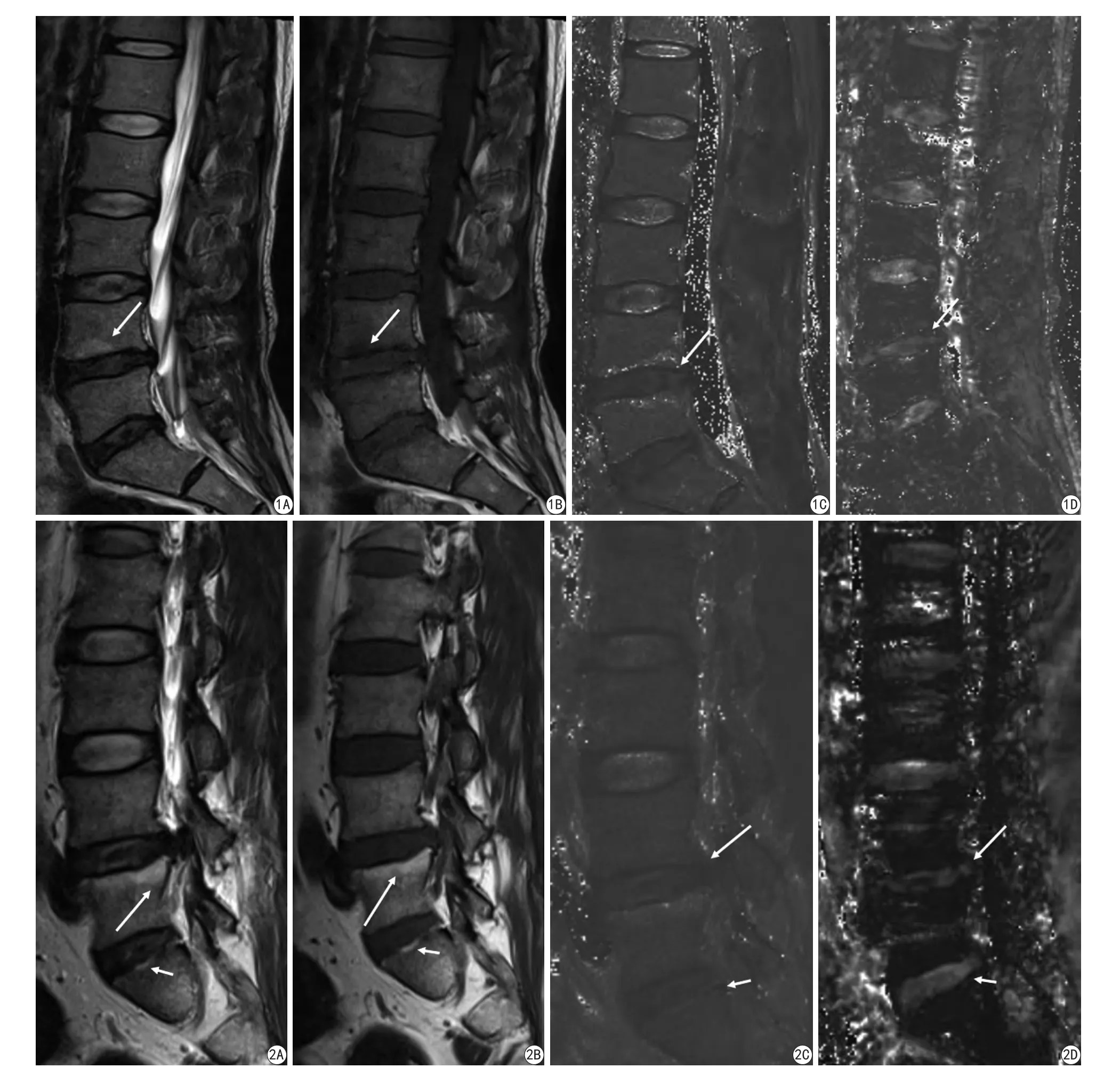
全部66例患者中,14例患者(21.2%)有19个椎间盘相邻的终板有Modic改变(34.8%),其中3例患者有2个椎间盘相邻的终板有Modic改变,1例患者有3个椎间盘相邻的终板伴有Modic改变,无一例患者有3个以上的椎间盘相邻的终板伴有Modic改变。19个椎间盘相邻终板Modic 改变中1型2个,2型17个,3型0个。330个椎间盘的相邻的终板Modic 改变与Pfirrmann评分、T2和T2*值见表2,不同Modic改变的相邻椎间盘Pfirrmann评分、T2和T2*值间差异存在统计学意义。由于与Modic 1型终板相邻的椎间盘仅2个和无Modic 3型终板,故未对这两型进行统计学处理。与正常终板(Modic 0型)相邻椎间盘比较,Modic 2型改变终板相邻的椎间盘的Pfirrmann评分升高、T2和T2*值下降(图1,2),差异有统计学意义(z值分别为5.436,4.328,2.754;P值分别为0.000,0.000,0.006)。

表1 T2和T2* mapping扫描参数Tab. 1 Parameters of T2 and T2* mapping

表2 终板Modic改变与相邻椎间盘Pfirrmann评分、T2和T2*值相关性Tab. 2 Association of endplate Modic changes with Pfirrmann scores, T2 and T2* values of the adjacent interveterbral discs
3 讨论
腰椎退变是引起下腰痛的最为常见原因,以往研究都关注椎间盘退变。在其他关节退变如膝关节的研究中发现,软骨、关节骨和周围软组织作为一个整体均会被累及[10]。膝关节退行性骨关节病中,伴随软骨损伤有软骨下骨硬化和MRI上出现水肿样信号,而且水肿样信号的增大与关节痛和软骨退变相关[10-11]。近年研究也表明,椎间盘、终板和脊旁肌肉也会作为一个整体会在脊椎退变中同时有改变[5-6]。在脊柱椎间盘关节,终板类似软管下骨,尽管终板改变也被认为是脊柱退变的征象之一,但其与椎间盘退变的关系尚不是十分明了。
与膝关节的软骨、软骨下骨、周围韧带和肌肉作为一个整体维系膝关节的功能处于一个动态变化一样,脊柱关节的椎间盘、终板和脊旁肌也作为一个整体处于一个相互影响的动态变化中。椎间盘作为承载人体轴向压力椎体关节的纤维软骨,特别是下腰椎的椎间盘,而且缺乏血管,营养主要靠终板弥散,因此较早出现退变[12-13]。退变的椎间盘2型胶原纤维含量增加和含水量降低,同时髓核的蛋白多糖降低,这样椎间盘的纤维含量增加,而且失去结构[12]。退变的椎间盘失去其应有的结构和功能,使椎体关节稳定性降低和缓冲轴向压力的能力降低,从而终板和脊旁肌受力增加,因此,退变的椎间盘也会使邻近的终板退变加剧。Modic等[9]根据椎体终板在MRI信号改变将终板分为3型。对照组织病理学改变,Modic 1型改变终板出现裂隙,邻近的骨髓内出现带血管的纤维组织,从而使得T1和T2延长;Modic 2型改变代表终板黄骨髓化,从而使得T1缩短和T2延长;Modic 3型改变代表骨质硬化,T1 和T2均缩短[13]。终板退变降低弥散,使椎间盘获得的营养减少,进而会加剧椎间盘退变。本研究结果也验证这一假设,终板Modic改变与椎间盘Pfirrmann评分、T2和T2*值有相关性,其中Modic 2型终板伴有椎间盘Pfirrmann评分升高、T2和T2*值降低,说明椎体终板改变常伴有或加剧相邻椎间盘退变。虽然有文献报道终板Modic改变与椎间盘Pfirrmann评分有相关性[6,14],但据笔者所知,终板Modic改变与椎间盘T2和T2*值相关性尚未见相关报道,因此,本研究为首次报道Modic改变与椎间盘T2和T2*值有相关性。
定量MRI对关节软骨和椎间盘退变的研究结果表明,T2和T2*值与关节软骨和椎间盘变性有较好的相关性,可以定量评价退变的程度,并且能较结构MRI早期发现退变[7,14-15]。早期发现椎间盘退变,可以指导临床及时干预,避免椎间盘进一步发展成为膨出或突出,引起严重的下腰痛和神经根症状,降低患者的劳动力,有重要的临床和社会意义。本研究初步报道Modic改变与椎间盘T2和T2*值有相关性,说明腰椎终板Modic改变是脊柱退变的征象之一,可以伴随椎间盘退变发生,并提示椎间盘退变的严重程度。
本研究存在的一些缺陷。由于对Modic改变还没有统一的分级标准,本研究未对Modic改变的程度进行评价,这样未能探讨Modic改变严重程度与椎间盘退变的程度相关性。其次,本研究未对病人进行随访,没有对终板改变和椎间盘退变随时间变化是否相关进行研究。本组资料样本量较少,以至于Modic改变中1型仅2个,没有3型改变。此外,文献报道终板Modic改变与下腰痛有相关性[16],由于本组资料缺乏相关临床资料,未能对两者相关性进行研究。

图1 男,35岁,L4终板Modic 1型改变。A:矢状面T2WI示L4椎体下缘终板斑片状高信号灶(箭)。B:矢状面T1WI示L4椎体下缘终板相应部位呈低信号(箭)改变。C:矢状面T2 map示L4-5椎间盘T2值降低(箭)(T2值:L1-2,150 ms;L2-3,140 ms;L3-4,99 ms;L4-5,53 ms;L5-S1,52 ms)。D:矢状面T2* map示L4-5椎间盘T2值降低(箭)(T2*值:L1-2,26 ms;L2-3,23 ms;L3-4,29 ms;L4-5,19 ms;L5-S1,21 ms)图2 男,49岁,L5和S1终板Modic 2型改变。A:矢状面T2WI显示L5椎体上缘终板长条状高信号灶(长箭)和S1上缘终板短条状高信号灶(短箭)。B:矢状面T1WI 终板相应部位呈高信号改变(长箭和短箭)。C:矢状面T2 map显示L4-5(长箭)和L5-S1(短箭)椎间盘T2值降低(T2值:L1-2,122 ms;L2-3,148 ms;L3-4,151 ms;L4-5,76 ms;L5-S1,51 ms)。D:矢状面T2* map显示L4-5 (长箭)和L5-S1 (短箭)椎间盘T2值降低(T2*值:L1-2,29 ms;L2-3,28 ms;L3-4,58 ms;L4-5,26 ms;L5-S1,25 ms)Fig. 1 A 35-year-old man with Modic type 1 change at the endplate of L4. A: Sagital T2WI shows that there is patch of high signal (arrow) at the lower endplate of L4. B: Sagital T1WI shows that it is of hypointense (arrow). C: Sagital T2 map demonstrates that the T2 values of L4-5 disc are decreased (arrow)(T2 values: L1-2, 150 ms. L2-3, 140 ms. L3-4, 99 ms. L4-5, 53 ms. L5-S1, 52 ms). D: Sagital T2* map shows the T2* values of L4-5 disc are decreased also(arrow) (T2* values: L1-2, 26 ms. L2-3, 23 ms. L3-4, 29ms. L4-5, 19 ms. L5-S1, 21 ms). Fig. 2 A 49-year-old man with Modic type 2 change at the endplates of L5 and S1. A: Sagital T2WI shows a hyperintense long strip (long arrow) at the upper endplate of L5 and a hyperintense short strip (short arrow) at the upper endplate of S1. B: Sagital T1WI shows that they are of hyperintense (long arrow and short arrow). C: Sagital T2 map reveals that the T2 values of L4-5 (long arrow) and L5-S1 (short arrow) discs are decreased (T2 values: L1-2, 122 ms. L2-3, 148 ms. L3-4, 151 ms. L4-5, 76 ms. L5-S1, 51 ms). D: Sagital T2* map shows the T2* values of L4-5 (long arrow) and L5-S1 (short arrow) discs (T2* values: L1-2, 29 ms. L2-3, 28 ms. L3-4, 58 ms. L4-5, 26 ms. L5-S1, 25 ms).
综上所述,本研究通过比较腰椎终板Modic改变与椎间盘退变的结构和定量MRI的相关性,结果表明,腰椎终板改变与椎间盘退变具有相关性,Modic 2型改变的终板相邻的椎间盘退变严重。腰椎终板改变可能是腰椎作为一个“整体-器官”在发生退变时的征象之一。在评价下腰痛患者MRI时,终板和椎间盘改变都应引起放射诊断医师的重视。
[References]
[1] Andersson GB. Epidemiological features of chronic low-back pain.Lancet, 1999, 354(9178): 581-585.
[2] Luoma K, Riihimaki H, Luukkonen R, et al. Low back pain in relation to lumbar disc degeneration. Spine, 2000, 25(4): 487-492.
[3] Hicks GE, Morone N, Weiner DK. Degenerative lumbar disc and facet disease in older adults: prevalence and clinical correlates. Spine(Phila Pa 1976), 2009, 34(12): 1301-1306.
[4] Powell MC, Wilson M, Szypryt P, et al. Prevalence of lumbar disc degeneration observed by magnetic resonance in symptomless women. Lancet,1986, 2(8520): 1366-1367.
[5] Farshad-Amacker NA, Hughes A, Herzog RJ, et al. The intervertebral disc, the endplates and the vertebral bone marrow as a unit in the process of degeneration. Eur Radiol, 2017, 27(6): 2507-2520.
[6] Teichtahl AJ, Urquhart DM, Wang Y, et al. Lumbar disc degeneration is associated with Modic change and high paraspinal fat content: a 3.0 T magnetic resonance imaging study. BMC Musculoskelet Disord,2016, 17(1): 439.
[7] Gong JS, Ling RN, Zhou YY, et al. T2 and T2*mapping to evaluate lumbar intervertebral disc degeneration in patients with low back pain using 3.0 tesla MR. Chin J CT MRI, 2016,14(10): 113-116.龚静山, 凌人男, 周阳泱, 等. 采用3.0 T MRI T2和T2*mapping评价下腰痛患者腰椎间盘退变. 中国CT和MRI杂志, 2016, 14(10):113-116.
[8] Pfirrmann CW, Metzdorf A, Zanetti M, et al. Magnetic resonance classification of lumbar intervertebral disc degeneration. Spine, 2001,26(17): 1873-1878.
[9] Modic MT, Masaryk TJ, Ross JS, et al. Imaging of degenerative disk disease. Radiology, 1988, 168(1): 177-186.
[10] Teichtahl AJ, Wluka AE, Davies-Tuck ML, et al. Imaging of knee osteoarthritis. Best Pract Res Clin Rheumatol, 2008, 22(6):1061-1074.
[11] Felson DT, Niu J, Guermazi A, et al. Correlation of the development of knee pain with enlarging bone marrow lesions on magnetic resonance imaging. Arthritis Rheum, 2007, 56(9): 2986-2992.
[12] Modic MT, Ross JS. Lumbar degenerative disk disease. Radiology,2007, 245(1): 43-61.
[13] Stokes IA, Iatridis JC. Mechanical conditions that accelerate intervertebral disc degeneration: overload versus immobilization.Spine, 2004, 29(23): 2724-2732.
[14] Cao GM, Zhao B. Quantitative assessment of lumber facet joints and intervertebral discs with axial MR T2 star mapping. J Med Imag,2014, 24(11): 1993-1997.曹观美, 赵斌. 轴面MR T2*mapping对腰椎小关节和椎间盘的定量研究. 医学影像学杂志, 2014, 24(11): 1993-1997.
[15] Huang YQ, Zhao XM, Wu QH, et al. Comparative study of MR T2 relaxation time and apparent diffusion coefficient for assessing lumbar intervertebral disc degeneration. Radiol Pract, 2016, 31(8):773-777.黄耀渠, 赵晓梅, 伍琼慧, 等. 磁共振 T2 弛豫时间和 ADC评估腰椎间盘退变的对比研究. 放射学实践, 2016, 31(8): 773-777.
[16] Mok FP, Samartzis D, Karppinen J, et al. Modic changes of the lumbar spine: prevalence, risk factors, and association with disc degeneration and low back pain in a large-scale population-based cohort. Spine J, 2016, 16(1): 32-41.
Quantitative MR imaging study of association between lumbar vertebral endplate Modic changes and intervertebral disc degeneration
GONG Jing-shan, MEI Dong-dong, ZHU Jin, ZHOU Yang-yang
Department of Radiology, Shenzhen People’s Hospital, the Second Clinical Medical College of Jinan University, Shenzhen 518020, China
Objective:To investigate the relationship between vertebral endplate Modical changes and adjacent intervertebral disc degeneration.Materials and Methods:Lumbar structure and quantitative MR images of 66 patients were retrospectively analyzed. Disc degeneration was semi-quantitatively assessed using Pfirrmann grading system and quantitatively assessed using T2 and T2*values.Association between Pfirrmann scores, T2 and T2*values of intervertebral discs with adjacent endplate Modic changes was analyzed.Results:Nineteen intervertebral discs with adjacent endplate Modic changes were detected in fourteen patients (14/66,21.2%), which included 2 discs with Modic type 1 changes and 17 discs with type 2 changes. Pfirrmann scores of the discs adjacent to normal endplates, Modic type 1 endplates and Modic type 2 endplates were 2.40±0.96、4±1.41 and 4.12±1.27,respectively. T2 values were (95.38±51.88) ms, (70.50±36.06) ms and (58.65±38.47) ms. T2*values were (34.43±19.16) ms, (24.00±1.41) ms and (28.65±23.00) ms.The differences of disc Pfirrmann scores, T2 and T2*values among different endplate changes were statistical significant withPvalues of 0.000, 0.000 and 0.023,respectively. Comparing to discs adjacent to normal endplates, discs adjacent to Modic type 2 endplates had severe degeneration with increased Pfirrmann scores and decreased T2 and T2*values. The differences were statistical significant (Pvalues were 0.000, 0.000 and 0.006, respectively).Conclusions:Modic changes of vertebral endplate are associated with adjacent intervertebral disc degeneration. Both of them should be regarded as a facet of the "whole-organ" pathologic conditions in the procession of spinal degeneration.
Intervertebral disc degeneration; Lumbar vertebrae; Magnetic resonance imaging
深圳市科技计划项目(编号:JCYJ2014041622811967)
深圳市人民医院(暨南大学第二临床医学院)放射科,深圳 518020
龚静山,E-mail:jshgong@sina.com
2017-04-13
接受日期:2017-05-03
R445.2;R681.53
A
10.12015/issn.1674-8034.2017.07.007
龚静山, 梅东东, 朱进, 等. 腰椎终板Modic改变与椎间盘退变的相关性的定量MRI研究. 磁共振成像, 2017, 8(7):514-518.*Correspondence to: Gong JS, E-mail: jshgong@sina.com
Received 13 Apr 2017, Accepted 3 May 2017
ACKNOWLEDGMENTSThis work was supported by Shenzhen Science &Technology Program (No. JCYJ2014041622811967).

