缺血性卒中后抑郁患者静息状态下默认网络功能连接的研究
张佩瑶,徐琴,王君,王晶,戴建平
缺血性卒中后抑郁患者静息状态下默认网络功能连接的研究
张佩瑶1,徐琴2,王君3,王晶4,戴建平5*

目的利用静息态功能磁共振成像(resting-state functional magnetic resonance imaging,rs-fMRI)技术,研究缺血性卒中后抑郁(post stroke depression,PSD)患者静息状态下默认网络(default mode network,DMN)功能连接的变化。材料与方法选取13例发病在两周内、汉密尔顿抑郁量表(hamilton depression scale,HAMD)评分>7分且为首发抑郁的PSD患者,同时选择13名正常被试作为对照组(control)分别进行静息态fMRI扫描。分别选取双侧楔前叶作为感兴趣区(region of interest,ROI),计算并比较两组被试默认网络功能连接的变化。结果与正常被试相比,PSD患者双侧顶下小叶、双侧额颞叶与右侧楔前叶功能连接减低;右侧顶下小叶、双侧额颞叶与左侧楔前叶功能连接减低,而未发现功能连接显著增高的脑区。结论默认网络功能连接的变化可能与卒中后抑郁的发生具有相关性。
卒中;抑郁症;默认网络;功能连接;磁共振成像,功能
抑郁是卒中幸存者最为常见的情绪障碍,有研究显示其发病率为33%,且抑郁症状会严重影响患者的康复[1]。以往对卒中后抑郁(post stroke depression,PSD)发病机制的研究主要集中在卒中本身的特性方面,主要包括病变位置、大小、体积等[2-4]。然而,研究者们却无法确定卒中病变与之后抑郁发生之间的相关性[5]。
近年来,静息态功能磁共振成像技术(restingstate functional magnetic resonance imaging,rsfMRI)为认知障碍性疾病的研究提供了新的方向。早期研究发现,在清醒闭眼的静息状态下人脑也存在着自发性低频震荡(low frequent fluctuation,LFF,0.01~0.08 Hz),这种低频振荡的时间相关性就定义为功能连接(functional connectivity,FC),同时具有功能连接的相关脑区就构成了特定的脑网络[6]。其中研究最广泛的是默认网络(default mood network,DMN),主要包括双侧楔前叶、内侧前额叶、前扣带、后扣带回及顶下小叶等区域,且与信息整合、情绪处理及自我意识相关[6-7]。以往有研究发现抑郁症患者的默认网络存在异常[8-9],但极少见到其在卒中后抑郁中的研究。
本试验旨在利用静息态功能磁共振技术研究缺血性卒中后抑郁患者默认网络功能连接变化,为PSD的早期诊断提供影像学依据。
1 材料与方法
1.1 研究对象
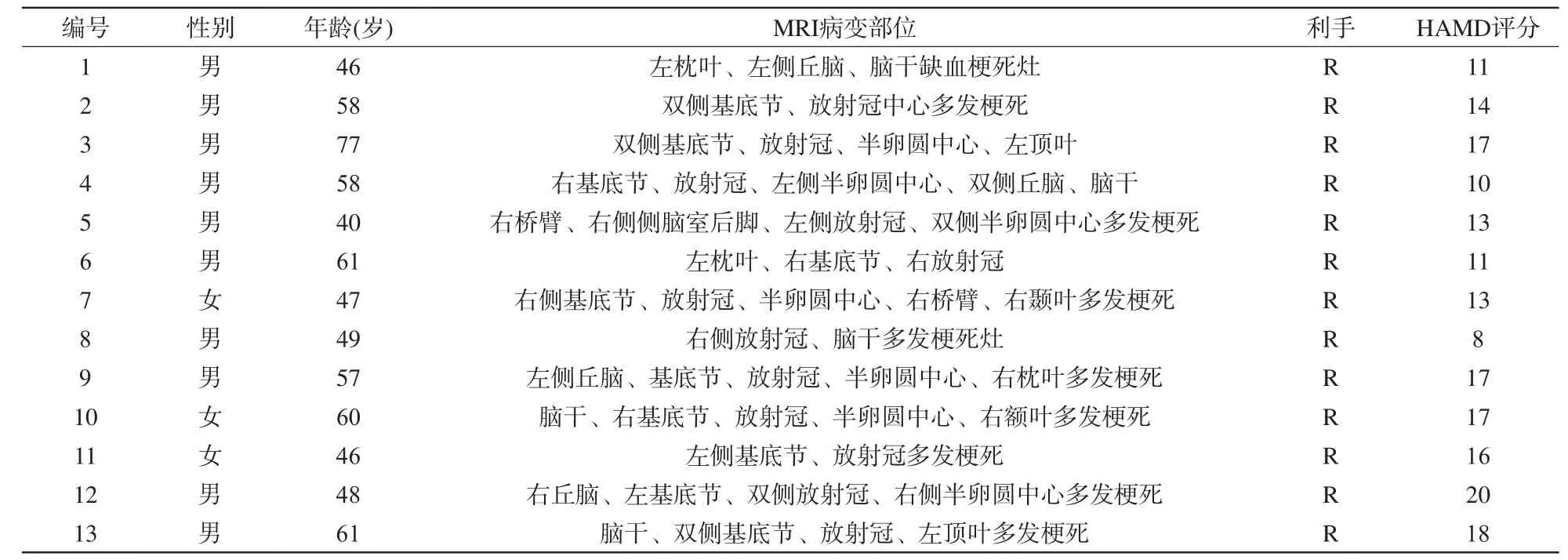
根据诊断和分类标准,选取2009年12月至2010年7月在北京天坛医院神经内科门诊就诊的发病在两周内的13例PSD患者。所有患者均为右利手,母语为汉语,其中男10例,女3例,平均年龄(54.0±9.7)岁,由两名神经内科医生依据美国精神疾病诊断与统计手册第4版(Diagnostic and Statistical Manual of Mental Disorders,fourth edition,DSM-IV)诊断为抑郁。汉密尔顿抑郁量表(hamilton depression scale,HAMD)评分>7分,且为抑郁首次发作。排除脑出血、在试验过程中再发卒中、酒精和药物成瘾,以及其他神经系统疾病和精神障碍或有MRI检查禁忌证的患者。所有病例均经两位工作5年以上的影像科医师对MRI片诊断为亚急性缺血性脑卒中。患者未接受抗抑郁治疗。患者信息见表1。
对照组13名健康志愿者,其中男8例,女5例,平均年龄(58.0±12.2)岁,均为右利手,母语为汉语,头颅MRI均显示正常,HAMD评分<7分,无癫痫、无神经精神性疾病、无脑血管异常、无幽闭恐惧症及外伤史。PSD患者和正常对照组基本信息见表2。
所有被试试验前均熟悉试验内容和要求,以取得良好的配合,均签署知情同意书。
1.2 研究方法
1.2.1 磁共振扫描及参数
本研究使用西门子3.0 T Trio磁共振扫描仪,标准正交头颅线圈。rs-fMRI序列扫描参数为:TR=2000 ms,TE=30 ms,Flip angle=90°,slices=31,resolution=64×64。然后用T1-MPRAG序列行176层覆盖全脑扫描,得到全脑高分辨率的解剖定位依据,以进行三维重建及空间配准,扫描参数为:TR=1900 ms,TE=2.13 ms,TI=900 ms,Flip angle=9°,resolution=256×256。整个扫描过程中,以枕部及双颞部海绵垫固定头部位置,限制头部运动,并要求受试者尽量保持头部及其他部位不动,减少头动伪影,以确保功能像与结构像的吻合。用耳塞降低噪音,嘱其放松、睁眼、平静呼吸,同时尽量不要进行思维活动。所有受试者均完成了试验要求,头动范围小于一个体素大小。
1.2.2 数据处理
对rs-fMRI数据进行格式转换后行预处理。由于初期图像的不稳定性以及试验对象需要一个扫描适应的过程,笔者剔除了静息态扫描前10个时间点的图像数据,进行层间时间校正;然后进行头动校正,剔除头动在X、Y、Z轴的平动超过2 mm或旋转角超过2°的受试者数据;进行空间标准化,将图像配准到蒙特利尔神经病学研究所(Montreal Neurological Institute,MNI)空间;平滑(8 mm×8 mm×8 mm,FWHM)以提高图像的信噪比;去除数据线性漂移,0.01~0.08 Hz带通滤波提取低频振荡信号部分,消除生理噪声。所有步骤使用REST工具包(http://restfmri.net/forum/)完成。
1.2.3 感兴趣区(region of interest,ROI)选择
根据以往对抑郁症默认网络的研究,使用MRCRO软件(http://www.cabiatl.com/mricro/)从自动解剖标记(automated anatomical labeling,AAL)模板选择双侧楔前叶作为ROI[10]。使用ROI分析法,首先通过计算双侧前扣带回所有体素的平均时间序列作为参考时间序列,然后计算该参考时间序列和全脑的所有体素对应时间序列的相关系数,使用费舍尔变换{z=0.5Ln[(1+r )/(1-r)]}将相关系数值转换为z分数值。

表1 缺血性卒中后抑郁患者基本临床资料Tab.1 Clinical characteristic of the PSD patients

表2 PSD患者和正常对照组的基本信息Tab.2 Demographic characteristic of the samples

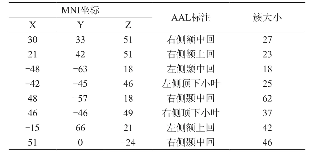
表3 与正常被试相比,PSD患者与右侧楔前叶功能连接减低的脑区Tab.3 Brain regions which showing decreased FCs with right precuneus between two groups

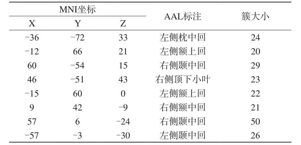
表4与正常被试相比,PSD患者与左侧楔前叶功能连接减低的脑区Tab.4Brain regions which showing decreased FCs with left precuneus between two groups
1.2.4 统计学分析
采用一般线性模型和高斯随机场理论进行统计分析,将绝对值化后的z值代入随机效应的单样本t检验,得到PSD患者及对照组静息状态下的功能连接值。PSD组与对照组间的功能连接值比较采用随机效应的独立样本t检验。统计分析采用SPSS 11.5统计软件包,P<0.05认为差异有统计学意义。
2 结果
2.1 PSD患者和正常对照组脑功能连接变化
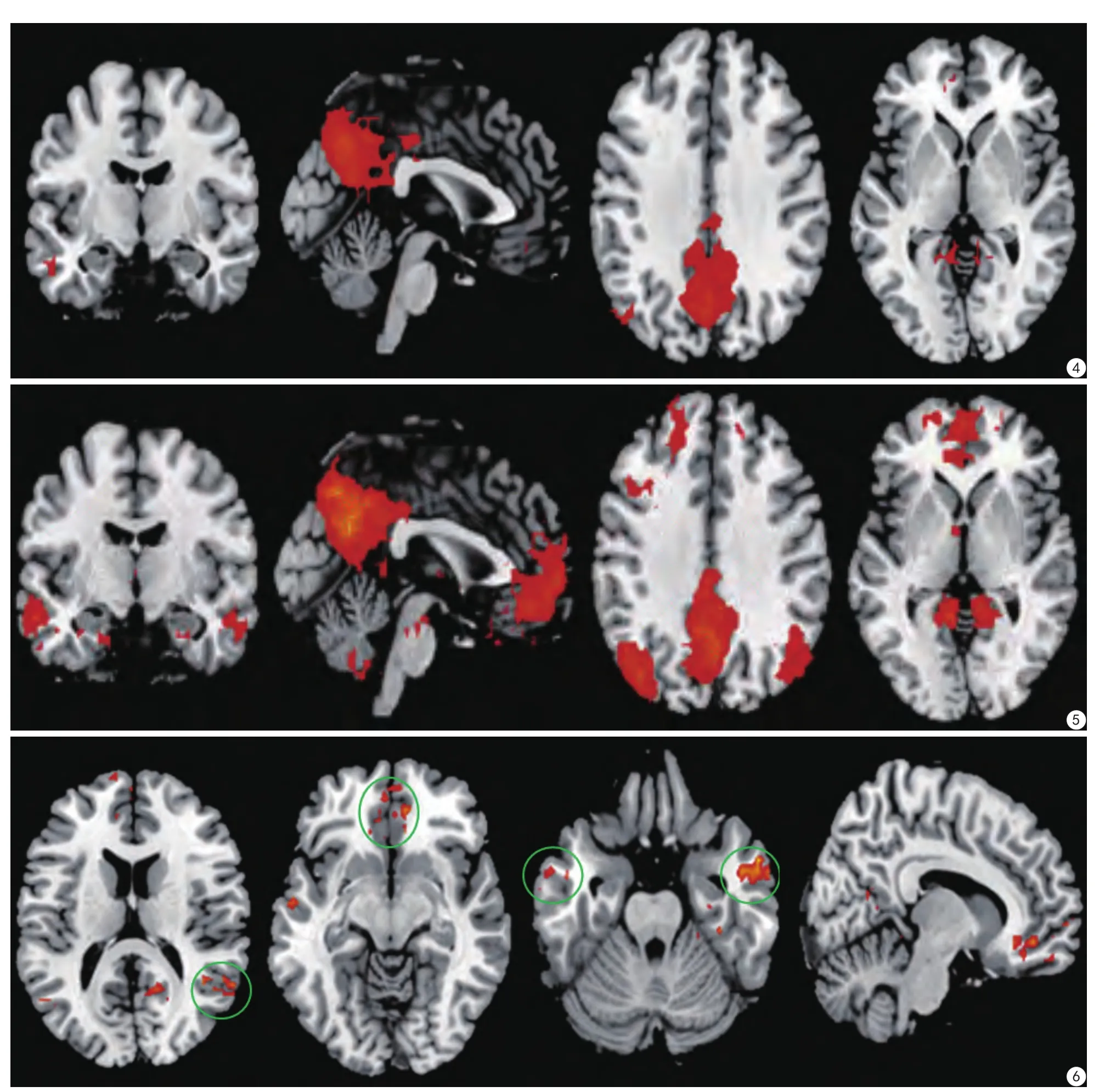
PSD患者组与正常对照组全脑功能连接的比较结果显示,PSD患者双侧顶下小叶、双侧额颞叶与右侧楔前叶功能连接减低(图1~3);右侧顶下小叶、双侧额叶、双侧颞叶与左侧楔前叶功能连接减低(图4~6),而未发现功能连接较对照组明显增高的脑区。计算结果具体数值如表3、4所示。
3 讨论
3.1 PSD与结构病变的相关性

图1 PSD患者全脑与右侧楔前叶存在显著功能连接的脑区 图2 正常被试全脑与右侧楔前叶存在显著功能连接的脑区 图3 与正常被试相比,PSD患者与右侧楔前叶功能连接减低脑区Fig. 1 Brain regions which showed significant functional connectivities (FCs) with the right precuneus in the PSD patients. Fig.2 Brain regions which showed significant functional connectivities (FCs) with the right precuneus in the healthy controls. Fig.3 Comparison of functional connectivities (FCs)with right precuneus between PSD patients and healthy controls.
以往对卒中后抑郁发病机制的研究主要集中在卒中病变自身的相关特性(位置、体积)方面,然而进一步的研究发现,这些特性与卒中后抑郁的发生并不存在明确相关性[5,11]。因此,尽管本试验中部分卒中病灶位于额颞叶等涉及默认网络的相关脑区,结构病变本身引发抑郁的可能性基本可以排除,同时进一步提示脑功能的异常对于卒中患者抑郁的发生起着更为决定性的作用。
3.2 PSD与边缘系统环路的相关性
本研究显示,PSD患者功能连接减弱的脑区与默认网络的部分脑区相吻合,主要集中在双侧额颞叶、双侧顶下小叶,同时这些脑区也是前额叶-边缘系统神经传导环路中重要的传导节点[12]。对抑郁症患者默认网络的研究发现,位于前额叶-边缘系统环路的额颞叶局部一致性(regional homogeneity,Reho)减低,导致该环路功能异常从而引发抑郁[13]。以往的研究也提出额叶-丘脑-边缘系统环路对情绪调控方面起重要作用,且与抑郁的发病机制有关[14]。另外,对帕金森患者伴发抑郁的研究发现,其抑郁和焦虑症状的发生可能与边缘系统的代谢异常有关[15]。因此本试验中,卒中后抑郁患者双侧额颞叶、双侧顶下小叶功能连接的改变可能导致前额叶-边缘系统神经传导环路功能的异常,从而进一步导致卒中后抑郁的发生。

图4 PSD患者全脑与左侧楔前叶存在显著功能连接的脑区 图5 正常对照组被试全脑与左侧楔前叶存在显著功能连接的脑区 图6 与正常被试相比,PSD患者与左侧楔前叶功能连接减低的脑区Fig.4 Brain regions which showed significant functional connectivities (FCs) with the left precuneus in the PSD patients. Fig.5 Brain regions which showed significant functional connectivities (FCs) with the left precuneus in the healthy controls. Fig.6 Comparison of functional connectivities (FCs)with left precuneus between PSD patients and healthy controls.
3.3 PSD与默认网络的相关性
额颞叶与人脑对内外环境信息的整合、情绪整合及情景记忆提取、控制人格和社会行为、感情和决策等功能有关[16],这也与默认网络的功能相一致[6];另有研究者提出抑郁的发生可能与默认网络异常导致患者自我意识障碍有关[17]。此外,对精神分裂症及焦虑症患者默认网络功能连接的研究也发现,功能连接减低的脑区多位于前额叶、顶下小叶[18-20]。Cullen等[21]发现青春期重度抑郁症患者右侧额中回、左侧额上回、额中回、左侧颞上回功能连接减低。Zhou等[22]报道,左侧前额叶背外侧和右侧额上回、额中回之间的功能连接与抑郁评分相关。最近对抑郁患者默认网络的研究还发现,与正常被试相比,抑郁患者左侧额中回、左侧顶下小叶Reho存在异常[13]。有研究者发现卒中后10 d内发生抑郁的PSD患者,抑郁严重程度与颞中回及前扣带回功能连接减低呈相关性[11]。本试验结果与以往的研究存在一定一致性,提示卒中后抑郁的发生可能是由于PSD患者默认网络相关脑区功能连接异常导致其认知能力降低,进而出现抑郁症状。且由于本试验选取的是发病在两周内的亚急性缺血性卒中患者,说明这些异常的功能连接在疾病早期即已存在,进一步证实卒中后抑郁的发生可能与默认网络的异常有关,而非药物治疗或疾病慢性化的神经毒性作用后果。
此外,有研究者提出,静息状态下抑郁患者前扣带回与丘脑、杏仁体及苍白球之间的功能连接减低[23]。之后,Greicius等[24]在对抑郁患者默认网络的研究中也提出,其膝下扣带回和丘脑功能连接增加。Bluhm等[25]对早期抑郁患者默认网络的研究发现,后扣带回与双侧尾状核功能连接减低。本研究结果与上述研究结果有所不同,原因一方面可能由于以往所研究的抑郁症患者一般不合并其他器质性病变,另一方面可能在于所选取的种子区不同。
3.4 本研究的局限性及改进
首先,静息态功能磁共振成像技术要求被试处于清醒、无思维活动的静息状态。很难保证所有被试在扫描全程完全没有意识活动,或者仅出现同类别的意识活动。尽管在扫描结束后,工作人员都会请每位被试回忆扫描过程中是否出现自发的想法,并对回答是的被试予以剔除,但仍然无法完全排除意识活动的存在及相异的可能。然而,本试验扫描时间较短,患者相对较易配合,因此意识水平的差异对本试验的影响并不显著。此外,生理水平的噪声可能会干扰静息态功能磁共振的低频信号,比如心脏跳动和呼吸运动造成的波动,从而使功能连接失真。这样的问题只能随着技术水平的提高得以改善,比如扫描时配以心脏运动和呼吸节律的记录仪等。除此之外,本研究主要针对默认网络功能连接的变化,而尚未关注大脑的其他功能网络,而且本研究病例数较少。
今后在扩大样本量的基础上,增加缺血性卒中后无抑郁的病例对照组,还将进一步研究缺血性卒中后抑郁患者全脑功能连接的变化,并尝试从脑结构-功能相结合的角度探讨缺血性卒中后抑郁脑神经机制。
4 结论
缺血性卒中后抑郁患者与正常对照组全脑功能连接的比较结果显示,PSD患者双侧顶下小叶、双侧额颞叶与右侧楔前叶功能连接减低;右侧顶下小叶、双侧额叶颞叶与左侧楔前叶功能连接减低,而未发现功能连接较对照组明显增高的脑区。提示卒中后抑郁的发生可能与默认网络功能连接的变化存在相关性。今后随着病例数的增多,缺血性卒中后无抑郁对照的加入,相信会得到更明确的结论。
[References]
[1] Hackett ML, Yapa C, Parag V, et al. Frequency of depression after stroke: a systematic review of observational studies. Stroke, 2005,36(6): 1330-1340.
[2] Tang WK, Chen YK, Lu JY, et al. Cerebral microbleeds and symptom severity of post-stroke depression: a magnetic resonance imaging study. J Affect Disord, 2011, 129(1-3): 354-358.
[3] Nys GM, van Zandvoort MJ, van der Worp HB, et al. Early cognitive impairment predicts long-term depressive symptoms and quality of life after stroke. J Neurol Sci, 2006, 247(2): 149-156.
[4] Bhogal SK, Teasell R, Foley N, et al. Lesion location and poststroke depression: systematic review of the methodological limitations in the literature. Stroke, 2004, 35(3): 794-802.
[5] Santos M, Kovari E, Gold G, et al. The neuroanatomical model of post-stroke depression: towards a change of focus. J Neurol Sci,2009, 283(1-2): 158-162.
[6] Greicius MD, Krasnow B, Reiss AL, et al. Functional connectivity in the resting brain: a network analysis of the default mode hypothesis.Proc Natl Acad Sci U S A, 2003, 100(1): 253-258.
[7] Raichle ME, MacLeod AM, Snyder AZ, et al. A default mode of brain function. Proc Natl Acad Sci U S A, 2001, 98(2): 676-682.
[8] Ho TC, Connolly CG, Henje BE, et al. Emotion-dependent functional connectivity of the default mode network in adolescent depression.Biol Psychiatry, 2015, 78(9): 635-646.
[9] Wu D, Yuan Y, Bai F, et al. Abnormal functional connectivity of the default mode network in remitted late-onset depression. J Affect Disord, 2013, 147(1-3): 277-287.
[10] Sheline YI, Price JL, Yan Z, et al. Resting-state functional MRI in depression unmasks increased connectivity between networks via the dorsal nexus. Proc Natl Acad Sci U S A, 2010, 107(24): 11020-11025.
[11] Lassalle-Lagadec S, Sibon I, Dilharreguy B, et al. Subacute default mode network dysfunction in the prediction of post-stroke depressionseverity. Radiology, 2012, 264(1): 218-224.
[12] Nauta WJ. Neural associations of the frontal cortex. Acta Neurobiol Exp (Wars), 1972, 32(2): 125-140.
[13] Liu CH, Ma X, Li F, et al. Regional homogeneity within the default mode network in bipolar depression: a resting-state functional magnetic resonance imaging study. PLoS One, 2012, 7(11): e48181.
[14] Jaracz J, Rybakowski J. Studies of cerebral blood flow in metabolism in depression using positron emission tomography (PET). Psychiatr Pol, 2002, 36(4): 617-628.
[15] Remy P, Doder M, Lees A, et al. Depression in Parkinson's disease:loss of dopamine and noradrenaline innervation in the limbic system.Brain, 2005, 128(Pt 6): 1314-1322.
[16] Ballmaier M, Toga AW, Blanton RE, et al. Anterior cingulate, gyrus rectus, and orbitofrontal abnormalities in elderly depressed patients:an MRI-based parcellation of the prefrontal cortex. Am J Psychiatry,2004, 161(1): 99-108.
[17] Sheline YI, Barch DM, Price JL, et al. The default mode network and self-referential processes in depression. Proc Natl Acad Sci U S A,2009, 106(6): 1942-1947.
[18] Zhao XH, Wang PJ, Li CB, et al. Altered default mode network activity in patient with anxiety disorders: an fMRI study. Eur J Radiol, 2007, 63(3): 373-378.
[19] Littow H, Huossa V, Karjalainen S, et al. Aberrant functional connectivity in the default mode and central executive networks in subjects with schizophrenia- a whole-brain resting-state ICA study.Front Psychiatry, 2015, 6: 26.
[20] Karbasforoushan H, Woodward ND. Resting-state networks in schizophrenia. Curr Top Med Chem, 2012, 12(21): 2404-2414.
[21] Drevets WC, Price JL, Furey ML. Brain structural and functional abnormalities in mood disorders: implications for neurocircuitry models of depression. Brain Struct Funct, 2008, 213(1-2):93-118.
[22] Zhou Y, Yu C, Zheng H, et al. Increased neural resources recruitment in the intrinsic organization in major depression. J Affect Disord,2010, 121(3): 220-230.
[23] Anand A, Li Y, Wang Y, et al. Activity and connectivity of brain mood regulating circuit in depression: a functional magnetic resonance study. Biol Psychiatry, 2005, 57(10): 1079-1088.
[24] Greicius MD, Flores BH, Menon V, et al. Resting-state functional connectivity in major depression: abnormally increased contributions from subgenual cingulate cortex and thalamus. Biol Psychiatry, 2007,62(5): 429-437.
[25] Bluhm R, Williamson P, Lanius R, et al. Resting state defaultmode network connectivity in early depression using a seed regionof-interest analysis: decreased connectivity with caudate nucleus.Psychiatry Clin Neurosci, 2009, 63(6): 754-761.
Resting-state functional magnetic resonance imaging study of ischemic post stroke depression
ZHANG Pei-yao1, XU Qin2, WANG Jun3, WANG Jing4, DAI Jian-ping5*1Department of Radiology, China-Japan Friendship Hospital, Beijing 100029, China
2Department of Medical Imaging, the First Affiliated Hospital of Guangzhou University of Traditional Chinese Medicine, Guangzhou 510405, China
3State Key Laboratory of Cognitive Neuroscience and Learning &IDG/Mc Govern Institute for Brain Research, Beijing Normal University, Beijing 100875, China
4Department of Neurology, Beijing Tiantan Hospital, Capital Medical University,Beijing 100050, China
5Department of Neuroimaging, Beijing Tiantan Hospital, Capital Medical University,Beijing 100050, China
Objective:To investigate the altered functional connectivity (FC) of the default mode network (DMN) in patients with post ischemic stroke depression.Materials and Methods:Thirteen PSD patients and 13 matched normal controls were recruited and the resting-state fMRI images were acquired on a 3.0 T Siemens MRI scanner. The datasets were analyzed using SPM8 and REST. The bilateral precuneus were selected as regions of interest (ROIs), and functional connectivity was calculated and compared between PSD patients and normal controls.Results:The FCs of the DMN was altered in PSD patients compared to normal controls. The brain areas which showed decreased FCs including the bilateral inferior parietal lobule, bilateral frontal lobule and bilateral temporal lobule. However, the increased FC has not been found in the current study.Conclusion:Dysfunction of the DMN may be associated with the development of PSD.
Stroke; Depression; Default mode network; Functional connectivity;Magnetic resonance imaging, functional
27 Feb 2017, Accepted 15 Apr 2017
作者单位:
1.中日友好医院放射科,北京 100029
2.广州中医药大学第一附属医院影像科,广州 510405
3.北京师范大学认知神经科学与学习国家重点实验室,北京 100875
4.首都医科大学附属北京天坛医院神经内科,北京 100050
5.首都医科大学附属北京天坛医院神经影像中心,北京 100050
戴建平,E-mail:djpbeijing@hotmail.com
2017-02-27
接受日期:2017-04-15
R445.2;R743.3
A
10.12015/issn.1674-8034.2017.06.001
张佩瑶, 徐琴, 王君, 等. 缺血性卒中后抑郁患者静息状态下默认网络功能连接的研究. 磁共振成像, 2017, 8(6):401-407.
*Correspondence to: Dai JP, E-mail: djpbeijing@hotmail.com

