信号传导与转录激活因子3在乳腺癌中的表达及其与人基质金属蛋白酶2、9的关系研究
胡慧 韦伟 易辛 冯瑰丽
·论著·
信号传导与转录激活因子3在乳腺癌中的表达及其与人基质金属蛋白酶2、9的关系研究
胡慧 韦伟 易辛 冯瑰丽
目的 观察乳腺癌组织中信号传导与转录激活因子3(signal transducer and activator of transcription 3,STAT3)的表达,分析其与患者临床特征的关系。方法 应用免疫组织化学法(EnVision)检测23例乳腺癌患者癌组织中STAT3、白细胞介素-6(interleukin,IL-6)、人基质金属蛋白酶(matrix metalloproteinase,MMP)-2和MMP-9的表达情况,并分析其与患者临床病理特征的关系。结果 浸润性癌组织中STAT3表达高于非浸润性癌。23例乳腺癌组织中IL-6表达阳性11例,阳性率47.83%;MMP-2阳性10例,阳性率43.48%;MMP-9阳性9例,阳性率39.13%;STAT3阳性表达与浸润型乳腺癌有统计学差异(χ2=7.738,P<0.05),两者呈正相关(r=0.161,P<0.05);STAT3阳性表达与IL-6阳性表达无相关性(r=0.032,P>0.05),与MMP-9(r=0.537,P<0.05)和MMP-2(r=0.649,P<0.05)阳性表达呈正相关。结论 STAT3与乳腺癌的浸润侵袭有关,其可能是通过调节MMP-2和MMP-9的表达控制乳腺癌的侵袭和转移。
乳腺癌; 信号传导与转录激活因子3; 侵袭; 基质金属蛋白酶-9; 基质金属蛋白酶-2
乳腺癌是女性最常见的恶性肿瘤,其发病率均居女性癌症第1位,手术、放化疗及靶向治疗与内分泌治疗是目前临床最有效的治疗手段[1-2],但仍有近17.0%的乳腺癌患者死于乳腺癌转移[3-4]。肿瘤干细胞假说认为,在肿瘤组织中存在一小部分具有自我更新和多向分化能力的细胞,并且生长不受控制。术后残留的静止期癌细胞可能为肿瘤干细胞,成了复发和转移的根源。研究表明,乳腺癌干细胞自我更新和增殖与信号转导和信号传导与转录激活因子3(STAT3)有密切关系[5]。研究发现,细胞外基质的破坏在肿瘤浸润和转移中扮演重要角色,而基质金属蛋白酶类(matrix metalloproteinases,MMPs)是参与破坏细胞外基质的主要蛋白酶类,特别是MMP-2和MMP-9,与肿瘤的浸润和转移关系密切[6-7]。我们对23例乳腺癌患者肿瘤组织中STAT3的表达进行检测,分析在乳腺癌中STAT3的表达与患者临床特征的相关性。
对象与方法
一、对象
2015年8月~2016年8月,我院收治的新发乳腺癌且有完整临床及病理资料的患者23例,均为女性,绝经后13例,年龄30~71岁,平均年龄(54.48±4.74)岁。其中T1 9例,T2 11例,T3 3例。N0 6例,N1 1例,N2 5例,N3 11例。分子分型:HER-2过表达型2例,Luminal A型4例,Luminal B型13例,三阴性型 3例。浸润性乳腺癌20例,非浸润性乳腺癌3例。标本采集符合北京大学深圳医院伦理委员会规定及赫尔辛基宣言。
二、方法
1.实验仪器与试剂:兔抗人单克隆抗体IL-6、STAT3、MMP-2和MMP-9均购自Proteintech(芝加哥,美国),抗兔/鼠通用型免疫组合试剂盒(REAL TMEnVision+/HRP RABBIT/MOUSE,Dako,丹麦)山羊血清、DAB显色液、苏木精均购自康为世纪(北京,中国)。
2.免疫组织化学分析乳腺癌组织中相关蛋白表达情况:组织4%多聚甲醛固定过夜,石蜡包埋,切片,常规脱蜡水化,抗原修复,正常山羊血清封闭20分钟后孵育IL-6(1∶200)、STAT3(1∶200)、MMP-2(1∶200)、MMP-9(1∶200)一抗过夜,PBS洗3次,每次5分钟,37度孵育二抗30分钟,PBS洗3次,每次5分钟,DAB显色5到10分钟,PBS洗10分钟,苏木精复染,盐酸酒精分化,自来水冲洗10分钟,脱水,透明,封片,镜检。
3.免疫组化染色阳性结果判断:IL-6、STAT3、MMP-2、MMP-9蛋白检测以细胞浆出现棕黄色颗粒为阳性,根据每张切片细胞的阳性强度按无着色、淡黄色、棕黄色、棕褐色分别打分0,1,2,3分;根据着色面积按无着色、着色<1/3、着色1/3~2/3、着色>2/3分别评0,1,2,3分。最终根据两项总分判定结果:<3分为阴性,≥3分为阳性。
三、统计学分析

结 果
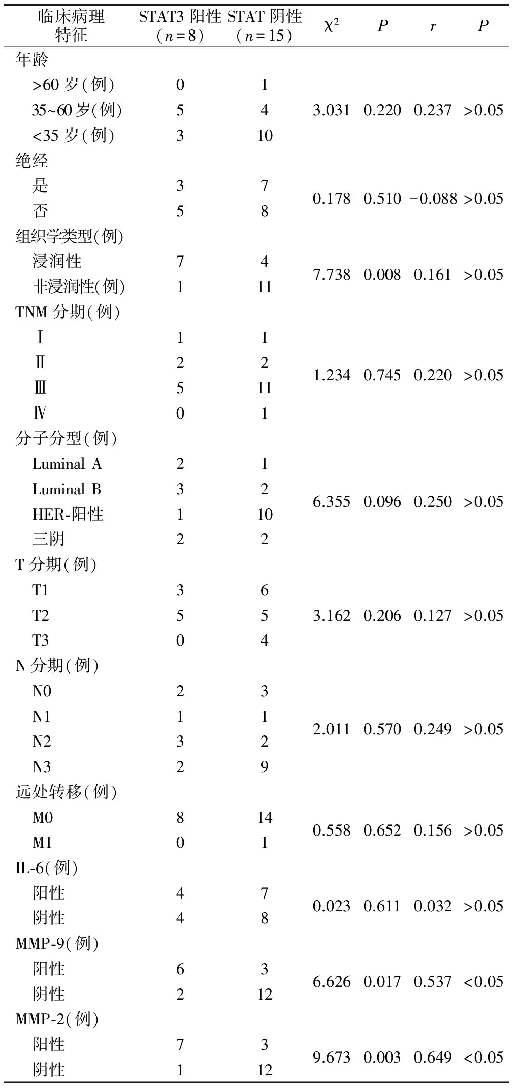
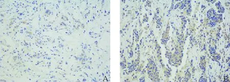
1.STAT3蛋白表达及患者临床病理特征分析:23例乳腺癌组织中STAT3阳性表达8例,其中浸润性癌7例,非浸润性癌1例,阳性率34.78%(图1)。非浸润性癌STAT3阳性表达与浸润型乳腺癌比较,差异有统计学意义(χ2=7.738,P<0.05),Spearman相关性分析显示,两者呈正相关(r=0.161,P<0.05)。与患者年龄、绝经与否、乳腺癌分期、乳腺癌分型、肿瘤大小、淋巴结转移和远处转移等比较均无统计学差异,亦无显著相关性(表1)。

表1 STAT3表达与患者临床特征的关系
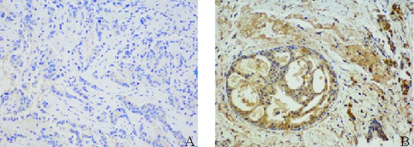
2.STAT3表达与IL-6、MMP-9、MMP-2表达相关性分析:23例乳腺癌患者肿瘤组织中,IL-6表达阳性11例,阳性率47.83%;MMP-2阳性10例,阳性率43.48%;MMP-9阳性9例,阳性率39.13%(图2、3、4)。使用Spearman相关系数进一步分析STAT3与IL-6、MMP-9、MMP-2的相关性,结果显示,STAT3阳性表达与IL-6阳性表达无显著相关性(r=0.032,P>0.05),与MMP-9(r=0.537,P<0.05)和MMP-2(r=0.649,P<0.05)阳性表达呈显著正相关(表1)。

A:STAT3表达阴性,细胞浆淡黄色,着色面积<1/3(× 200倍,DAB染色);B:STAT3表达阳性:细胞浆棕褐色,着色面积>2/3(× 200倍,DAB染色)图1 STAT3在乳腺癌组织中表达

A:IL-6表达阴性,细胞浆淡黄色,着色面积<1/3(× 200倍,DAB染色);B:IL-6表达阳性:细胞浆棕褐色,着色面积>2/3(× 200倍,DAB染色)图2 IL-6在乳腺癌组织中表达

A:MMP-2表达阴性,细胞浆淡黄色,着色面积<1/3(× 200倍,DAB染色)B:MMP-2表达阳性:细胞浆棕褐色,着色面积>2/3(× 200倍,DAB染色)图3 MMP-2在乳腺癌组织中表达

A:MMP-9表达阴性,细胞浆淡黄色,着色面积<1/3(× 200倍,DAB染色)B:MMP-9表达阳性:细胞浆棕褐色,着色面积>2/3(× 200倍,DAB染色)图4 MMP-9在乳腺癌组织中表达
STAT是一种转录因子家族的蛋白质,在肿瘤的发生发展中扮演着重要角色,特别是STAT3的异常激活在癌症发生机制中处于核心位置[8]。在正常细胞中,STAT3的激活在时间和程度上均受到严格控制。但是在许多肿瘤细胞中,STAT3被持续激活,导致靶基因连续表达,促进肿瘤细胞增殖、存活、侵袭和血管生成[9]。在恶性肿瘤中,驱动STAT3活化的主要驱动因子是IL-6,它可通过一种复杂的IL-6受体ɑ和gp130激活两面激酶(Janus kinases,JAK),最终磷酸化STAT3[10]。IL-6驱动STAT3活化的重要性已在多种肿瘤中被验证,包括乳腺癌、肺癌、肝癌、前列腺癌、胰腺癌、结肠癌、黑色素瘤等[11]。有报道认为,在三阴型乳腺癌中通过抑制IL-6,能进一步抑制STAT3而达到抑制肿瘤的目的;而在HER-2阳性乳腺癌中则有可能通过IL-6的高表达激活STAT3通路而导致肿瘤对抗HER-2治疗耐药[12-13]。我们利用免疫组化的方法检测23例乳腺癌患者癌组织中IL-6和STAT3的阳性表达,IL-6阳性率为47.83%,STAT3阳性率为34.78%。杨洁等[14]采用同样方法检测了126例乳腺癌患者组织中STAT3的表达,阳性率达到82.5%。杨增等[15]检测了36例乳腺癌组织中STAT3的表达,阳性率为58.3%。齐凤杰等[16]的研究阳性率为65%。杨林等[17]通过检测三阴性乳腺癌组织中IL-6的表达,发现阳性率达72.6%。本研究中三阴型乳腺癌例数少,HER-2阳性例数多,因此,是否本研究样本量少,以及由于肿瘤异质性导致在不同分子分型和不同生物学类型的乳腺癌中表达有差异还有待研究。
本研究发现,STAT3阳性表达与浸润型乳腺癌有正相关性,说明STAT3的高表达与乳腺癌的浸润侵袭有着密切的关系。为了进一步探究STAT3与乳腺癌侵袭转移的关系,我们利用免疫组化观察了乳腺癌组织中MMPs的表达。MMPs是一种锌依赖的肽链内切酶家族,在各种组织细胞外环境中均有表达[18]。研究发现,MMPs不仅在正常生理过程中发挥着重要作用,同时也与肿瘤的侵袭和转移有着极其密切的关系[19-20]。肿瘤的侵袭和转移过程复杂,其中就包括细胞外基质(extracellular matrix,ECM)的异常破裂[21]。而MMPs及其抑制剂,如TIMPs和膜相关MMP抑制剂(RECK)是ECM降解的重要调节因子[22]。MMP-9和MMP-2作为IV胶原酶,能够破坏基底膜,特别是降解IV胶原,从而加速肿瘤在体外的侵袭。在乳腺癌中,MMP-9循环水平显著升高,MMP-9和/或MMP-2的释放在乳腺肿瘤的侵袭和转移中扮演着重要的角色[23]。在本研究中的23例乳腺癌组织中MMP-2阳性率为43.48%,MMP-9阳性率为39.13%,均与STAT3阳性表达呈正相关,提示STAT3可能是通过调节MMP-2和MMP-9的表达控制乳腺癌转移侵袭的。有研究显示,在结肠癌中,IL-6/STAT3/Fra-1信号通路能够通过调节MMP-2和MMP-9等上皮-间质转化(epithelial-mesenchymal transition,EMT)促进因子的表达,促进EMT和结肠癌侵袭[24]。祝立和等[25]通过研究乳腺浸润性导管癌组织中STAT3与MMP-2和MMP-9表达的相关性,发现其呈显著正相关,并认为STAT3基因可能通过调节MMP-2、MMP-9 的表达介导 EMT 从而促进乳腺浸润性导管癌的侵袭转移。
我们采用免疫组化检测了23例乳腺癌患者肿瘤组织中STAT3、IL-6、MMP-2和MMP-9的表达,分析STAT3表达与患者临床病理特征的关系,结果表明,STAT3与乳腺癌的浸润、侵袭有关,其可能通过调节MMP-2和MMP-9的表达实现控制乳腺癌的侵袭和转移。但由于本研究样本量少,具体机制还有待进一步阐明。
[1] DeSantis CE,Fedewa SA,Goding Sauer A,et al.Breast cancer statistics,2015:Convergence of incidence rates between black and white women[J].CA Cancer J Clin,2016,66(1):31-42.
[2] 殷茜,张汝超,杨筱嵬,等.中电导钙激活钾离子通道在乳腺癌中的表达及其与临床病理指标的关系[J].临床外科杂志,2016,24(9):666-668.
[3] 李会,李兴睿.乳腺癌靶向治疗进展及趋势[J].临床外科杂志,2012,9(24):656-659.
[4] Polvak K,Weinberg RA.Transitions between epithelial and mesenchymal states:acquisition of malignant and stem cell traits[J].Nat Rev Cancer,2009,9(4):265 - 273.
[5] Su F,Ren F,Rong Y,et al.Protein tyrosine phosphatase Meg2 dephosphorylates signal transducer and activator of transcription 3 and suppresses tumor growth in breast cancer[J].Breast Cancer Res,2012,14(2):R38.
[6] 李靖若,李盂圈,鲍俊涛.乳腺癌患者肿瘤转移相关基因和基质金属蛋白酶9的表达及意义[J].中华医学杂志,2008,88(32):2278-2280.
[7] Li HC,Cao DC,Liu Y.Prognostic value of matrix metalloproteinases(MMP-2 and MMP-9)in patients with lymph node-negative breast carcinoma[J].Breast Cancer Res Treat,2004,88(1):75-85.
[8] Frank DA.STAT3 as a central mediator of neoplastic cellular transformation[J].Cancer Lett,2007,251(2):199-210.
[9] Rodriguez-Barrueco R,Yu J,Saucedo-Cuevas LP,et al.Inhibition of the autocrine IL-6-JAK2-STAT3-calprotectin axis as targeted therapy for HR-/HER2+breast cancers[J].Genes Dev,2015,29(15):1631-1648.
[10]Sansone P,Bromberg J.Targeting the interleukin-6/Jak/stat pathway in human malignancies[J].J clin oncol,2012,30:1005-1014.
[11]Hedvat M,Huszar D,Herrmann A,et al.The JAK2 inhibitor AZD1480 potently blocks Stat3 signaling and oncogenesis in solid tumors[J].Cancer cell,2009,16:487-497.
[12]Liang S,Chen Z,Jiang G,et al.Activation of GPER suppresses migration and angiogenesis of triple negative breast cancer via inhibition of NF-κB/IL-6 signals[J].Cancer Lett,2017,1(386):12-23.
[13]Huang WC,Hung CM,Wei CT,et al.Interleukin-6 expression contributes to lapatinib resistance through maintenance of stemness property in HER2-positive breast cancer cells[J].Oncotarget,2016,7(38):62352-62363.
[14]杨洁,叶双梅,蒋学锋,等.STAT3与Enolase-1在乳腺癌组织中的表达及意义[J].肿瘤防治研究,2012,39(12):1451-1455.
[15]杨增,严丽,蔡建辉.TAT3与pSTAT3在乳腺癌中的表达及其临床意义[J].中国肿瘤临床与康复,2011,18(1):24-27.
[16]齐凤杰,赵树鹏,朱培.TAT3和CyclinD1蛋白在乳腺癌中的表达及意义[J].山东医药,2010,50(52):106-108.
[17]杨林,韩思奇.三阴性乳腺癌中SOCS3和IL-6表达的相关性及临床意义[J].华中科技大学学报,2015,44(1):82-86.
[18]Back M,Ketelhuth DF,Agewall S.Matrix metalloproteinases in athero-thrombosis[J].Prog Cardiovasc Dis,2010,52(5):410-428.
[19]Chen PS,Shih YW,Huang HC,et al.Diosgenin,a steroidal saponin,inhibits migration and invasion of human prostate cancer PC-3 cells by reducing matrix metalloproteinases expression[J].PLoS One,2011,6(5):e20164.
[20]Gonzalez-Arriaga P,Pascual T,Garcia-Alvarez A,et al.Genetic polymorphisms in MMP 2,9 and 3 genes modify lung cancer risk and survival[J].BMC Cancer,2012,28(12):121.
[21]Chambers AF.The metastatic process:basic research and clinical implications[J].Oncol Res,1999,11(4):161-168.
[22]Radisky ES,Radisky DC.Matrix metalloproteinase-induced epithelial- mesenchymal transition in breast cancer[J].J Mammary Gland Biol Neoplasia,2010,15(2):201-212.
[23]Lu P,Takai K,Weaver VM,et al.Extracellular matrix degradation and remodeling in development and disease[J].Cold Spring Harb Perspect Biol,2011,3(12):pii:a005058.
[24]Liu H,Ren G,Wang T,et al.Aberrantly expressed Fra-1 by IL-6/STAT3 transactivation promotes colorectal cancer aggressiveness through epithelial- mesenchymal transition[J].Carcinogenesis,2015,36(4):459-468.
[25]祝立和,卢洪胜,周凯敏,等.STAT3基因与MMP-2、MMP-9在乳腺浸润性导管癌中的表达及其相互关系[J].临床研究,2012,50(29):58-64.
(本文编辑:杨泽平)
欢迎赐稿 欢迎订阅
邮发代号:38-184
Relationship research between expression of STAT3 in breast cancer with MMP-2/9
HUHui,WEIWei,YIXin,etal.
(DepartmentofBreastandThyroidSurgery,PekingUniversityShenzhenHospital,Shenzhen518036,China)
Objective To observe the expression of STAT3(signal transducer and activator of transcription 3,STAT3)in breast cancer and explore the relationship between the expression and clinical features.Methods The expression of STAT3,IL-6(interleukin,IL-6),MMP(matrix metalloproteinase,MMP)-2 and MMP-9 in 23 cases of breast cancer were observed by immunohistochemistry(EnVision)and positive rates were calculated.Chi Square and Spearman correlation coefficients were used to further analyze the correlation between STAT3 expression and clinical pathological features.Results The expression of STAT3 in invasive carcinoma was higher than that in non-invasive carcinoma.11 cases of IL-6,10 cases of MMP-2,9 cases of MMP-9 were positive in 23 cases of breast cancer,positive rate were 47.83%,43.48% and 39.13% respectively.There was a significant difference between STAT3 positive expression and invasive breast cancer(χ2=7.738,P<0.05),and there was a significant positive correlation between them(r=0.161,P<0.05).There was no significant correlation between STAT3 positive expression and IL-6 positive expression(r=0.032,P>0.05),while significant correlations were with MMP-2(r=0.649,P<0.05)and MMP-9(r=0.537,P<0.05)respectively.Conclusion STAT3 is closely related to invasion of breast cancer,which may be to controll the invasion and metastasis of breast cancer by regulating the expression of MMP-2 and MMP-9.
breast cancer; STAT3; invasion; MMP-2; MMP-9
10.3969/j.issn.1005-6483.2017.07.012
广东省医学科学技术研究基金资助项目(A2015545)
518036 深圳,北京大学深圳医院乳腺甲状腺外科
韦伟,Email:rxwei1123@163.com
2017-03-14)

