体外诱导磁控胶囊内镜进入小肠的临床研究
叶玲,徐玫丽,谭攀,龙利民,王海琴,郭永红
(中南大学湘雅二医院 老年病科,湖南 长沙 410011)
体外诱导磁控胶囊内镜进入小肠的临床研究
叶玲,徐玫丽,谭攀,龙利民,王海琴,郭永红
(中南大学湘雅二医院 老年病科,湖南 长沙 410011)
目的 探讨体外诱导磁控胶囊内镜(MCE)进入小肠的方法及应用价值。方法 将40例接受MCE检查的患者随机分为两组:对照组在胃部检查结束后停止控制胶囊运动,使其随患者消化道蠕动进入小肠;研究组胃部检查结束后患者取右侧卧位,检查医生将胶囊移至幽门口,并调控磁球位置使其靠近患者右下腹,借助磁力诱导胶囊进入小肠。对比分析两组MCE胃部检查时间、胃部滞留时间、小肠通过时间及全小肠检查完成率的差异。结果 对照组平均胃部检查时间为(32.50±11.71)min,与研究组(31.75±9.12)min比较差异无统计学意义(P >0.05)。胃部检查结束后,对照组的胃部滞留时间为(40.60±21.43)min,全小肠检查完成率为40%;研究组胃部滞留时间为(13.55±9.62)min,全小肠检查完成率为75%,两组比较差异有统计学意义(P <0.05)。对照组小肠通过时间为(329.25±90.00)min,与研究组(342.00±89.80)min比较差异无统计学意义(P >0.05)。结论 体外调控磁球位置结合患者体位改变可显著缩短MCE胃部滞留时间,提高全小肠检查完成率。
磁控胶囊内镜;临床应用;体外诱导;全小肠检查完成率
胶囊内镜是检查小肠疾病的重要手段[1]。磁控胶囊内镜(magnetic capsule endoscopy,MCE)在第一代胶囊无线传输功能的基础上增加体外磁场控制功能[2],可同时对胃和小肠进行检查。MCE电池寿命有限,胃部检查结束后如果胶囊长时间滞留于胃部,小肠检查势必受到影响,从而降低该技术的诊断价值。本研究旨在探讨能否通过体外调控磁球位置及改变患者体位诱导胶囊通过幽门,从而提高全小肠检查完成率。
1 资料与方法
1.1 一般资料
2015年11 月-2016年8月本院共40例患者接受MCE检查。其中,8例患者表现为消化道出血(均因高龄、疾病等无法耐受普通胃肠镜检查),另32例患者表现为不同程度的腹痛、腹胀、腹泻等。将患者随机分为两组:对照组20例,其中男15例,女5例,平均年龄(58.46±20.25)岁;研究组20例,其中男13例,女7例,平均年龄(60.18±17.32)岁。两组性别比、年龄等一般情况比较差异无统计学意义(P >0.05)。MCE检查的排除标准如下[3]:绝对禁忌证:无手术条件或拒绝接受任何腹部手术者;相对禁忌证:①已知或怀疑胃肠道梗阻、狭窄及瘘管者;②心脏起搏器或其他电子仪器植入者;③吞咽障碍者;④孕妇。所有患者检查前均签署知情同意书。
1.2 仪器设备
MCE检查系统由上海安翰医疗技术有限公司和安翰光电技术(武汉)有限公司联合生产,由MCE控制系统、NaviCam磁控胶囊内镜、便携记录器、胶囊定位器及操作软件系统等5部分组成。胶囊控制系统包括检查床、磁球、平移旋转台及控制台4部分。通过控制台的左右控制杆,可以完成胶囊的水平、垂直旋转,亦可以完成胶囊的潜水、跳跃、悬浮及掉头等动作。磁控胶囊内镜长27.0 mm,直径11.8 mm,头端视角140°。
1.3 方法
受检者在检查前1天低渣饮食,检查前6 h服用复方聚乙二醇电解质散溶液(68.56 g/袋×2袋,溶于2 L温开水)清洁肠道,检查前2 h饮清水约1 000 ml冲洗胃内黏液及气泡,检查前30 min口服二甲硅油(规格为5.00∶0.30 g,溶于50 ml温开水),吞服胶囊前5 min饮500~750 ml清水充盈胃,穿戴便携记录器后向左侧卧于检查床。操作者通过体外磁场结合患者体位改变控制胶囊运动,进行全方位的胃黏膜观察。胃部检查结束后对照组停止控制胶囊,使其随受检者消化道蠕动自然进入小肠;研究组患者取右侧卧位,检查医生将胶囊移至幽门口,并调控磁球位置使其靠近患者右下腹,借助磁力诱导胶囊通过幽门。对照组在胃部检查结束后6 h进食少许清淡食物;研究组在确认胶囊进入小肠后4 h进食少许清淡食物。检查小肠期间受检者可在医院自由走动,但需避免剧烈运动及进入强磁场区域,以防图像信号受到干扰[3]。告知患者出现任何腹痛、呕吐等胃肠道不适时,及时告知医护人员。
1.4 观察指标
胃部检查时间定义为胶囊通过贲门至检查者结束胃黏膜观察的时间。胃部滞留时间定义为检查者结束胃黏膜观察至胶囊进入小肠的时间。小肠通过时间定义为胶囊进入小肠至通过回盲瓣进入大肠的时间。全小肠检查完成率是指完成全小肠检查的病例数除以全部病例数得到的百分率。
1.5 统计学方法
采用SPSS 18.0统计软件进行数据分析,计量资料以均数±标准差(±s)表示,采用独立样本t检验;计数资料用率表示,采用χ2检验。P <0.05为差异有统计学意义。
2 结果
2.1 两组受检者MCE胃部检查时间、胃部滞留时间及小肠通过时间对比
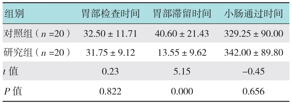
两组胃部检查时间、胃部滞留时间及小肠通过时间见附表。统计学分析显示,对照组平均胃部检查时间与研究组比较差异无统计学意义(P >0.05);胃部观察结束后,研究组胶囊胃部滞留时间明显短于对照组(P <0.05)。两组胶囊小肠通过时间比较差异无统计学意义(P >0.05)。
2.2 两组受检者全小肠检查完成率的比较
每组20例受检者中,对照组有8例完成全小肠检查,研究组有15例完成全小肠检查。对照组全小肠检查完成率为40%,研究组全小肠检查完成率为75%,两组比较差异有统计学意义(χ2=5.01,P =0.025,P <0.05)。
附表 两组MCE胃部检查时间、胃部滞留时间及小肠通过时间对比 (min,±s)Attached table Comparison of gastric inspection time,gastric residence time and small intestine transit time between the two groups (min,±s)

附表 两组MCE胃部检查时间、胃部滞留时间及小肠通过时间对比 (min,±s)Attached table Comparison of gastric inspection time,gastric residence time and small intestine transit time between the two groups (min,±s)
组别 胃部检查时间 胃部滞留时间 小肠通过时间对照组(n =20) 32.50±11.71 40.60±21.43 329.25±90.00研究组(n =20) 31.75±9.12 13.55±9.62 342.00±89.80 t值 0.23 5.15 -0.45 P值 0.822 0.000 0.656
3 讨论
胶囊内镜具有操作简便、无创安全和图像清晰等优点,已经成为小肠疾病的一线检查方法[1,4]。随着科技的进步,胶囊内镜得以不断完善,并且出现了一批新型胶囊内镜如食管胶囊内镜、结肠胶囊内镜等。2013年我国自主研制了NaviCam磁控胶囊内镜,通过磁控技术实现主动控制胶囊,弥补了传统胶囊内镜对胃部检查不足的缺陷。一项大样本、多中心、自身对照的临床研究证实MCE对胃部疾患的诊断准确率可与电子胃镜媲美[5]。与传统胶囊内镜一样,MCE也是检查小肠的重要手段。MCE能否在有限的电池寿命内完成全小肠检查对诊断小肠疾病极为重要。据报道,接受胶囊内镜检查的患者中,约20%无法完成全小肠检查[6-7]。胶囊本身重量及大小对全小肠检查完成率无明显影响[8]。既往腹部手术史、住院及胶囊胃通过时间(胶囊进入胃部至通过幽门的时间)过长等是胶囊内镜不能完成全小肠检查的危险因素[9-10]。其中,胶囊胃内滞留或胃排空延迟等导致胶囊胃部通过时间延长是最主要的影响因素[9,11]。因此,缩短胶囊内镜胃通过时间对于提高胶囊内镜的全小肠检查完成率具有重要的临床意义。
为缩短胶囊内镜胃通过时间,研究者们探索了许多干预措施,包括胃镜辅助下实施胶囊内镜检查[12-13]、应用促胃肠动力药物[14]以及改变患者体位等[15]。采用胃镜下推送胶囊进入小肠的方法直接、有效,但是该方法削弱了胶囊内镜检查简单、方便及无创的优势,难以被患者接受。促胃肠动力药物的应用虽然可缩短胶囊胃通过时间,但是胶囊内镜在小肠内通过时间也明显缩短,可能影响小肠病变的观察。吞服胶囊后让患者取右侧卧位能否缩短胃通过时间、提高全小肠检查完成率,目前尚存争议[16]。由于可通过体外磁场主动控制胶囊运动,本研究探讨体外诱导胶囊进入小肠的方法并评估其应用价值。既往研究中,胃通过时间是评估干预措施的重要指标。然而,MCE检查尚包括胃部检查,不同类型病变可能导致患者之间胃部检查时间不一致。为了减少该因素的影响,本研究将胃通过时间分为胃部检查时间和胃部滞留时间,并采用胃部滞留时间作为评估干预措施是否有效的指标之一。研究结果显示,在胃部检查结束后,对照组胶囊胃部滞留时间为(40.60±21.43)min,全小肠检查完成率为40%,而研究组胶囊胃部滞留时间为(13.55±9.62)min,全小肠检查完成率为75%,两组相比差异有统计学意义(P <0.05)。研究结果证实通过体外调控磁球位置及改变患者体位可有效诱导磁控胶囊进入小肠,显著缩短胶囊胃部滞留时间,提高全小肠检查完成率。目前国内外尚未见类似文献报道。
综上所述,MCE继承了传统胶囊内镜无创安全、图像清晰等优点,弥补了传统胶囊内镜对胃部观察不全面的缺陷。在胃部检查结束后通过体外调控磁球位置及改变患者体位可缩短胶囊胃部滞留时间,提高全小肠检查完成率,具有较好的临床应用价值。
[1] 王海红, 金鹏, 赵晓军, 等. 1415例胶囊内镜在消化道疾病诊断中的应用体会[J]. 中国内镜杂志, 2015, 21(2): 121-126.
[1] WANG H H, JIN P, ZHAO X J, et al. 1415 cases of gastrointestinal diseases diagnosis under capsule endoscopy[J]. China Journal of Endoscopy, 2015, 21(2): 121-126. Chinese
[2] 刘希双. 磁控胶囊内镜的临床应用[J]. 中华临床医师杂志: 电子版, 2016, 10(4): 461-463.
[2] LIU X S. Clinical application of magnetically guided capsule endoscopy[J]. Chinese Journal of Clinicians: Electronic Edition,2016, 10(4): 461-463. Chinese
[3] 中华医学会消化内镜学分会. 中国胶囊内镜临床应用指南[J].中国实用内科杂志, 2014, 34(10): 984-991.
[3] Chinese Medical Association of Digestive Endoscopy. Guideline for clinical application of capsule endoscopy in China[J]. Chinese Journal of Practical Internal Medicine, 2014, 34(10): 984-991.Chinese
[4] 王晓艳, 沈守荣, 肖定华, 等. 胶囊内镜在小肠疾病诊断中的应用[J]. 中南大学学报(医学版), 2010, 35(9): 995-999.
[4] WANG X Y, SHEN S R, XIAO D H, et al. Capsule endoscopy in the diagnosis of intestinal diseases[J]. Journal of Central South University (Med Sci), 2010, 35(9): 995-999. Chinese
[5] LIAO Z, HOU X, LIN-HU E Q, et al. Accuracy of magnetically controlled capsule endoscopy, compared with conventional gastroscopy, in detection of gastric diseases[J]. Clin Gastroentero lHepatol, 2016, 14(9): 1266-1273.
[6] LIAO Z, GAO R, XU C, et al. Indications and detection,completion, and retention rates of small-bowel capsule endoscopy:a systematic review[J]. Gastrointest Endosc, 2010, 71(2): 280-286.
[7] HÖÖG C M, BARK L A, ARKANI J, et al. Capsule retentions and incomplete capsule endoscopy examinations: an analysis of 2300 examinations[J]. Gastroenterol Res Pract, 2012, 2012: 518718.
[8] 高良庆, 韩泽龙, 陈振煜, 等. 两种不同重量及大小的胶囊内镜之间胃肠通过时间及全小肠检查完成率的对比[J]. 中国内镜杂志, 2016, 22(2): 1-6.
[8] GAO L Q, HAN Z L, CHEN Z Y, et al. Comparison of gastrointestinal transit time and completion rates of two kinds of capsule endoscopy with different size and weight[J]. China Journal of Endoscopy, 2016, 22(2): 1-6. Chinese
[9] WESTERHOF J, WEERSMA R K, KOORNSTRA J J. Risk factors for incomplete small-bowel capsule endoscopy[J]. Gastrointest Endosc, 2009, 69(1): 74-80.
[10] LEE M M, JACQUES A, LAM E, et al. Factors associated with incomplete small bowel capsule endoscopy studies[J]. World J Gastroenterol, 2010, 16(42): 5329-5333.
[11] RONDONOTTI E, HERRERIAS J M, PENNAZIO M, et al.Complications, limitations, and failures of capsule endoscopy: a review of 733 cases[J]. Gastrointest Endosc, 2005, 62(5): 712-716.
[12] ALMEIDA N, FIGUEIREDO P, LOPES S, et al. Capsule endoscopy assisted by traditional upper endoscopy[J]. Rev Esp Enferm Dig, 2008, 100(12): 758-763.
[13] 康艳, 陈星, 赵丹瑜, 等. 胃镜下经幽门将胶囊内镜送入小肠的应用研究[J]. 中华消化杂志, 2011, 31(10): 706-707.
[13] KANG Y, CHEN X, ZHAO D Y, et al. Study on pushing capsule endoscopy into small intestine through pylorus by gastroscopy[J].Chinese Journal of Digestion, 2011, 31(10): 706-707. Chinese
[14] 卢筱洪, 丁一娟, 罗和生. 术前口服伊托必利在胶囊内镜检查中的作用分析[J]. 中华消化内镜杂志, 2012, 29(3): 141-143.
[14] LU X H, DING Y J, LUO H S. Effects of itopride prior to capsule endoscopy[J]. Chinese Journal of Digestive Endoscopy, 2012,29(3): 141-143. Chinese
[15] 刘敏芝, 李运泽, 梁昌宇, 等. 右侧30°半卧位在胶囊内镜检查中的作用研究[J]. 中国内镜杂志, 2014, 20(7): 775-777.
[15] LIU M Z, LI Y Z, LIANG C Y, et al. Clinical value of right lateral position of 30° in capsule endoscopy inspection[J]. China Journal of Endoscopy, 2014, 20(7):775-777. Chinese
[16] APARICIO J R, MARTLNEZ J, CASELLAS J A. Right lateral position does not affect gastric transit times of video capsule endoscopy: a prospecfive study[J]. Gastrointest Endosc, 2009,69(1): 34-37.
(吴静 编辑)
Clinical study on extracorporal induction of magnetic capsule endoscopy into small intestine
Ling Ye, Mei-li Xu, Pan Tan, Li-min Long, Hai-qin Wang, Yong-hong Guo
(Department of Geriatrics, the Second Xiangya Hospital, Central South University,Changsha, Hunan 410011, China)
Objective To explore and evaluate an extracorporal method for inducing magnetic capsule endoscopy into small intestine. Methods 40 patients receiving magnetic capsule endoscopy were randomly divided in two groups: the control group: doctors stopped manipulating capsule after the examination of stomach, and the capsule entered small intestine by the natural gastrointestinal motility; and the study group: after the examination of stomach, the patient lay on the right side, doctors moved the capsule to the pylorus, and then moved magnetic ball to induce capsule into small intestine. Gastric inspection time, gastric residence time, small intestine transit time and the completion rate were compared between the two groups. Results The average time for checking stomach was (32.50 ± 11.71) min in control group and (31.75 ± 9.12) min in study group respectively, and the difference was not signifi cant (P > 0.05). After the observation of stomach, the gastric residence time in the control group was(40.60 ± 21.43) min, and the completion rate was 40%, while the average gastric residence time in the study group was (13.55 ± 9.62) min, and the completion rate was 75%. The difference between the two groups was statistically signifi cant (P < 0.05). Small intestine transit time was (329.25 ± 90.00) min in the control group and (342.00 ± 89.80)min in the study group, and the difference was not signifi cant (P > 0.05). Conclusion By doctors moving magnetic ball and the patient lying on the right side after the observation of stomach, gastric residence time could be reduced and the completion rate could be elevated obviously.
magnetic capsule endoscopy; clinical application; extracorporal induction; completion rate
R574.5
A
2016-10-31
郭永红,E-mail:guoyhong@sina.cn;Tel:0731-85295277
10.3969/j.issn.1007-1989.2017.06.006
1007-1989(2017)06-0026-04

