无菌和带菌松材线虫对赤松的致病性*
林 丽 周 蕾 潘 珺 康李鹏 叶建仁 朱丽华
(南方现代林业协同创新中心 南京林业大学林学院 南京 210037)
无菌和带菌松材线虫对赤松的致病性*
林 丽 周 蕾 潘 珺 康李鹏 叶建仁 朱丽华
(南方现代林业协同创新中心 南京林业大学林学院 南京 210037)
【目的】 探讨无菌和带菌松材线虫对赤松的致病性,明确松材线虫在松树萎蔫病中的地位,为致病机制研究及病害防控提供依据。【方法】 以赤松胚性愈伤组织为材料,每块组织接种无菌松材线虫250条,培养12天后,利用2,3,5-三苯基氯化四氮唑(TTC)染色法对愈伤组织进行染色,分析无菌松材线虫对赤松愈伤组织细胞活性的影响。以赤松组培苗和4年生盆栽实生苗为对象,每株接种无菌松材线虫和带菌松材线虫(组培苗250条,4年生苗3 000条),分别于18天和35天后统计2种类型线虫接种所致松苗萎蔫率,并对植株体内线虫进行再分离,分析2种类型松材线虫对赤松幼苗的致病能力。【结果】 无菌松材线虫接种导致赤松胚性愈伤组织严重水渍化,TTC染色为米黄色或淡粉色,证明愈伤组织已失去活性。无菌松材线虫和带菌松材线虫接种均能导致赤松组培苗萎蔫,接种18天后,萎蔫率分别为70%和60%; 萎蔫植株中均可再分离到松材线虫,其每株平均分别为599±567条和365+240条,二者差异显著(P<0.01)。剩余外观健康的赤松组培苗中也分离到线虫,但数量较少(仅10~20条)。2种类型松材线虫接种引起赤松盆栽苗同等程度的萎蔫,接种35天后的萎蔫率均达80%,无菌和带菌松材线虫致萎蔫植株中均可再分离到松材线虫,其每株平均分别为34 733±34 162条和25 057±21 410条,但差异不显著(P=0.508)。其余外观尚健康的松苗也进行线虫分离,2株接种无菌松材线虫的植株体内线虫分别为486条和22条; 接种带菌松材线虫的2株松苗中,1株分离到646条线虫,另1株没有分离到线虫。【结论】 无菌和带菌松材线虫均对赤松具有致病性,松材线虫是赤松萎蔫的直接原因,伴生细菌并非赤松萎蔫的必须因子。
松材线虫病; 松材线虫; 无菌; 致病性; 赤松
松材线虫病又称松树萎蔫病(pine wilt disease),是松树的一种毁灭性病害。该病涉及寄主松树、线虫、天牛、真菌、细菌及环境等多个方面(Rorizetal., 2011; Niuetal., 2012),迄今为止其致病机制尚未清楚,因此给该病害的防治工作带来极大困难(杨宝君, 2002; 谈家金等, 2003; 王敏敏等, 2006)。明确病原是进行病害防控的最基本问题,而松材线虫病的病原迄今尚存在较大争议。大多数学者认为松材线虫(Bursaphelenchusxylophilus)是该病害的唯一病原(杨宝君, 2002; 叶建仁等, 2012)。由于松材线虫种群大量增长前,感病寄主松树已在生理和组织上出现变化,Oku等(1979)推测松材线虫以外的因素参与病害发生发展过程。随后,Oku(1980)提出松树萎蔫与松材线虫携带细菌的代谢产物有关。Kawazu等(1997; 1999)经过一系列研究提出该病的真正病原是松材线虫携带的致病细菌。洪英娣等(2003)和Han等(2003)亦报道松苗萎蔫与线虫种类无关,而是由线虫伴生细菌的种类所决定。也有学者认为该病害是由松材线虫及其伴生细菌引起的复合侵染性病害(郭道森等, 2000; 赵博光, 2004; 陈守常, 2010)。为了弄清松材线虫在松树萎蔫过程中的地位,本研究以赤松(Pinusdensiflora)胚性愈伤组织、赤松组培苗及赤松4年生盆栽苗为试验材料,探讨无菌松材线虫的致病性。
1 材料与方法
1.1 供试材料
供试松材线虫为强毒松材线虫虫株AMA3的近交系后代AMA3c1(本文称带菌松材线虫),使用前采用单异活体法培养,即培养于灰葡萄孢(Botrytiscinerea)上,4 ℃保存。无菌松材线虫(代号为在带菌虫株前加BF,即BF AMA3c1)的制备及无菌检验参照朱丽华等(2011)的方法。
供试赤松胚性愈伤组织参照吴静等(2015)的方法培养,于增殖培养基中生长14天后备用。
供试赤松组培苗的培养参照朱丽华等(2010)的方法,将外植体切成0.5~0.8 cm大小茎段,接种于GD+2 mg·L-16-BA+0.5 mg·L-1NAA培养基上,25 ℃条件下培养5周后,将外植体连同已分化的丛生芽转移至添加0.5 mg·L-1活性炭的DCR培养基上生长,1个月后,将伸长的丛生芽单个切下,接种于DCR培养基上,每个月继代1次,连续6个月,备用。
赤松4年生盆栽实生苗培养于南京林业大学南大山温室,备用。
1.2 无菌松材线虫对赤松愈伤组织的影响及愈伤组织的活力测定
无菌条件下将无菌松材线虫悬浮液接种到生长良好的赤松愈伤组织上,每块组织接种100 μL(约含250条线虫),共3皿(每皿6块),接种等量无菌水为对照(1皿)。接种后,将其置于25 ℃培养箱中暗培养,7天后观察其形态变化。
接种12天后,参照刘华等(2001)的方法,利用2,3,5-三苯基氯化四氮唑(TTC)染色检测愈伤组织活力,具体方法稍加改动。取0.1 g愈伤组织放入离心管中,加入0.1 mol·L-1磷酸二氢钠(NaH2PO4)缓冲液(pH 7.0)和0.4%TTC溶液各0.5 mL,25 ℃暗培养,12 h后观察其染色情况。
1.3 松材线虫对赤松组培苗的致病力测定及线虫再分离
赤松组培苗的接种采用截顶法,具体参照朱丽华等(2011)的方法。从超净工作台中将组培苗取出,截去顶端后,将其接种于新鲜培养基中,再将无菌小棉球置于伤口上,随后将无菌松材线虫和带菌松材线虫分别接种到棉球上,每虫株接种赤松组培苗10株,接种量为每株250条,无菌水为对照。接种后,于28 ℃光照培养箱中培养,接种后第2天开始观察松苗萎蔫情况,持续观察18天,统计松苗枯萎率。枯萎率=枯萎数/接种数×100%。
接种18天后,将感病植株及健康植株取出,用剪刀将针叶和茎段剪成2~4 mm长短的小段,采用贝尔曼漏斗进行线虫的再分离,24 h后收集线虫至离心管中,离心去上清后保留1 mL,显微镜下统计线虫数量。其中,无菌线虫接种植株体内线虫的再分离于无菌条件下完成,并对所分离线虫进行无菌检测,以检验接种过程是否污染细菌,检验条件参照朱丽华等(2011)的方法。
1.4 松材线虫对温室赤松盆栽实生苗的致病力测定及线虫再分离
赤松4年生盆栽实生苗致病力测定采用皮接法(张治宇等, 2002),将无菌松材线虫和带菌松材线虫分别接种于伤口处的棉球上,每虫株接种赤松苗10株,接种量为每株3 000条,无菌水为对照。接种后,于28 ℃温室条件下培养,观察松苗发病情况,统计松苗枯萎率。
接种5周后,对所有植株分别进行线虫分离。将植株从基部剪断,针叶与枝条分开后,用剪刀将枝条剪成长度为5~6 mm小段,采用贝尔曼漏斗进行线虫分离,24 h后将线虫收集至离心管中,离心去上清后保留1 mL,显微镜下统计线虫数量。
1.5 数据统计与分析
处理数据采用SPSS软件进行统计和分析。
2 结果与分析
2.1 无菌松材线虫对赤松胚性愈伤组织的影响
赤松胚性愈伤组织接种无菌松材线虫(BF AMA3c1)后,未发现明显褐变现象,但水渍化现象严重。接种7天后,愈伤组织水渍化程度达100%(图1A),而接种无菌水的对照愈伤组织依然呈紧密的团块状,表面颗粒状,生长正常(图1B)。接种无菌松材线虫12天后的愈伤组织经TTC染色后呈现米黄色或淡粉色,表明愈伤组织已丧失活力或活力极弱(显色越淡,活力越弱); 而对照愈伤组织呈鲜艳的红色,表明其活力很强(图1C)。
2.2 松材线虫对赤松组培苗的致病性
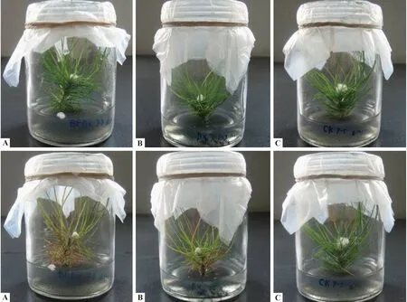
接种5天后,开始观察到针叶褪色、黄化,最后枯萎变成红褐色,无菌松材线虫和带菌松材线虫所接种赤松苗的萎蔫症状一致,18天后,接种无菌松材线虫及带菌松材线虫的赤松组培苗致萎率分别达70%和60%(表1),接种无菌水的赤松组培苗始终保持健康状态(图2)。
线虫再分离结果表明,无菌松材线虫和有菌松材线虫接种发病的赤松组培苗中均分离到线虫。其中无菌松材线虫接种萎蔫的每株松苗中线虫平均为599±567条,经牛肉膏蛋白胨液体培养基检验72 h后未发现有任何细菌生长; 带菌松材线虫接种致萎的每株松苗中线虫平均为365±240条(表1)。剩余外观健康的赤松组培苗中也分离到线虫,但数量较少,为10~20条。
2.3 松材线虫对赤松4年生盆栽实生苗的的致病性
温室接种试验表明,无菌松材线虫及带菌松材线虫均能使赤松4年生盆栽实生苗发病,接种5周后,2种类型线虫的致萎率均达80%(表2),且所致萎蔫症状一致,均为针叶先褪色、黄化,进而枯萎变成红褐色,而对照松苗针叶依然保持健康的绿色(图3)。

表1 赤松组培苗接种松材线虫的萎蔫率及再分离线虫数量(接种18天后)①Tab.1 Wilting ratios of tissue-cultured shoots of P. densiflora inoculated with aseptic and non-aseptic pine wood nematode and number of nematodes recovered(18 d after inoculation)
① 数值表示平均值±标准误,不同字母表示差异显著,t检验(P< 0.01)。Values represent the mean ± SD. Different letters are significantly different according tottest (P< 0.01). N代表此处未研究。 “N” means not done.下同。The same below.

表2 赤松4年生盆栽苗接种松材线虫的萎蔫率及再分离线虫数量(接种后35天)Tab.2 Wilting ratios of 4-year-old P. densiflora seedlings inoculated with aseptic and non-aseptic pine wood nematode and number of nematodes recovered(35 d after inoculation)

图1 无菌松材线虫对赤松胚性愈伤组织活力的影响Fig.1 Effect of aseptic pine wood nematode on the viability of the embryogenic calli of P. densifloraA. 接种无菌松材线虫的赤松胚性愈伤组织(7天后); B. 接种无菌水的赤松胚性愈伤组织(第7天); C. 接种无菌松材线虫12天后愈伤组织TTC染色结果(前3管为接种无菌松材线虫的愈伤组织,后3管为对照)。A. Embryogenic calli inoculated with aseptic pine wood nematode(7 d later); B. Embryogenic calli inoculated with non-aseptic pine wood nematode(7 d later); C. Embryogenic calli staining with TTC solution(the first three tubes were aseptic PWNs inoculation and the last three tubes were CK).

图2 接种无菌松材线虫和带菌松材线虫对赤松组培苗的影响Fig.2 Symptoms of tissue-cultrued shoots of P. densiflora after inoculated with aseptic and non-aseptic pine wood nematodes上排: 接种1天后。Up: 1 d after inoculation; 下排: 接种18天后。Down: 18 d after inoculation.A. 无菌松材线虫; B. 带菌松材线虫; C. 对照(无菌水)。A. Aseptic pine wood nematode; B. Non-aseptic pine wood nematode; C. CK(sterile water).

图3 接种无菌松材线虫、带菌松材线虫对赤松盆栽实生苗的影响Fig.3 Symptoms of potted seedlings of P.densiflora after inoculated with aseptic and non-aseptic pine wood nematodes
线虫再分离表明,2类线虫接种萎蔫的赤松苗体内均能再分离到松材线虫,其数量差异不显著,其中无菌线虫接种萎蔫植株平均每株线虫为34 733±34 162条,带菌线虫接种萎蔫植株平均每株线虫为25 057±21 410条(表2)。对其余外观尚健康的松苗也进行线虫分离,2株接种无菌松材线虫的健康植株体内线虫分别为486条和22条; 接种带菌松材线虫的2株松苗中,1株分离到646条线虫,1株没有分离到线虫。
3 讨论
松材线虫病是涉及多个因素的复杂病害,关于该病害的病原国际上存在较大争议。各国学者采用多种寄主材料对松材线虫及其携带细菌在松树萎蔫中的作用进行探讨。郭道森等(2001)和洪英娣等(2003)曾利用愈伤组织验证细菌与松材线虫病的关系,发现无菌松材线虫接种不造成愈伤组织褐变,而携带细菌的线虫可导致愈伤组织褐变,因此认为松材线虫所携带细菌在病害发生过程中起至关重要的作用。由于植物愈伤组织对培养基成分、激素种类及含量、pH值等要求比较严格,条件稍有变化,常容易引起褐变坏死,很难区别其褐变坏死是由于细菌分泌的毒素所致,还是因为细菌分泌了某些物质引起培养条件改变所致。Tamura(1983)利用无菌赤松苗进行试验,发现无菌松材线虫单独接种能使松苗萎蔫,Zhu (2012)和Faria (2015)等进一步证实Tamura的结果; 而Kawazu等(1997)、Zhao (2003)、洪英娣等(2003)以及Han (2003)等却得到相反的结果,即无菌化处理使松材线虫失去致病性。Bolla (1982)和Zhao (2013)温室条件下的接种试验结果显示,无菌松材线虫能使松苗萎蔫,而Zhao (2003)、池树友等(2006; 2008)和谈家金等(2008)的试验结果与之相反。
4 结论
本研究利用赤松胚性愈伤组织、赤松组培苗以及温室赤松盆栽实生苗为材料,在完全无菌和开放的环境条件下,证明无菌松材线虫和带菌松材线虫一样,均具有致病性。无菌松材线虫接种赤松愈伤组织7天后,导致其水渍化现象严重; 接种12天后,赤松愈伤组织虽然未明显褐变,但TTC染色显示愈伤组织已完全失去活性或活性极低,从细胞水平证实无菌松材线虫接种对寄主细胞造成极大影响甚至死亡。无菌松材线虫与带菌松材线虫接种均可致赤松组培苗萎蔫,且萎蔫率相当; 2类线虫致萎蔫植株体内均再分离到松材线虫,无菌松材线虫接种萎蔫植株中分离到的线虫数量显著高于带菌线虫接种植株,且无菌检验证实无菌接种过程未感染任何细菌,进一步证实无菌化处理并未使松材线虫失去致病性。在温室开放环境中,无菌及带菌松材线虫接种同样导致赤松4年生盆栽实生苗萎蔫枯死,且致萎蔫率相当,说明无菌化处理后的松材线虫依然保持其致病性。线虫再分离结果显示,在2种类型松材线虫接种致萎蔫的植株中均分离到松材线虫,带菌线虫接种植株体内再分离到的线虫数量相对较少,但与无菌线虫接种植株体内再分离线虫数量差异不显著。此外,对针叶也进行了分离(结果未列出),其中,无菌线虫接种萎蔫植株的针叶中,有6株分离到线虫,数量7~1 000条不等; 而带菌线虫接种萎蔫植株的针叶中,有5株分离到线虫,数量相对较少,仅3~50条。温室条件下的接种试验再次验证松材线虫能单独导致松苗萎蔫。因此,松材线虫是导致赤松萎蔫的的直接原因。
陈守常.2010.松材线虫病病原与致病机理研究进展.四川林业科技,31(1): 18-25.
(Chen S C. 2010. Advance in researches on of pathogens and pathogenenic mechanism of pine wood nematode disease. Journal of Sichuan Forestry Science and Technology, 31(1): 18-25. [in Chinese])
池树友,韩正敏,何月秋.2006.无菌松材线虫对10年生黑松致病性的研究.林业科学,42(10): 71-73.
(Chi S Y, Han Z M, He Y Q. 2006. Studies on the pathogenicity of 10 years old black pine inoculated with aseptic pine wood nematode. Scientia Silvae Sinicae, 42 (10): 71-73. [in Chinese])
池树友,何月秋,韩正敏,等.2008.松材线虫及携带细菌对黑松复合的侵染.福建林学院学报,28(1): 92-96.
(Chi S Y, He Y Q, Han Z M,etal. 2008. Studies on complex infecting of pine wood nematodes and the bacterium carried by it. Journal of Fujian College of Forestry, 28 (1): 92-96. [in Chinese])
郭道森,赵博光,高 蓉.2001.利用愈伤组织验证细菌分离物B619与松材线虫病的关系.南京林业大学学报:自然科学版,25(5): 71-74.
(Guo D S, Zhao B G, Gao R. 2001. Experiments on the relationship between the bacterium isolate B619 and the pine wilt disease by using calli ofPinusthunbergii. Journal of Nanjing Forestry University:Natural Science Edition, 25(5): 71-74. [in Chinese])
郭道森,赵博光,高 蓉,等.2000.细菌分离物B619与松材线虫病关系的初步研究.南京林业大学学报:自然科学版,24(4): 72-74.
(Guo D S, Zhao B G, Gao R,etal. 2000. A preliminary study on the relationship between the bacterium isolate B619 and pine wilt disease. Journal of Nanjing Forestry University:Natural Science Edition, 24(4): 72-74. [in Chinese])
洪英娣,赵博光,曹 越. 2003.松材线虫携带细菌的致病性.南京林业大学学报: 自然科学版,27(2): 45-48.
(Hong Y D, Zhao B G, Cao Y. 2003. Pathogenicity of bacteria carried by pine wood nematodes. Journal of Nanjing Forestry University: Natural Sciences Edition, 27(2): 45-48. [in Chinese])
刘 华,梅兴国.2001.TTC法测定红豆杉细胞活力.植物生理学通讯,37(6): 573-539.
(Liu H, Mei X G. 2001. Examining cell viability ofTaxuschinensiswith TTC (2,3,5-triphenyl-2H-tet-razolium chloride). Plant Physiology Communications, 37(6): 573-539. [in Chinese])
谈家金,向红琼,冯志新. 2008.松材线虫伴生细菌的分离鉴定及其致病性.林业科技开发,22(2): 23-26.
(Tan J J, Xiang H Q, Feng Z X. 2008. A preliminary study on isolation, identification and pathogenicity of the bacterium accompanyingBursaphelenchusxylophilus. China Forestry Science and Technology, 22(2): 23-26. [in Chinese])
谈家金,叶建仁.2003.松材线虫致病机理的研究进展.华中农业大学学报,22(6): 613-617.
(Tan J J, Ye J R. 2003. Advance in research of pathogenetic mechanism of pine wood nematode. Journal of Huazhong Agricultural University, 22(6): 613-617.[in Chinese])
王敏敏,叶建仁,潘宏阳. 2006.松材线虫病致病机理和防治技术研究进展.南京林业大学学报: 自然科学版,30(2): 103-107.
(Wang M M, Ye J R, Pan H Y. 2006. Research advances of pathogenic mechanism and controlling technology for pine wood nematode disease. Journal of Nanjing Forestry University: Natural Sciences Edition, 30(2): 103-107. [in Chinese])
吴 静,朱丽华,许建秀,等.2015.抗松材线虫病赤松胚性愈伤组织的诱导及增殖.南京林业大学学报: 自然科学版,39(1): 17-21.
(Wu J, Zhu L H, Xu J X,etal. 2015. Inductionand proliferation of embryogenic callus in nematode-resistantPinusdensiflora. Journal of Nanjing Forestry University: Natural Sciences Edition, 39(1): 17-21. [in Chinese])
杨宝君.2002.松材线虫病致病机理的研究进展.中国森林病虫,21(1): 27-31.
(Yang B J. 2002. Advance in research of pathogenetic mechanism of pine wood nematode. Forest Pest and Disease, 21(1): 27-31.[in Chinese])
叶建仁,黄 麟.2012.松材线虫病病原学研究的几个问题.中国森林病虫,31(5): 13-21.
(Ye J R, Huang L. 2012. Some aspects of the pathogen of the pine wilt disease. Forest Pest and Disease, 31(5): 13-21.[in Chinese])
张治宇, 张克云, 林茂松, 等. 2002. 不同松材线虫群体对黑松的致病性测定. 南京农业大学学报, 25(2): 43-46.
(Zhang Z Y, Zhang K Y, Lin M S,etal. 2002. Pathogenicity determination ofBursaphelenchusxylophilusisolates toPinusthunbergii. Journal of Nanjing Agricultural University, 25(2): 43-46.[in Chinese])
赵博光,郭道森.2004.松材线虫携带的一株细菌分离及其致病性.北京林业大学学报,26(1): 57-61.
(Zhao B G, Guo D S. 2004. Isolation and pathogenicity of a bacterium strain carried by pine wood nematode. Journal of Beijing Forestry University, 26(1): 57-61. [in Chinese])
朱丽华,季锦衣,吴小芹,等.2011.一种制备无菌线虫的方法.东北林业大学学报,39(6): 65-67.
(Zhu L H, Ji J Y, Wu X Q,etal. 2011. A method for obtaining aseptic pine wood nematode. Journal of Northeast Forestry University, 39(6): 65-67. [in Chinese])
朱丽华,吴小芹,戴培培,等.2010.赤松离体培养植株再生体系的建立.南京林业大学学报: 自然科学版,34(2): 11-14.
(Zhu L H, Wu X Q, Dai P P,etal. 2010. In vitro plantlet regeneration from seedling explants ofPinusdensiflora. Journal of Nanjing Forestry University: Natural Sciences Edition, 34(2): 11-14. [in Chinese])
Bolla I, Jordan W. 1982. Cultivation of the pine wilt nematode,Bursaphelenchusxylophilus, in axenic culture media. Journal of Nematology, 14(3): 377-381.
Faria J M, Sena I, da Silva I V,etal. 2015. In vitro co-cultures ofPinuspinasterwithBursaphelenchusxylophilus: a biotechnological approach to study pine wilt disease. Planta, 241(6): 1325-1336.
Han Z M, Hong Y D, Zhao B G. 2003. A study on pathogenicity of bacteria carried by pine wood nematodes. J Phytopathol, 151(11/12): 683-689.
Kawazu K, Kaneko N. 1997. Asepsis of the pine wood nematode isolate OKD-3 causes it to lose its pathogenicity. Jpn Nematology, 27(2): 76-80.
Kawazu K, Kaneko N, Hiranka K. 1999. Reisolation of the pathogens from wilted red pine seedlings inoculated with the bacterium-carrying nematode isolates. Scientific Reports of Faculty of Agriculture, Okayama University,88: 1-5.
Niu H T, Zhao L, Lu M,etal. 2012. The ratio and concentration of two monoterpenes mediate fecundity of the pinewood nematode and growth of its associated fungi. PLoS ONE, 7(2): e31716.doi: 10.1371/journal.pone.0031716.
Oku H, Shiraishi T, Kurozumi S. 1979. Participation of toxin in wilting of Japanese pines caused by a nematode. Naturwissenschaften, 66(4): 210.
Oku H. 1980. Pine wilt toxin, the metabolite of a bacterium associated with a nematode. Naturwissenschaften, 67(4): 198-199.
Roriz M, Santos C, Vasconcelos M W. 2011. Population dynamics of bacteria associated with different strains of the pine wood nematodeBursaphelenchusxylophilusafter inoculation in maritime pine (Pinuspinaster). Experimental Parasitology, 128(4): 357-364.
Tamura H. 1983. Pathogenicity of asepticBursaphelenchusxylophilusand associated bacteria to pine seedlings. Jpn Nematology, 13: 1-5.
Zhao B G, Wang H L, Han S F,etal. 2003. Distribution and pathogenicity of bacteria species carried byBursaphelenchusxylophilusin China. Nematology, 5(6): 899-906.
Zhao H, Chen C, Liu S,etal. 2013. AsepticBursaphelenchusxylophilusdoes not reduce the mortality of young pine tree. Forest Pathology, 43(6): 444-454.
Zhu L H, Ye J, Negi S,etal. 2012. Pathogenicity of asepticBursaphelenchusxylophilus. PLoS ONE, 7(5): e38095.doi: 10.1371/journal.pone.0038095.
(责任编辑 王艳娜 郭广荣)
Pathogenicity of Aseptic and Germ-CarryingBursaphelenchusxylophilusonPinusdensiflora
Lin Li Zhou Lei Pan Jun Kang Lipeng Ye Jianren Zhu Lihua
(Co-Innovation Center for Sustainable Forestry in Southern China College of Forestry, Nanjing Forestry University Nanjing 210037)
【Objective】The aim of this study is to explore the pathogenicity of asepticBursaphelenchusxylophilus(pine wood nematode, PWN) and non-aseptic PWN onPinusdensiflora, to better understand the role of PWN in pine wilt disease development, and to provide useful information on pathogenicity mechanism and disease control. 【Method】 The embryogenic calli ofP.densiflorawere inoculated with aseptic PWNs under aseptic conditions and cultured for 5 days. The effect of aseptic PWNs on the activity of embryogenic callus cells were evaluated by staining the embryogenic calli with the triphenyltetrazoliumchloride (TTC) solution. Tissue-cultured microshoots and 4-year-old seedlings cultured in green house were inoculated with aseptic PWNs and non-aseptic PWNs, respectively. In 18 and 35 days after inoculation, the pine wilt ratio was recorded and the PWNs were isolated from inoculated seedlings. The pathogenicity of aseptic PWN and non-aseptic PWN onP.densiflorawas analyzed. 【Result】The inoculation by aseptic PWNs caused severe water stain in embryogenic calli ofP.densiflora, and the TTC assay showed beige/light pink of embryogenic calli, revealing that the embryogenic cells lost viability. The embryogenic calli of control treatment remained healthy and showed bright red in TTC assay. Both the aseptic PWN and non-aseptic PWN wilted the tissue-cultured microshoots ofP.densiflora. The wilting rates of the microshoots were 70% and 60% in 18 days after inoculation with aseptic PWNs and non-aseptic PWNs, respectively. PWNs were recovered from all wilted microshoots with average number of PWNs 599+567/microshoot for aseptic PWNs inoculation and 365+240/microshoot for non-aseptic PWNs. The number of nematodes recovered from wilted microshoots showed significant difference (P< 0.01) between aseptic PWN and non-aseptic PWN treatments. Fewer PWNs (10-20/microshoot) were also recovered from the remaining healthy-looking microshoots. The aseptic PWN could induce wilting of potted-seedlings ofP.densiflora, as the same as non-aseptic PWN, with average of 80% wilting ratio in 35 days after inoculations. PWNs were recovered from wilted seedlings in both treatments. The number of recovered PWNs per seedling was 34 733±34 162 and 25 057±21 410 for aseptic PWNs and non-aseptic PWNs inoculations, respectively. No significant difference was found in number of recovered PWNs between the two treatments (P= 0.508). The number of recovered PWNs in two healthy-looking seedlings of aseptic PWNs treatment was 486 and 22, while only one healthy-looking seedlings of non-aseptic PWNs treatment contained 646 PWNs. 【Conclusion】 The aseptic and non-aseptic PWN could cause wilting ofP.densiflora. The PWN is the main factor causing wilt of Japanese red pine, while the bacteria carried by the PWN are not necessary for the development of PWD.
pine wilt disease;Bursaphelenchusxylophilus; asepsis; pathogenicity;Pinusdensiflora
10.11707/j.1001-7488.20170510
2016-09-23;
2016-12-11。
江苏省高校自然科学研究重大项目(15KJA220003); 江苏高校优势学科建设工程资助项目(PAPD)。
S763.11
A
1001-7488(2017)05-0082-06
*朱丽华为通讯作者。

