人肠道病毒D68型在儿童急性呼吸道感染中的临床特征与分子生物学特征分析
郝金斗 李明珠 刘培辉
人肠道病毒D68型在儿童急性呼吸道感染中的临床特征与分子生物学特征分析
郝金斗 李明珠 刘培辉★
目的 了解人肠病毒D68(EV-D68)在儿童急性呼吸道感染(acute respiratory tract infection,ARTI)中的临床特征及分子流行病学特征。 方法 采集2014年3月至2016年3月年深圳市妇幼保健院住院及门诊年龄1月至14岁符合急性呼吸道感染(ARTI)患儿共13 472份咽拭子分泌物,使用EV通用核酸试剂盒进行肠道病毒检测,EV病毒通用核酸阳性标本通过RT-PCR扩增并测序分型。 结果 在13 472份鼻咽部拭子标本中,EV阳性病例1 222例,阳性率为9.10%,EV-D68感染患儿7例,阳性率0.05%,其VP1扩增序列分离的5株D68均与进化簇1病毒序列具有高同源性,2例与进化簇2有高同源性。 结论 EV-D68可能是急性呼吸道感染的一种新的主要病原体,感染高峰季节可能在秋冬季节,以急性下呼吸道感染(acute lower respiratory tract infections,ALRTI)为主要表现。
肠道病毒;急性呼吸道感染;肠道病毒D68
人类肠道病毒(human enterovirus,HEV)属小RNA病毒科肠道病毒属,可分为脊髓灰质炎病毒、埃可病毒、柯萨奇病毒和新型肠道病毒。目前基于对VP1基因序列的进化研究,又可分为EV-A、 EV-B、EV-C、EV-D4组[1]。近年来,肠道病毒尤其新型肠道病毒引起的急性呼吸道感染越来越引起关注,有研究表明在病因明确的上呼吸道感染疾病中,大约15%由HEV感染造成,其中肠道病毒D68型(EV-D68)是最主要型别[2-3]。EV-D68为正向单链RNA病毒,与D70、D94、D111、D120同属于EV-D组。1962年,EV-D68首次被分离发现,2014年在美国的密苏里州和伊利诺伊州引起严重呼吸系统病例,并引发全国范围内的流行[4]。
在开展新型肠道病毒在急性呼吸道感染的研究中,我们发现了EV-68感染的急性呼吸道感染儿童,总结其临床特点及分子生物学特征如下。
1 材料与方法
1.1 研究对象
选取2014年3月至2016年3月年深圳市妇幼保健院住院及门诊年龄1月至14岁符合急性呼吸道感染(acute respiratory tract infection,ARTI)患儿13 472例作为研究对象,性别不限。ARTI包括上呼吸道感染及下呼吸道感染。排除标准:①近3月反复呼吸道感染或免疫缺陷者;②病程≥7 d。急性呼吸道感染诊断标准及疾病谱按照第八版《褚福棠实用儿科学》[5]。
1.2 标本与病例信息采集
(1)咽拭子标本采集:入院48 h内,常规消毒后通过抽吸器取患儿的鼻咽深部分泌物,采集后置入-20℃低温冰箱保存备用。(2)在采集符合标准的住院儿童呼吸道标本后,医生或护士使用统一标准问卷,收集病例的基本人口统计学信息、临床症状、体征、血常规、临床生化检查、细菌培养结果、影像学检查、主要诊断、治疗和预后等信息。采用EpiData3.0软件录入相关数据,建立数据库。
1.3 研究方法
使用EV通用核酸检测试剂盒(PCR-荧光探针法)(中山大学达安基因股份有限公司)由深圳市妇幼保健院检验科对所采集标本统一检测。EV病毒通用核酸阳性标本统一由广州云诺生物公司进一步测序分型。方法采用传统RT-PCR和测序。RTPCR采用222:5′-CICCIGGIGGIAYRWACAT-3’和292:5’-MIGCIGYIGARACNGG-3′引物[7],扩增VP1基因部分片段,长度为358 bp。产物以Sanger法测序后,所得序列以BLAST工具在GenBank数据库中进行序列对比分析得到分型结果。
1.4 系统进化分析
测序所得核苷酸VP1部分序列使用BioEdit程序进行比对,系统进化树由MEGA程序(Version6.0)构建。采用neighbor-joining方法,bootstrap值为1 000[8]。

图1 7例EV-D68部分VP1基因序列测序结果与Fermon原型株及国内分离株的的比对Figure 1 Partial VP1 gene from the seven EV-D68 were compared with EV-D68 isolated from China(Shenzhen and Beijing)and the original Strain Fermon

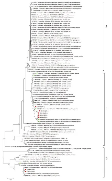
图2 7例分离株VP1部分基因序列系统进化分析图Figure 2 Phylogenetic analysis of partial VP1 gene sequence of enterovirus D68 isolated from the seven samples
1.5 统计学方法
采用EpiData3.0软件录入相关数据,建立数据库。
2 结果
2.1 EV检测及EV-D68的鉴别
在13 472份鼻咽部拭子标本中,EV阳性病例1 222例,阳性率为9.10%,发现EV-D68感染患儿7例,阳性率0.05%,7例EV-D68病例其VP1扩增序列与Fermon原型株及国内分离株的比对情况见图1所示。本次研究分离到的VP1序列基本与2015年所报道的深圳分离株序列一致,与Fermon株序列有较多差异。
2.2 EV-D68的系统进化分析
此前已有研究者对EV-D68进行了系统进化树分析,并分为群1、2、3三个进化簇,如图2所示。此次分离的7株D68中5株与进化簇1病毒序列具有更高同源性,2株与进化簇3有更高同源性。
2.3 临床特征及流行病学分析
7例EV-D68感染患者均系住院病人,男女比例4∶3,发病年龄7月~6岁1月,中位数年龄2岁6月,主要症状包括:咳嗽伴喘息(3例)、咳嗽伴腹泻(2例)、发热伴喘息(1例)、咳嗽(1例);上呼吸道感染2例(喘息性支气管炎及急性支气管炎各1例),下呼吸道感染5例(肺炎4例,毛细支气管炎1例)。2014 年9月及10月各1例,2014年11月2例,2015年10月及12月2016年1月各发现1例。由于感染例数有限,不足以做临床特征的统计学分析。
3 讨论
EV-D68是1962年在美国加尼福尼亚发现的一种肠道病毒,2014年在美国及加拿大引起严重呼吸道疾病的爆发,从而引起人们关注。美国疾病预防控制中心当年共计检测2 600个标本,其中36%测试为EV-D68阳性,所有确诊感染病例均为儿童,共计14例死亡病例报告,但目前仍不确定病毒的致病途径[9]。此后,欧洲、亚洲等国家均有EV-D68的病例报道[10-13]。根据病毒流行情况及研究人群不同,在流行爆发区域收集的呼吸道样本中EV-D68阳性率可达44%[4]。国内目前报道的EV-D68阳性率在0.2%~1%[14-15]。有研究[14]发现在北京7 945份咽拭子标本中肠道病毒阳性率6.99%(555/7 945),EVD68阳性率0.2%(12/7 945)。本研究发现肠道病毒阳性率9.1%(1 222/13 472),EV-D68阳性率0.05% (7/13 472),EV-D68阳性率较北京地区的研究结果低,可能与地区差异气候、研究对象不同及标本来源不同有关。EV-D68引起的急性呼吸道感染表现不同,既可以引起轻微的呼吸道感染,也可以引起严重的呼吸道感染,甚至死亡。在本研究中7例患者均表现为咳嗽、喘息,以下呼吸道感染多见,与既往报道一致[4,15]。但是由于EV-D68阳性病例少,其感染与临床特征之间的关系还需进一步研究。Xiao等[15]研究也发现2014年9~10月重庆EVD68感染较前增多,本研究7例EV-D68患者均在发生秋冬季节,2014年美国EV-68爆发流行在9~10月份[4],与本研究结果一致,提示秋冬季节可能是EV-D68感染高峰季节。虽然目前研究EV-D68感染率低,但还需要长时间检测人群中EV-D68的流行特征,以应对其可能流行爆发情况。
本次鉴别出的5例EV-D68病毒VP1区域基因序列与2015年度在深圳市疾病控制中心分离到的病毒株[17]具有高度同源性(94%~96%),同属于EV-D68病毒进化树中的进化簇1;2014年检出2例序列同属于病毒进化树种的群3,与美国2014年分离株大多数属于进化簇3在基因组上具有高度同源性,同源性为89%~91%,提示可能与美国EV-D68的流行有关。深圳市EV-D68目前感染率低,仍处于散发,但是该病毒的进化和变异明显,且在国外曾形成局部爆发流行,需进一步研究。但本研究由于样本例数和临床信息有限,不足做回顾性研究。还需进一步收集多中心更多样本的研究,包括不同年度样本、样本完整临床信息、病例临床症状及严重程度,以更好地掌握EV-D68的感染季节规律,以及不同基因谱的毒株与疾病严重程度的相关性。
[1] Palacios G,Oberste MS.Enteroviruses as agents of emerging infectious diseases[J].J Neurovirol,2005,11(5):424-433.
[2] Lu QB,Wo Y,Wang HY,et al.Detection of enterovirus 68 as one of the commonest types of enterovirus found in patients with acute respiratory tract infection in China[J].J Med Microbiol,2014,63(3):408-414.
[3] Xiang Z,Gonzalez R,Wang Z,et al.Coxsackievirus A21,enterovirus 68,and acute respiratory tract infection,China[J].Emerging Infectious Disease,2012,18(5):821-824.
[4] BA Brown,WA Nix,M Sheth,et al.Seven strains of Enterovirus D68 detected in the United States during the 2014 severe respiratory disease outbreak[J].Genome Announcements,2014,2(6):1201-1214.
[5] 江载芳,申昆玲,沈颖.诸福棠实用儿科学[M].8 版.北京:人民卫生出版社,2015:1247-1287.
[6] 中华人民共和国卫生部.手足口病诊疗指南(2010年版)[J].国际呼吸杂志,2010,30(24):1473-1475.
[7] Oberste MS,Maher K,Flemister MR,et al.Comparison of classic and molecular approaches for the identification of untypeable enteroviruses[J].Journal of Clinical Microbiology,2000,38:1170-1174.
[8] Tamura K,Peterson D,Peterson N,et al.MEGA5:molecular evolutionary genetics analysis using maximum likelihood,evolutionary distance,and maximum parsimony methods[J].Molecular Biology Evolution,2011,28:2731-2739.
[9] Centers for Disease Control and Prevention.Enterovirus D68[EB/OL].http://www.cdc.gov/non-polio-enterovirus/outbreaks/EV-D68-outbreaks.html.2016-07-19/2017-02-08.
[10] Imamura T,Suzuki A,Lupisan S,et al.Molecular evolution of enterovirus 68 detected in the Philippines [J].PloS One,2013,8:74221.
[11] Lu QB,Wo Y,Wang HY,et al.Detection of enterovirus 68 as one of the commonest types of enterovirus found in patients with acute respiratory tract infection in China[J].Journal of Medical Microbiology,2014,63:408-414.
[12] Ikeda T,Mizuta K,Abiko C,et al.Acute respiratory infections due to enterovirus 68 in Yamagata,Japan between 2005 and 2010[J].Microbiol Immunol,2012,56(2):139-143.
[13] Piralla A,Girello A,Grignani M,et al.Phylogenetic characterization of enterovirus 68 strains in patients with respiratory syndromes in Italy[J].Journal of Medical Virology,2014,86:1590-1593.
[14] Zhang T,Li A,Chen M,et al.Respiratory infections associated with enterovirus D68 from 2011 to 2015 in Beijing,China[J].Journal of Medical Virology,2016,88(9):1529-1534.
[15] Xiao Q,Ren L,Zheng S,et al.Prevalence and molecular characterizations of enterovirus D68 among children with acute respiratory infection in China between 2012 and 2014[J].Scientific Reports,2015,5:16639.
[16] Chen L,Shi L,Yang H,et al.Identification and wholegenome sequencing of four Enterovirus D68 strains in southern China in Late 2015[J].Genome Announcements.2016,4(5):1014-1016.
The clinical and molecular characterizations of enterovirus D68 among children with acute respiratory infection
HAO Jindou,LI Mingzhu,LIU Peihui★
(Shenzhen maternity and children's hospital affiliated Southern medical university,Shenzhen,Guangdong,China,518028)
Objective To explore the clinical and molecular epidemiological characteristics of children with enterovirus D68 in acute respiratory tract infection(ARTI). Methods 13 472 cases which were diagnosed with acute respiratory tract infection in the outpatient and ward from March 2014 to March 2016 were screened for enterovirus(EV).The EV positive cases of enterovirus were collected to identify the types of enterovirus by RT-PCR and sequencing. Results 1 222 EV positive cases were found in 13 472 nasopharyngeal swab specimens,the positive rate was 9.10%;and 7 EV-D68 infection were detected,the positive rate was 0.05%.Compared to the Fermon prototype strain of the VP1 sequence,5 strains of EV-D68 have higher homology with group 1 virus sequence,while 2 with group 2 virus sequence. Conclusions EVD68 may be a new predominant pathogen in acute lower respiratory tract infection(ARTI).The infection peak season may be in autumn and winter season.ALRTI may be a clinical feature of EV-D68 infection.
Enterovirus;Acute respiratory infection;EV-D68
深圳市科创委项目资助(深圳市新型HEVs在小儿ARTI中的临床及分子流行病学特征)(JCYJ20140414145619894)
南方医科大学附属深圳妇幼保健院,广东,深圳518028
★通讯作者:刘培辉,E-mail:lph521521@126.com

