沉积物中X射线衍射物相定量分析中的两种方法对比研究
2017-03-15 10:37:04林伟伟宋友桂
地球环境学报 2017年1期
林伟伟,宋友桂 ,
1.中国科学院地球环境研究所 黄土与第四纪地质国家重点实验室,西安710061
2.全球变化研究协同创新中心,北京 100875
沉积物中X射线衍射物相定量分析中的两种方法对比研究
林伟伟1,宋友桂1,2
1.中国科学院地球环境研究所 黄土与第四纪地质国家重点实验室,西安710061
2.全球变化研究协同创新中心,北京 100875
沉积物中X射线衍射(XRD)物相定量方法的选择和如何提高定量分析的精度一直是地学XRD定量研究的难点问题。本文针对沉积物中常见矿物组合特征,设计了一系列条件实验,比较了MacDiff的面积积分法(PAI法)和Highscore的强度参比法(RIR法)定量分析方法的优缺点。结果表明两种方法计算结果基本一致,但也各有优缺点。其中RIR法对衍射峰矮而宽的矿物定量更准确,PAI法则更适用于衍射峰尖锐的矿物以及含四种以上矿物、衍射峰重叠的样品,MacDiff的PAI方法分析过程不需要其他参数,误差较少,重现性好,而且全谱拟合还能部分消除择优取向的影响,可为沉积物XRD矿物相定量分析提供可靠数据。
X射线衍射;定量分析;MacDiff面积积分法; Highscore强度参比法
X射线衍射(XRD)物相定量研究中如何提高精确度一直是地学XRD矿物相定量分析研究的难题。X射线衍射物相定量分析的理论成熟之后,一直不断衍生出新的定量分析方法(Moore and Reynolds,1997;Chipera and Bish,2002,2013;Petschick,2002;Eberl,2003;曾蒙秀和宋友桂,2013)。常规方法有内标法(Popović and Gržeta-Plenković,1979)、增量法(储刚,1998)、绝热法(Chung,1974b) 、外标法(Chu,1994)、无标样法(刘仕子,1994)、基体冲洗法(K值法,RIR法)(Chung,1974a)和Rietveld方法(马礼敦,1996)等。这些方法各有优缺点,前3种方法都需要在待测样品中加入标样(标准物相)并绘制标准工作曲线,当样品物相种类较多时,标样反而增加衍射谱线的重叠机会,进一步给定量分析带来困难。外标法虽然不需要在样品中加入标准物相,但需要用纯的待测相物质制作工作曲线,而纯的待测相物质比较难提取或获得,这在实际应用中也是极为不便。在地学研究中,大部分地质样品成分复杂,很难找到合适的标样或者标样价格非常昂贵,加标样的定量分析方法应用很有限 (房俊卓和徐崇福,2010)。在实际应用中迫切需要一种简便、高效的普适性多物相无标样定量分析方法。而基体冲洗法、无标样法和Rietveld方法等分析方法不需要配制一系列标样和绘制工作曲线,但需要烦琐的数学计算,而且Rietveld方法对样品物相组成、结晶程度等要求较严,其实际应用也受到了一定限制(Madsen et al,2013;Chipera and Bish,2013)。
近些年,针对X射线衍射的定量分析,不同的仪器厂家和科研工作者也开发了一系列的软件或分析方法,诸如荷兰帕纳科公司、德国布鲁克、日本理学、美国伊诺斯、日本岛津、国内丹东方园等仪器自带软件以及面向大众开发的Jade,MacDiff,Rockjock等软件。仪器自带的软件侧重于定性分析即物相的鉴定,而开发的软件侧重于定量分析。Jade由美国材料数据公司(Materials Data Ltd.)开发,是应用最广泛的XRD分析工具软件,既可物相定性定量分析,也可以进行图谱拟合、晶体结构、晶相、晶胞精修、全谱拟合、残余应力、图谱模拟等,广泛应用于化学、材料学、生物学、医学、地学等领域。 Rockjock是一个Excel软件(Eberl,2003),比较简单,主要应用于地学矿物分析,功能有限。MacDiff是基于苹果系统开发的软件(Petschick,2002),在粘土矿物定量分析方面应用较多(Arnalds et al,2007;Liu et al,2007;Liu et al,2010;Wu et al,2012;Tudryn et al,2013),并取得很好的效果,但对全岩矿物的报道较少,其精度和可靠性仍不清楚(Ehrmann,1998)。
本文设计了系列对比实验,比较了MacDiff面积积分法(Peak area integration,PAI法)和最常用的HighScore强度参比法(Reference intensity ratio,RIR法)在矿物学定量分析的优缺点,以期找到一种计算简单,精确度高的普适定量方法,从而为地学研究提供便利。
1 研究方法简介
两种方法基本原理见下文。
1.1 RIR法
参比强度RIR是通过添加内标测量或计算得到的一个与仪器无关的常数。当标准物相是刚玉时:
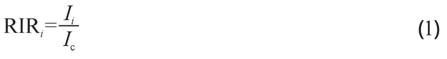
RIR值可以在PDF卡片库中进行搜索查询。基体清洗法的工作方程为:

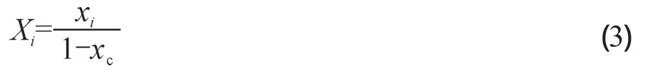
xc为加入样品中内标物质刚玉的重量分数(已知),Ic为刚玉的强度,Ii为待测物相的强度。因此只需要测出i相及内标物质的最强衍射峰强度即可算出i相在混合样品中的质量分数xi,而i相在原始样品中的质量分数Xi,则为:
1.2 PAI法
X射线在晶体上发生的衍射效应可以看成是平行或基本平行于样品板平面的晶面对入射的X射线产生对称性的选择性反射的物理现象(林西生,1990)。粉晶衍射谱图是无数微小晶粒各衍射面产生衍射叠加的结果。一个晶粒不止一组晶面,但是只有平行于试样表面的晶面(hkl),才对衍射起作用。样品制备完成时,基本上可以理想化认为晶面数等同于晶粒数。而对于混合物中的i相的某一衍射线强度:
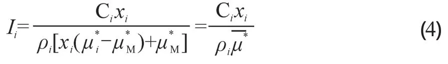
其中Ci是与角因子、温度因子、多重性因子、结构因子等有关的常数;xi为i相在混合物中的重量百分数;为i相的质量吸收系数;为基体(混合物中的除i相的其余部分)的质量吸收系数;为整个混合物的质量吸收系数;ρi为i相的密度。由此公式可知多晶粉末中i相某一衍射晶面(hkl)强度:
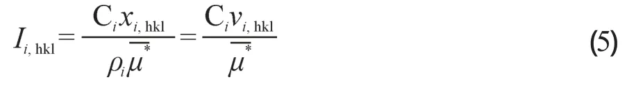
其中vi,hkl为i相晶面(hkl)在平行于样品表面且满足布拉格方程的晶粒在混合物中的体积百分数。I相所有衍射晶面的强度:
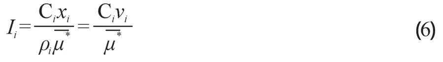
vi为i相在混合物中的体积百分数。由此可知,忽略角因子、多重性因子、结构性因子、温度因子的影响,某一相所有衍射线强度之和与衍射晶粒体积成正比。
2 实验设计与方法
2.1 实验材料
为了考虑研究方法的实用性,论文选择第四纪沉积物常见的石英(Qtz)、长石(Fsp)、方解石(Cal)、云母(Mca)和自然界不易见的刚玉(Crn)作为对比矿物,X衍射标准样品来自美国Chemplex industries公司。先用X衍射仪分别扫描其X射线定性衍射谱并与粉末衍射数据库PDF2004的图谱进行比较,确定与标样最匹配的PDF卡片。最终确定Qtz、Fsp、Cal、Mca、Crn最匹配的卡片号分别为01-083-2465,01-078-0434,01-086-2334,00-042-1399, 01-074-1081。用精度为0.01 mg的电子天平进行样品称量,将矿物标样按4种不同组合、4种不同比例混合配制成16个已知配比的实验样品。样品配比见表1。将称量好的物相粉末在研钵中轻轻研磨5分钟充分混合均匀,放入样品槽中压片制样。

表1 16个实验样品的成分配比Tab.1 The proportion of the 16 experimental samples
2.2 实验方法
测试工作在黄土与第四纪地质国家重点实验室环境矿物室完成,测试仪器为荷兰帕纳科公司生产的X’Pert Pro MPD 多晶X射线衍射仪。测试条件:Cu-Kα辐射;工作电压和电流分别为40 kV、40 mA;发散狭缝与散射狭缝均为1°,接收狭缝0.2 mm;采用连续扫描方式,扫描范围:5—70°(2θ),扫描时间19.685 s;扫描步长0.0167°(2θ)。为了最大减少仪器精度和样品制备引起的误差。每个样品重复测定3次,每次都重新压片。
测试数据分别使用帕纳科仪器自带的X’Pert HighScore软件以及面向大众开发的MacDiff软件对其进行定性和定量分析。具体分析过程:
(1)HighScore软件——RIR法
在HighScore软件中打开上述四种不同组合的混合矿物XRD图谱,依次进行以下操作:寻找基线、剥离Kα2、平滑、寻峰。寻峰结果与PDF2004卡片数据库进行矿物比对,选择得分高并含有RIR值的矿物,得出定性和半定量分析结果。
(2)MacDiff软件——PAI法
在MacDiff软件中打开XRD图谱,依次进行Kα2剥离、平滑、寻找基线、扣除背底、平滑基线、峰校正、寻峰、鉴定矿物,得到定性结果。根据定性结果编写峰分析程序,使图谱中每一衍射峰都与物相衍射晶面一一对应。按此程序对每个衍射峰的峰形参数进行精修,得到衍射峰拟合面积。背景采用四次多项式近似,数据用最小二乘法处理。最后将单一物相的峰面积数据进行加和,得到正比于该物相体积分数的总衍射峰面积。根据公式(6),乘以各物相密度得到正比质量分数的峰面积,最后进行100%归一化,得到PAI法半定量分析结果。
3 结果与讨论
图1罗列了4种不同矿物组合的2号样品的定性结果。由图1可以看出,不同的矿物衍射峰形不同。像石英、方解石这类常见的造岩矿物,其化学组成简单,晶系对称性高(庞小丽等,2009;薛治国等,2013),其X射线衍射峰少而且尖锐,不与其他物相的峰重叠。相反,化学组成复杂且晶系对称性低的矿物,如长石类矿物,其多层解理致使成分多变,微观结构共生和产生择优取向(Chipera and Bish,2013),其衍射峰多,矮而宽,常与其他物相衍射峰重叠,尤其是高角度区。等量的石英和长石均匀混合,测量发现石英衍射峰峰强明显要比长石高很多。这间接验证了只通过一个或几个衍射峰计算物相含量会有明显的误差,特别是当有择优取向存在时(于学峰和刘钊,2010)。本实验曾尝试选取前三强峰或前五强峰面积来计算相对含量,结果偏差非常大,尤其是长石类矿物。这就是为什么当仅用特征峰或几个强峰进行定量时,往往需要引用强度因子(龚键等,2013)、参比强度(杨波等,2014)或匹配强度(储刚等,2004)等。相反PAI法对所有衍射峰进行(分裂)拟合,就会大大减少因择优取向、峰重叠而导致的定量误差。
通过PAI法和RIR法实验值和配比值的回归分析,发现含量较高的物相(石英、长石)两种方法的计算结果与配比值比较接近。其中结晶质矿物石英通过两种方法得到的数据拟合为同一条曲线,与配比值相比,当含量≤60%时,值偏高(图2a)。图2b显示RIR法得到的长石半定量结果与配比值非常接近,PAI法的结果偏低,这种偏差随着含量的增加而增大。可能原因是长石衍射峰形不明显,峰重叠性较高导致分峰不准确。从图2c、d可以看出用PAI法对含量较低的方解石和云母定量结果与配比值比较接近,RIR法定量结果值偏低。其中图2d可以看出RIR法对低含量(≤10%)的云母不敏感,定量结果偏低程度很大,相对偏差达62%。根据公式(2),可知云母RIR值偏大(13.8)是造成定量结果偏低的原因。值得一提的是云母虽然不是粘土矿物,但是可以代表粘土级别的矿物,其RIR值并不是按定义去测量的(Hillier,2000)。总体来说RIR法的定量结果还是比较粗的。而PAI法对云母的定量结果稍稍偏高,原因是云母层理特别发育,在实际情况中存在非层面衍射(徐钿和王冠鑫,2003),PAI法将其理想化了,误差就不可避免了。从图1还可以看出云母的衍射线稀少,且相距甚远无叠加现象,说明制样过程中确实存在择优取向。刚玉通常被认为是标准物相(Hubbard et al,1976),图2e的定量结果说明RIR法和PAI法的定量结果是基本一致的,但都比真实值偏低。
实际配比过程中,组分种类越多的样品其相应的含量就越低,图2可以看出五种矿物含量越低,与真实值就越接近。这反映了当样品含有较多矿物种类时,尤其是含有四种以上矿物时,PAI法更能显示出优势。
图3比较了PAI法和RIR法对五种物相的定量绝对误差。PAI法(图3a)的绝对误差在10%以内,而少量石英的RIR法测量结果绝对误差在10% — 15%(图3b)。石英两种方法的定量结果绝对误差在各个区间都有,其中在≤3%区间内最多。长石误差基本属于平均分布,RIR法则主要集中在3%以内。相反,方解石的PAI法定量结果绝对误差都≤3%。推出PAI法适合结晶性较好,衍射峰高而尖的物相,RIR法则适合结晶性一般,衍射峰矮而宽的物相。这是因为RIR法主要侧重于强峰,而PAI法对于结晶度差、晶粒细、有缺陷的矿物,因其衍射图谱宽化而无法用峰面积准确的定量。同时从图3还可以看出PAI法对低含量的云母和刚玉的定量结果也比RIR法好,除了2种组合的云母绝对误差在3% — 5%,其余均小于3%。结合图2,说明PAI法对低含量的物相定量结果可靠,而物相的较低含量正是造成的RIR法结果偏低的原因之一(曲高生, 1990)。

图1 四种不同矿物种类混合样品的典型X射线衍射谱(图注括号内为相应矿物含量)Fig.1 The typical X-ray diffraction patterns of 4 kinds of different mineral mixed samples (Corresponding mineral contents are shown in legend brackets)

图2 PAI法(蓝线)和RIR法(红线)得到的石英(a)、长石(b)、方解石(c)、云母(d)、刚玉(e)实验值与配比值(黑线)的相关关系Fig.2 Relationship of matched value and calculated value of Qtz (a),Fsp (b),Cal (c),Mca(d),Crn (e) by PAI and RIR quantitative methods,respectively

图3 PAI法(a)和RIR法(b)和实验值与配比值绝对误差频数分析Fig.3 The absolute error frequency distribution between matched values and computed results by PAI (a) and RIR (b) quantitative methods,respectively
图2和图3 共同说明PAI法与RIR法两种定量方法应用于矿物定量结果基本一致,同时两种方法也各有优缺点。RIR法快速、方便但并不适用于所有矿物,对于结构复杂的物相困难较大(赖振宇等,2014)。误差主要来源于两个方面:(1)RIR值的选择。RIR值受限于PDF卡片库,选择不同的RIR值,就得到不同的结果(房俊卓和徐崇福,2010)。当一种物相的RIR值与其他物相相差很大时,由RIR值计算导致的误差比由峰重叠导致的误差要明显的多。(2)衍射峰的强度。RIR定义用的是峰高强度。峰高强度=1/2 ×峰高×峰宽。对于低含量物相,峰形不明显,峰强小造成峰高强度偏低。PAI法直接对整个衍射谱内所有衍射峰进行拟合,既不需要强度因子,也不需要晶体结构信息。但PAI法因须扣除背景,无法获取非晶态物质的定量结果(胡秀荣等,2005;李响和蔡元峰,2014),而且编写分析程序时比较繁琐,用时长。PAI法误差来源包括三个方面:(1)劈裂峰的不准确。因为矿物相较多,衍射峰互相重叠,矿物类质同象的影响使衍射峰的起点和终点发生漂移,造成积分强度较大的误差(曲高生,1990)。(2)单纯的对整个衍射谱峰进行面积积分,忽略晶体结构的影响,理想化了晶面衍射,结果是值偏高。(3)根据布拉格方程,θ取值范围为0 — 45°。本实验局限于PDF 定性结果,θ范围仅取5.5 — 35°。当然样品预处理过程也会对实验误差造成很大的影响(陈涛等,2013;李艳丽等,2014)。
采用积分面积有利于提高X射线衍射物相定量分析的准确度(李真等,2006;房俊卓等,2008)。运用PAI 法进行沉积物矿物定量,可以更好地展现积分强度的优越性。实验证明用峰的位置、高度、宽度、形态系数和不对称性5要素描述衍射峰形态比仅用峰的位置、高度、宽度3要素描述衍射峰形态更准确、全面、完整(Shi et al,2002)。本文采用的MacDiff软件不但可以提供六种不同的“钟罩形”函数来完成峰形拟合,多有重叠峰,尤其是在高角度区,而且可以根据衍射不相干原理——重叠峰的积分强度等于各物相积分强度的代数和(苗春省,1988)——将矿物X射线衍射谱重叠峰分离开来,部分消除物相质量吸收、择优取向、仪器稳定性误差和多重性因子的影响(储刚等,2004;Chipera and Bish,2013)。这两者正是MacDiff与一般常规峰分析软件的特别之处。
4 结论
实验结果说明PAI法和RIR法在物相定量分析上是基本一致的。RIR法作为目前最常用的物相定量法,其优点是简单,快速,能有效的对结晶性不好的矿物进行分析,缺点是RIR值的选取十分受限于卡片库,而且含量较低物相RIR法定量结果偏低。而PAI法忽略了晶体微观结构,理想化了晶粒中晶面衍射的情况,单纯用峰面积计算矿物含量。虽然该方法分析过程稍微繁琐,但是对于结晶性较好的物相和含矿物种类较多的样品定量结果准确(绝对误差多数情况下≤3%)、重现性好,可部分消除择优取向的影响,是理想的实验方法,可为沉积物矿物定量分析提供可靠数据。
陈 涛,宋友桂,曾蒙秀,等.2013.影响XRD衍射谱形态的实验条件分析[J].地球环境学报,4(2):1249 – 1254.[Chen T,Song Y G,Zeng M X,et al.2013.Analysis on the experimental conditions affecting the shape of the XRD spectrum [J].Journal of Earth Environment,4(2):1249 – 1254.]
储 刚.1998.含非晶相样品的X射线衍射增量法定量相分析[J].物理学报,47(7):1143 – 1148.[Chu G.1998.A doping method for quantitative X-ray diffraction phase analysis of samples containing amorphous material [J].Acta Physica Sinica,47(7):1143 – 1148.]
储 刚,翟秀静,符 岩,等.2004.X射线衍射多谱峰匹配强度比定量相分析方法[J].分析测试学报,23(1):48 – 51.[Chu G,Zhai X J,Fu Y,et al.2004.The multipeak match intensity ratio method for X-ray diffraction quantitative phase analysis [J].Journal of Instrumental Analysis,23(1):48 – 51.]
房俊卓,徐崇福.2010.三种X射线物相定量分析方法对比研究[J].煤炭转化,33(2):88 – 91.[Fang J Z,Xu C F.2010.Study on three kinds of XRD quantitative analysis methods [J].Coal Conversion,33(2):88 – 91.]
房俊卓,张 霞,徐崇福.2008.实验条件对X射线衍射物相定量分析结果的影响[J].岩矿测试,27(1):60 – 62.[Fang J Z,Zhang X,Xu C F.2008.Effect of experimental conditions on X-ray diffractometric quantitative phase analysis [J].Rock and Mineral Analysis,27(1):60 – 62.]
龚 键,李福春,马 芳,等.2013.土壤中常见黏土矿物定量方法的改进研究[J].土壤通报,44(4):884 – 888.[Gong J,Li F C,Ma F,et al.2013.A study for the improvement of quantitative determination of main clay minerals in soil [J].Chinese Journal of Soil Science,44(4):884 – 888.]
胡秀荣,吕光烈,顾建明,等.2005.天然膨润土中蒙脱石丰度的定量方法研究[J].矿物学报,25(2):59 – 63.[Hu X R,Lü G L,Gu J M,et al.2015.Three methods for quantification of montmorillontte abundances in natural bentonttes [J].Acta Metallurgica Sinica,25(2):59 – 63.]
赖振宇,邹秋林,卢忠远,等.2014.Rietveld 全谱拟合方法对磷酸镁水泥水化产物的定量分析研究[J].计量学报,35(4):398 – 402.[Lai Z Y,Zou Q L,Lu Z Y,et al.2014. Quantitative analysis of hydration products in magnesium phosohate cement with Rietveld whole pattern fi tting [J].Acta Metallurgica Sinica,35(4):398 – 402.]
李 响,蔡元峰.2014.沉积物中铁氧化物的定量方法及其在白垩纪大洋红层中的应用[J].高校地质学报,20(3):433 – 444.[Li X,Cai Y F.2014.The quantitative analysis methods for iron oxides in sediment and their application in cretaceous oceanic red beds [J].Geological Journal of China Universities,20(3):433 – 444.]
李艳丽,刘志飞,赵玉龙,等.2014.预处理过程对沉积物中粘土矿物半定量分析的影响[J].第四纪研究,34(3):635 – 644.[Li Y L,Liu Z F,Zhao Y L,et al.2014.On the influences of pretreatment on semi-quantitative determination of clay minerals in sediment [J].Quaternary Sciences,34(3):635 – 644.]
李 真,尹 琳,熊 飞,等.2006.关于《凹凸棒粘土中坡缕石的内标法X 衍射定量分析研究》一文的修正[J].高校地质学报,12(3):410 – 412.[Li Z,Yin L,Xiong F,et al.2006.Comment on “quantitative analysis of X-ray diffraction for palygorskite within attapulgite clay” [J].Geological Journal of China Universities,12(3):410 – 412.]
林西生.1990.X射线衍射分析技术及其地质应用[M].北京:石油工业出版社:11 – 23.[Lin X S.1990.X-ray diffraction analysis technique and application in geology [M].Beijing:Petroleum Industry Press:11 – 23.]
刘仕子.1994.一种实用的X射线无标定量相分析方法[J].岩石矿物学杂志,13(3):268 – 277.[Liu S Z.1994.A practical method of X-ray quantitative phase analysis without standards [J].Acta Petrologica et Mineralogica,13(3):268 – 277.]
马礼敦.1996.X射线粉末衍射的新起点—— Rietveld全谱拟合[J].物理学进展,16(2):251 – 271.[Ma L D.1996.The new starting point of X-ray powder diffraction —Rietveld full pattern fi tting [J].Progress in Physics,16(2):251 – 271.]
苗春省.1988.X射线定量分析方法及应用[M].沈阳:辽宁科技出版社:31 – 34.[Miao C S.1988.X-ray quantitative analysis method and application [M].Shenyang:Liaoning Science and Technology Press:31 – 34.]
庞小丽,刘晓晨,薛 雍,等.2009.粉晶X射线衍射法在岩石学和矿物学研究中的应用[J].岩矿测试,28(5):452 – 456.[Pang X L,Liu X C,Xue Y,et al.2009.Application of powder X-ray diffraction in petrology and mineralogy [J].Rock and Mineral Analysis,28(5):452 – 456.]
曲高生.1990.西沙群岛琛航岛碳酸盐沉积物 X 射线定量分析方法研究[J].矿物学报,10(4):74 – 83.[Qu G S.1990.Quantitative X-ray diffraction analysis of carbonate sediments from Chenhang Island,Xisha Islands,China:a methodological study [J].Acta Metallurgica Sinica,10(4):74 – 83.]
徐 钿,王冠鑫.2003.云母族矿物八面体化学成分与层面X射线衍射特征[J].分析测试学报,22(3):54 – 56.[Xu D,Wang G X.2003.Relation between octahedral composition and X-ray diffraction characterization projected onto lay for mica [J].Journal of Instrumental Analysis,22(3):54 – 56.]
薛治国,刘子琦,陈 浒,等.2013.喀斯特洞穴晶体沉积速率的气候响应[J].地球环境学报,4(1):1191 – 1196.[Xue Z G,Liu Z Q,Chen H,et al.2013.Climatic response from modern crystal sediments in karstic cave [J].Journal of Earth Environment,4(1):1191 – 1196.]
杨 波,刘兴起,王永波.2014.湖泊沉积物碳酸盐含量的XRD半定量分析[J].湖泊科学,26(4):637 – 640.[Yang B,Liu X Q,Wang Y B.2014.Carbonate contents of lake sediments determined by XRD method [J].Journal of Lake Sciences,26(4):637 – 640.]
于学峰,刘 钊.2010.红原泥炭颗粒物表面形态与矿物组成研究[J].地球环境学报,1(2):122 – 125.[ Yu X F,Liu Z.2010.Surface texture and mineralogical characters of the peat in Hongyuan swamp [J].Journal of Earth Environment,1(2):122 – 125.]
曾蒙秀,宋友桂.2013.麦夸特算法在X射线衍射物相定量分析中的应用[J].地球科学:中国地质大学学报,38(2):431 – 440.[ Zeng M X,Song Y G.2013.Application of the Levenberg-marquardt algorithm to X-ray diffraction quantitative phase analysis [J].Earth Science:Journal of China University of Geosciences,38(2):431 – 440.]
Arnalds Ó,Óskarsson H,Bartoli F,et al.2007.Soils of volcanic regions in Europe [M].Berlin Heidelberg:Springer.Doi:10.1007/978-3-540-48711-1.
Chipera S J,Bish D L.2002.FULLPAT:a full pattern quantitative analysis program for X-ray powder diffraction using measured and calculated patterns [J].Journal of AppliedCrystallography,35(6):744 – 749.
Chipera S J,Bish D L.2013.Fitting full X-Ray diffraction patterns for quantitative analysis:A method for readily quantifying crystalline and disordered phases [J].Advances in Materials Physics amp; Chemistry,3(1):47 – 53.
Chu G.1994.An external standard method of quantitative phase analysis of the sample containing an amorphous phase by X-ray diffraction [J].Acta Metallurgica Sinica,7(3):179 – 182.
Chung F H.1974a.Quantitative interpretation of X-ray diffraction patterns of mixtures.Ⅰ.Matrix-flushing method for quantitative multicomponent analysis [J].Journal of Applied Crystallography,7(6):519 – 525.
Chung F H.1974b.Quantitative interpretation of X-ray diffraction patterns of mixtures.Ⅱ.Adiabatic principle of X-ray diffraction analysis of mixtures [J].Journal of Applied Crystallography,7(6):526 – 531.
Eberl D D.2003.User’s guide to RockJock—A program for determining quantitative mineralogy from powder X-ray diffraction data:Open-File Report 03-78 [R].US Geological Survey:47.
Ehrmann W.1998.Mineralogy of sediments from CRP 1 as revealed by X-ray diffraction [J].Terra Antartica,5(3):627 – 632.
Hillier S.2000.Accurate quantitative analysis of clay and other minerals in sandstones by XRD:comparison of a Rietveld and a reference intensity ratio (RIR) method and the importance of sample preparation [J].Clay Minerals,35(1):291 – 302.
Hubbard C R,Evans E H,Smith D K.1976.The reference intensity ratio,I/Ic,for computer simulated powder patterns [J].Journal of Applied Crystallography,9(2):169 – 174.
Liu Z F,Colin C,Huang W,et al.2007.Clay minerals in surface sediments of the Pearl River Drainage Basin and their contribution to the South China Sea [J].Chinese Science Bulletin,52(8):1101 – 1111.
Liu Z F,Colin C,Li X J,et al.2010.Clay mineral distribution in surface sediments of the northeastern South China Sea and surrounding fluvial drainage basins:source and transport [J].Marine Geology,277(1 /2 /3 / 4):48 – 60.
Madsen I C,Scarlett N V Y,Riley D P,et al.2013.Modern diffraction methods:chapter 10 quantitative phase analysis using the Rietveld method [M].Weinheim:Wiley-VCH Verlag GmbH amp; Co.:285 – 320.
Moore D M,Reynolds R C.1997.X-ray diffraction and the identi fi cation and analysis of clay minerals [M].Oxford:Oxford University Press.
Petschick R.2002.MacDiff 4.2.6 (Free X-ray powder diffractometry analysis tool) [M/OL].[2015-10-10].http:// www.geol.uni-erlangen.de/html/software/soft.html.
Popović S,GržetaR-Plenković B.1979.The doping method in quantitative X-ray diffraction phase analysis [J].Journal of Applied Crystallography,12(2):205 – 208.
Shi N C,Bai W J,Ma Z S,et al.2002.A study of X-ray diffraction of diamond inclusions from Luobusha,Tibet [J].Acta Geologica Sinica,76(4):497 – 500.
Tudryn A,Tucholka P,Özgûr N,et al.2013.A 2300-year record of environmental change from SW Anatolia,Lake Burdur,Turkey [J].Journal of Paleolimnology,49(4):647 – 662.
Wu J W,Liu Z F,Zhou C.2012.Late Quaternary glacial cycle and precessional period of clay mineral assemblages in the western Pacific warm pool [J].Chinese Science Bulletin,57(28 / 29):3748 – 3760.
A comparative study on X-ray diffraction mineral quantitative analysis of two methods in sediments
LIN Weiwei1,SONG Yougui1,2
1.State Key Laboratory of Loess and Quaternary Geology,Institute of Earth Environment,Chinese Academy of Sciences,Xi’an,710061,China
2.Joint Center for Global Change Studies,Beijing 100875,China
Background,aim,and scopeHow to select suitable X-ray diffraction (XRD) phase analysis methods and improve the accuracy have been an important problem in quantitative analysis of sediments.Many new methods for quantitative analysis have been derived since the theory of XRD phase quantitative analysis was mature.The requirement of these methods is too high either for the standard or for sample,which make its application restricted.In recent years,in view of XRD quantitative analysis,different instrument manufacturers and researchers have also developed a series of analysis software.MacDiff has been currently widely applied in clay mineral rather than the whole rock mineral,and its precision and reliability are still unclear.In this paper,we compare the advantagesand disadvantages of MacDiff with Peak Area Integration (PAI) and HighScore with Reference Intensity Ratio (RIR),which is the most commonly used in the quantitative analysis of mineralogy,hoping to fi nd a kind of simple calculation,high precision and universal quantitative method,so as to provide convenience for geological research.Materials and methodsConsidering the practice of research method,we choose common quaternary sediments of quartz (Qtz),feldspar (Fsp),calcite (Cal),mica (Mca) and uncommon mineral corundum (Crn) as materials.According to four different combinations and four different mixing ratios,16 known content experimental samples are prepared,conducted XRD testing,analyzed using the PAI and RIR quantitative analysis method,respectively.Based on the theory of powder diffraction spectrum is the diffraction stack results of countless small grain diffraction planes and diffraction intensity is proportional to the diffraction grain volume,PAI method is made by integrating all diffraction peak areas with MacDiff,and then through Normalization method to calculate all mineral contents.The key of the RIR method is the Reference Intensity RIR,which is a constant and independent of the instrument,is measured or calculated the strongest diffraction peak intensity by adding the internal standard.The mineral semiquantitative results can be got immediately if the matched RIR value is researched in HighScore PDF database.ResultsThe Relationship of matched value and calculated value of PAI and RIR quantitative methods (Fig.2) shows that with high crystallinity and high content,the calculated value of quartz in two methods are very close to the matched value.For poor crystallinity,high content of feldspar,the semi-quantitative results of RIR method closer to matched value,while PAI result is on the low side and the deviation increases with the known content increasing.For the low content of minerals,such as calcite and mica,PAI quantitative result is better than RIR.Low content of corundum is usually considered a standard phase,both RIR and PAI methods quantitative results are basically consist,lower than the real value.Fig.2 also shows that PAI method can show more advantages on the samples containing more than four kinds of minerals.By comparing the RIR and PAI method of fi ve phase quantitative absolute error (Fig.3),PAI method quantitative results of quartz,calcite,mica obtained with less error,and feldspar,in contrast,RIR method error is less.The absolute errors of corundum got with two methods are within 3%.DiscussionBecause of the difference of mineral crystallinity,such as Qtz and Fsp,provided that only selecting top three or fi ve peak areas to calculate the relative content,the results deviate largely,especially when preferred orientation exists.So PAI method fitting all diffraction peaks,will greatly reduce quantitative errors leading by preferred orientation,overlapping peaks.The results are in agreement with those provided by PAI and RIR methods,and each method has its own advantage and shortage.More speci fi cally,the PAI method is more practical in minerals with the sharp diffraction peaks and samples containing more than four minerals whose diffraction peaks are overlapped.But for mica,the PAI quantitative results are slightly on the high side cause of mica fl ourish bedding leading to the existence of non-plane diffraction in the actual situation.On the contrary,the RIR method is suitable for the minerals with some short and wide diffraction peaks,such as feldspar,which the diffraction peak shape is not obvious,higher peak overlaps maybe lead to inaccurate splitting peaks.Unfortunately,the RIR method is not sensitive signi fi cantly to low content (10%) or less.According to the principle of RIR method,larger RIR value (eg.RIR(mica)=13.8) causes lower quantitative results.So the errors of RIR method are mainly derived from the RIR value and the diffraction peak intensity,while the PAI method errors source from three aspects:inaccurate peak splitting,idealized crystal plane diffraction and ? range.ConclusionsExperimental results show that the PAI and RIR method on the phase quantitative analysis is the basically consist.By far,RIR method is the most commonly used phase quantitative method.Its advantage is simple,rapid,especially for bad crystalline minerals,while the disadvantage is that the selection of RIR value is restricted by PDF database,and the quantitative result of low content phase trend to less.Although the analysis process of PAI method is a little complicated,but for better crystalline materials or samples contain more mineral species,the method owns accurate quantitative results (absolute error in most cases ≤3%),good reproducibility,and can partly eliminate the effect of preferred orientation.Recommendations and perspectivesThe PAI analysis method does not need other parameters with small error and good reproducibility,and the full pattern fi tting can partially eliminate thein fl uence of preferential orientation,therefore,PAI method can provide reliable data for mineral quantitative analysis in sediments.
Key Deployment Project of Chinese Academy of Sciences (KZZD-EW-04-02); National Natural Science Foundation of China (41290253,41572162); Fund of Xi’an Center of Geological Survey,China Geological Survey (Water[2016](4))
SONG Yougui,E-mail:syg@ieecas.cn
X-ray diffraction; quantitative analysis; MacDiff area integration; Highscore reference intensity ratio method
2016-12-03;录用日期2017-01-23
Received Date:2016-12-03;Accepted Date2017-01-23
中国科学院重点部署项目(KZZD-EW-04-02);国家自然科学基金项目(41290253,41572162);中国地质调查局西安地质调查中心项目(水[2016](4))
宋友桂,E-mail:syg@ieecas.cn
林伟伟,宋友桂.2017.沉积物中X 射线衍射物相定量分析中的两种方法对比研究[J].地球环境学报,8(1):78 – 87.
: Lin W W,Song Y G.2017.A comparative study on X-ray diffraction mineral quantitative analysis of two methods in sediments [J].Journal of Earth Environment,8(1):78 – 87.
10.7515/JEE201701010
猜你喜欢
中学生数理化·八年级物理人教版(2023年10期)2023-11-30 01:57:54
机电安全(2022年5期)2022-12-13 09:22:26
世界科学技术-中医药现代化(2020年2期)2020-07-25 02:06:06
世界有色金属(2020年4期)2020-05-16 05:55:44
科学(2020年1期)2020-01-06 12:21:34
中成药(2018年12期)2018-12-29 12:25:44
中成药(2017年6期)2017-06-13 07:30:35
上海金属(2015年6期)2015-11-29 01:08:49
医学研究杂志(2015年4期)2015-06-10 06:42:43
石油炼制与化工(2014年10期)2014-09-16 03:52:04