
First Description of the Male of BaalzebubyouyiensisZhao & Li (Araneae, Theridiosomatidae)
2016-12-12 10:04:39ZHANGTianyiLINYucheng
四川动物 2016年2期
ZHANG Tianyi, LIN Yucheng
(Key Laboratory of Bio-resource and Eco-environment of Ministry of Education,College of Life Sciences, Sichuan University, Chengdu 610064, China)
DOI:10.11984/j.issn.1000-7083.20150367
First Description of the Male ofBaalzebubyouyiensisZhao & Li (Araneae, Theridiosomatidae)
ZHANG Tianyi, LIN Yucheng*
(Key Laboratory of Bio-resource and Eco-environment of Ministry of Education,College of Life Sciences, Sichuan University, Chengdu 610064, China)
The male ofBaalzebubyouyiensisZhao & Li, 2012 is described for the first time and newly recorded from Laos. Morphological description, diagnosis and comparative photos were given for both genders of this species.
taxonomy; spider; diagnosis characteristic; male supplement; new distribution
The theridiosomatid genusBaalzebubwas erected by Coddington (1986) to accommodate the type species,B.bauboCoddington, 1986 and other two,TheridiosomaalbonotatumPetrunkevitch, 1930 andT.brauniWunderlich, 1976. At present, 6 species were included in this genus (World Spider Catalog, 2015), while their distribution is very wide and still “circumtropical”, as summarized by Coddington (1986). The type species andB.albonotatuswere from Latin America (Coddington, 1986),B.braunifrom Queensland of Australia (Davies, 1988) and the other three species,B.nemesisMiller, Griswold & Yin, 2009,B.rastrariusZhao & Li, 2012 andB.youyiensisZhao & Li, 2012, were from the subtropical area of China: Yunnan, Guizhou and Guangxietc. (Milleretal., 2009; Zhao & Li, 2012). The genusBaalzebubcan be distinguished by elongate tegulum, a small, triangular median apophysis, a long, sharp paracymbium, and complicated branching embolus covered by broad conductor, and by the spermathecae connected with each other, broad epigynal plate with a central sclerotized pit, and forming a backward-pointed scape.
The species,B.youyiensisZhao & Li, 2012 was originally described based on four female specimens collected from a cave of Guangxi. When we checked the theridiosomatid materials from Laos, aBaalzebubspecies containing 3 male and 17 female specimens caught our attention. The females wereB.youyiensis. The male specimens have habitus and marking similar to the females, and the palpal structures show the characters ofBaalzebub(Coddington, 1986; Davies, 1988; Zhao & Li, 2012). Therefore, we considered both males and females conspecific, and the males ofB.youyiensiswere described for the first time.
All measurements are given in mm. Specimens are preserved in 95% alcohol and were examined and measured under a Leica M205C stereomicroscope, and photographed with a Canon EOS 60D digital camera (18.0 megapixels) mounted on an Olympus BX43 compound microscope. The images were mounted using Helicon Focus 3.10.3 software (Khmeliketal., 2006). Map was created using Arcview 3.3. Specimens are deposited in the Museum of Sichuan University (SCUM) in Chengdu.
BaalzebubyouyiensisZhao & Li, 2012 (Figs 1~17)
BaalzebubyouyiensisZhao & Li, 2012: 17, figs 9A~E, 10 A~B (female).
Material examined: 4 females (holotype and paratypes, IZCAS): China, Guangxi: Pingxiang county-level city, Youyi town, Bantou village, Niuyan cave, 22°05.666′ N, 106°45.439′ E, elevation ca 251 m, 18 January 2011, Z.G. Chen & Z.W. Zha leg.; 3 males and 17 females (SCUM): Laos, Bolikhamsai: Lak Sao, Transiten, in Lang cave, 18°23.318′ N, 104°32.675′E, elevation ca 318 m, August 2012, P. Jäger. leg.
Diagnosis: Male of this species is similar toB.albonotatus(see Coddington, 1986: 74, fig. 164) andB.brauni(see Davies, 1988: 326, fig. 43) in having a cluster of setae of cymbium and a long and pointed paracymbium, but can be distinguished from otherBaalzebubspecies by the presence of a broad lateral lobe (Fig. 14), the slender median apophysis (Figs 7, 11), the securiform conductor (Fig. 13) and the multiramose embolus (Figs 7, 10~12). Female distinguished by the semi-transparent epigynal scape with a blunt tip, which is proportionately shorter (Fig. 16). Spermathecae contrasted with short, long spermathecae in otherBaalzebubspecies (Fig. 17). The relatively shorter, smaller spermathecae surrounding by copulatory ducts made up of ovoid loops (Fig. 17).
Description: Habitus as in Figs 1~6. Dorsal shield of prosoma broad, nearly pear-shaped, orange. Cephalic area slightly evelated. Eyes with black base. Anterior eye row procurved, posterior eye row recurved. Cervical groove distinct, thoracic fovea unconspicuous. Sternum heart-shaped. Legs yellow, brown from patella to tarsus (except leg Ⅰ). Opisthosoma sub-elliptic in dorsal view, rugose in laterals, pale yellow in male, glum yellow in female, with darkish pigmentation patches in laterals and venter.
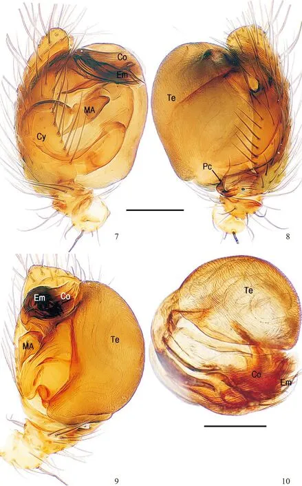
Male palp (Figs 7~14): Patella with a strong macroseta (Fig. 7). Tibia with three trichobothria and macrosetae (Figs 9, 14). Paracymbium L-shaped, with a long, sharp, distal end (Figs 8, 14). Lateral lobe broad, semi-transparent (Fig. 14). Tegulum large, surface with delicate texture (Figs 8~10). Median apophysis relatively small, sub-triangular, with a sharp corner (Figs 7, 11). Conductor securiform, translucent, with distinct texture and grains (Fig. 13). Embolic end multiramose, with a broad affiliated lobe (Fig. 12).
Female copulatory organ (Figs 15~17): see Zhao & Li (2012).
Male: Total length 1.52, carapace 0.86 long, 0.88 wide, clypeus 0.18 high, sternum 0.41 long, 0.45 wide, coxae Ⅳ separated by 1.00 time their width. Posterior median eyes separated by less than half of their diameter. Macrosetae: Leg Ⅰ: femur r 1, patella d 1, tibia p 2, r 3; Leg Ⅱ: patella d 1, tibia r 1, metatarsus p 1; Leg Ⅲ: tibia d 1; Leg Ⅳ: patella d 1, tibia d 2. Metatarsal trichobothria: Tm Ⅰ: 0.15; Tm Ⅱ: 0.29; Tm Ⅲ: 0.17; Tm Ⅳ: 0.30. Leg measurements: Ⅰ 3.61 (1.05, 0.42, 0.98, 0.73, 0.43); Ⅱ 2.96 (0.88, 0.38, 0.72, 0.59, 0.39); Ⅲ 2.13 (0.63, 0.30, 0.42, 0.42, 0.36); Ⅳ 2.45 (0.81, 0.29, 0.51, 0.53, 0.31).
Female: Total length 1.95, carapace 1.08 long, 0.99 wide, clypeus 0.18 high, sternum 0.49 long, 0.51 wide, coxae Ⅳ separated by 1.00 time their width. Posterior median eyes separated by less than half of their diameter. Macrosetae: Leg Ⅰ: patella d 1, tibia d 1, r 1; Leg Ⅱ: patella d 1, tibia d 2, r 1; Leg Ⅲ: patella d 1, tibia r 1; Leg Ⅳ: patella d 1, p 1; tibia d 1. Metatarsal trichobothria: Tm Ⅰ: 0.22; Tm Ⅱ: 0.33; Tm Ⅲ: 0.33; Tm Ⅳ: 0.32. Leg measurements: Ⅰ 3.95 (1.29, 0.47, 0.96, 0.79, 0.44); Ⅱ 3.40 (1.11, 0.42, 0.78, 0.67, 0.42); Ⅲ 2.32 (0.71, 0.34, 0.48, 0.48, 0.31); Ⅳ 2.73 (0.88, 0.33, 0.62, 0.59, 0.31).
Distribution: south China (Guangxi), Laos (Fig. 18).
Acknowledgements: Thanks to Dr. Peter Jäger (Senckenberg Research Institute, Senckenberganlage, Frankfurt am Main, Germany) for providing valuable specimen materials. We are grateful to anonymous referees for their comments on the manuscript.
Reference:
Coddington JA. 1986. The genera of the spider family Theridiosomatidae[J]. Smithsonian Contributions to Zoology, 422: 1-96.
Davies VT. 1988. An illustrated guide to the genera of orb-weaving spiders in Australia[J]. Memoirs of the Queensland Museum, 25: 273-332.
Khmelik VV, Kozub D, Glazunov A. 2006. Helicon focus 3.10.3[DB/OL]. [2013-08-20]. http://www.heliconsoft.com/heliconfocus.html.
Miller JA, Griswold CE, Yin CM. 2009. The symphytognathoid spiders of the Gaoligongshan, Yunnan, China (Araneae, Araneoidea): systematics and diversity of micro-orbweavers[J]. ZooKeys, 11: 9-195.
World Spider Catalog. 2015. World Spider Catalog. Natural History Museum Bern[DB/OL]. [2015-11-15]. http://wsc.nmbe.ch, version 16.5.
Zhao QY, Li SQ. 2012. Eleven new species of theridiosomatid spiders from southern China (Araneae, Theridiosomatidae)[J]. ZooKeys, 255: 1-48.

Figs. 1~6 Baalzebub youyiensis Zhao & Li, 2012, male habitus (1~3) and female habitus (4~6) 1, 4. Dorsal view; 2, 5. Ventral view; 3, 6. Lateral view; Scale bars=0.50 mm.
Figs. 7~10 Baalzebub youyiensis Zhao & Li, 2012, male left palp (7~9) and palpal bulb (10, lactic acid-treated)
7. Male left palp, prolateral view, 8. Ditto, retrolateral view, 9. Ditto, ventral view, 10. Palpal bulb, apical view; Abbs: Co=conductor, Cy=cymbium, Em=embolus, MA=median apophysis, Pc=paracymbium, Te=tegulum; Scale bars=0.20 mm.
2015-11-20 接受日期:2016-01-07
Q959.226
A
1000-7083(2016)02-0221-06
友谊巴力蛛(蜘蛛目:球体蛛科)的雄性新发现
张天毅, 林玉成*
(四川大学生命科学学院,生物资源与生态环境教育部重点实验室,成都610064)
本文首次报道了友谊巴力蛛BaalzebubyouyiensisZhao & Li, 2012的雄性,标本采自老挝洞穴。标本保存于四川大学自然博物馆(SCUM)。
分类; 蜘蛛; 鉴别特征; 雄性补充; 新分布
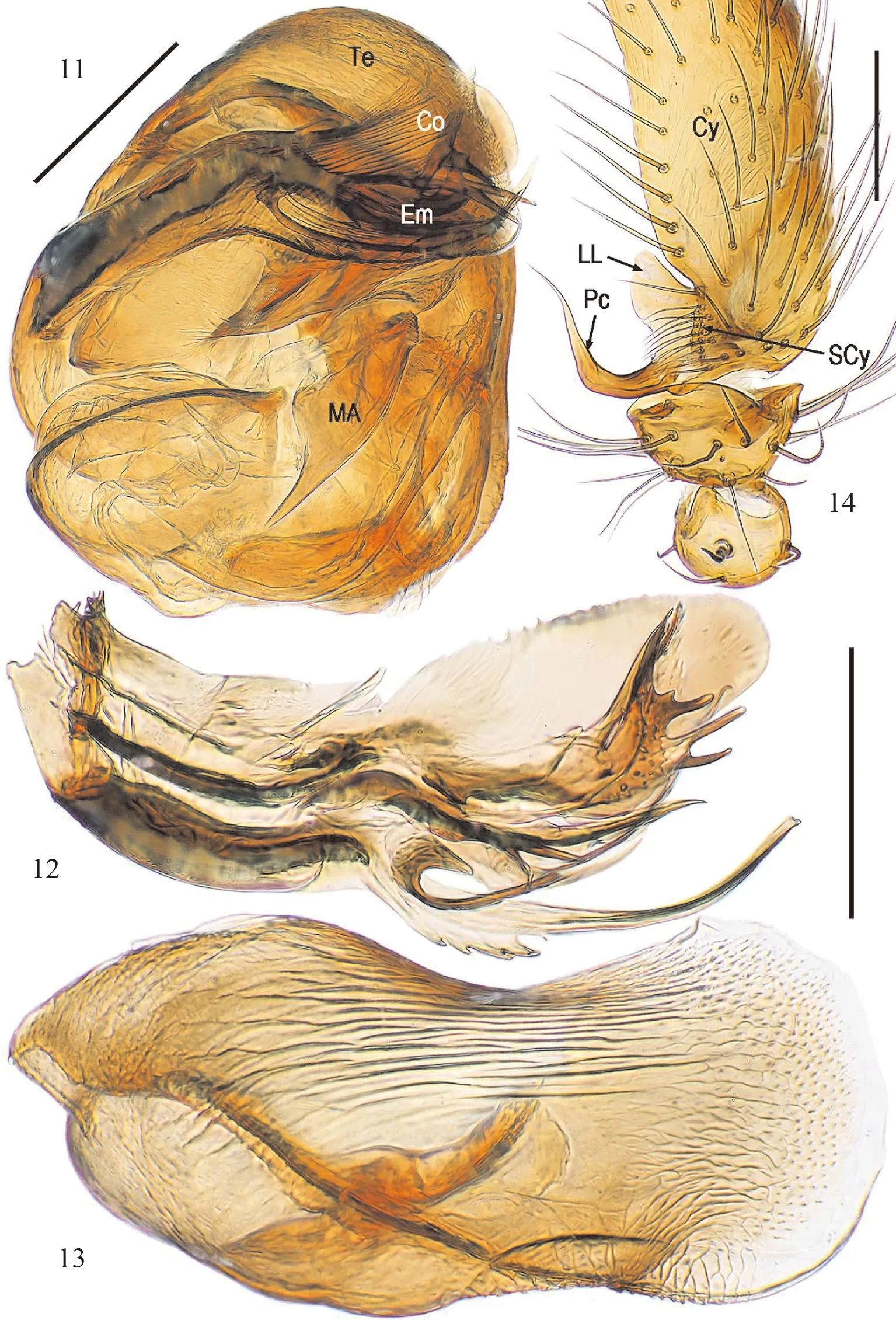
Figs. 11~14 Baalzebub youyiensis Zhao & Li, 2012, palpal pars (lactic acid-treated)11. Palpal bulb, prolateral view, 12. Embolus, ventral view, 13. Conductor, dorsal view, 14. Cymbium, retrolateral view; Abbs: Co=conductor, Cy=cymbium, Em=embolus, LL=lateral lobe, MA=median apophysis, Pc=paracymbium, SCy=setae of cymbium, Te=tegulum; Scale bars=0.10 mm.
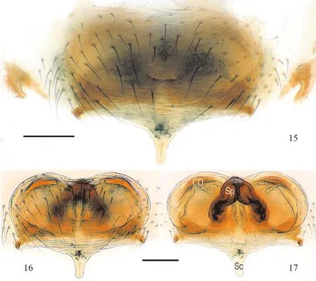
Figs. 15~17 Baalzebub youyiensis Zhao & Li, 2012, female copulatory organ 15. Epigyum (untreated), ventral view, 16. Ditto (lactic acid-treated), ventral view, 17. Vulva, dorsal view; Abbs: CD=copulatory duct, Sc=scape, Sp=spermatheca; Scale bars=0.10 mm.
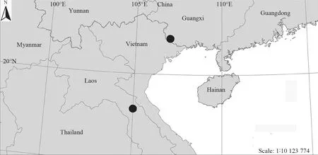
Fig. 18 Distribution records of Baalzebub youyiensis Zhao & Li, 2012
*通信作者Corresponding author, E-mail:linyucheng@scu.edu.cn
友谊巴力蛛BaalzebubyouyiensisZhao & Li, 2012(图1~图17)
鉴别特征:该种蜘蛛雄性近似于B.albonotatus(Petrunkevitch, 1930)和B.brauni(Wunderlich, 1976),均具有一簇跗舟刺毛和尖长的副跗舟,但可根据副跗舟旁具一宽阔侧膜、中突修长、斧状引导器和多分枝的插入器等特征与该属其他种区分。
猜你喜欢
垂钓(2023年11期)2024-01-21 16:07:04
垂钓(2023年9期)2023-12-10 19:39:30
中小学校长(2021年9期)2021-10-14 14:36:16
小朋友·快乐手工(2018年3期)2018-04-22 11:48:52
小学阅读指南·低年级版(2017年6期)2017-06-12 08:22:47
中国眼镜科技杂志(2016年9期)2016-12-01 06:37:33
飞碟探索(2016年5期)2016-05-10 23:44:30
饲料博览(2016年7期)2016-04-05 14:20:34
生物骨科材料与临床研究(2016年6期)2016-03-06 08:20:12
小朋友·快乐手工(2015年1期)2015-03-13 00:05:24
