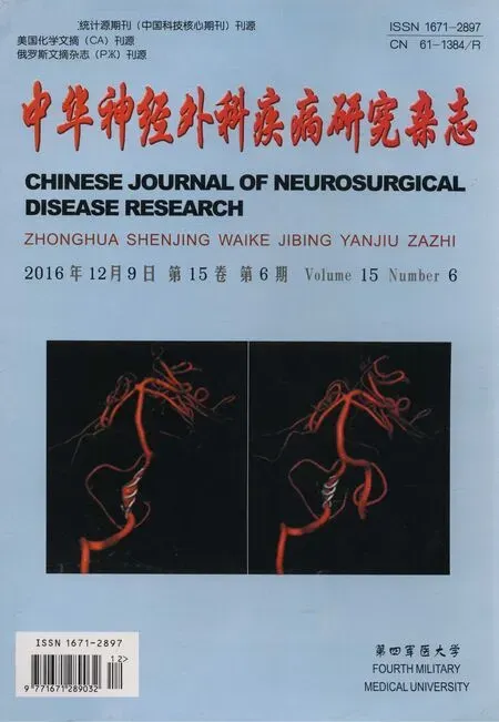
低氧条件下BAVM动物模型VSMC中TGF-β表达及细胞功能改变
潘俊 赵晓勇 张晓丽
(1海南省儋州市人民医院神经外科,海南 儋州 571700;2南方医科大学附属花都医院神经外科,广东 广州 510800)
·论著·
低氧条件下BAVM动物模型VSMC中TGF-β表达及细胞功能改变
潘俊1赵晓勇2*张晓丽2
(1海南省儋州市人民医院神经外科,海南 儋州 571700;2南方医科大学附属花都医院神经外科,广东 广州 510800)
目的探讨分析缺氧对脑动静脉畸形(BAVM)血管内膜平滑肌细胞(VSMC)中转化生长因子-β(TGF-β)表达及VSMC细胞功能改变。方法建立稳定的BAVM实验用猪动物模型,分离脑底微血管网(RM)的VSMC后行原代培养。分组:①对照组采用正常实验用猪VSMC:(A组)21%O2浓度;②实验组采用BAVM模型实验用猪VSMC:(B组)21%O2浓度、(C组)1%O2浓度。细胞免疫荧光检测VSMC密度、实时定量-多聚酶链反应(RT-PCR)和蛋白印记(Western blot)法验证VSMC中TGF-β mRNA及蛋白差异、末端脱氧核苷酸转移酶介导的生物素脱氧尿嘧啶核苷酸缺口末端标记(TUNEL)法检测VSMC的调亡、Transwell测定VSMC的侵袭能力。结果VSMC对缺氧敏感,实验组与对照组比较及实验组组间比较,TGF-β的mRNA及蛋白差异有统计学意义(Plt;0.01)、72 h VSMC凋亡及细胞侵袭数目差异有统计学意义(Plt;0.01)。结论缺氧对动物模型VSMC中TGF-β表达有显著的影响且促使VSMC凋亡及侵袭能力增加,加速畸形血管团的形成。在细胞分子生物学水平分析能为BAVM临床治疗提供一种有益的思路。
缺氧; 猪模; 血管内膜平滑肌细胞; 血管内皮生长因子; 细胞功能
脑动静脉畸形(brain arteriovenous malformations,BAVM)是中枢神经系统最为常见的先天性脑血管异常[1]。猪的脑底微血管网(rete mirabile,RM)结构与人BAVM畸形血管团高度相似[2],本研究是先建立稳定型实验用猪BAVM模型基础上,用缺氧因素干预分离培养好RM的原代血管内膜平滑肌细胞(vessels smooth muscle cells,VSMC),分析缺氧因素对于VSMC中转化生长因子-β(transforming growth factor-β,TGF-β)的影响和VSMC的细胞功能作用,为BAVM畸形血管团的加速形成机制提供可能的基础实验研究理论。
材料与方法
一、实验组建立稳定的BAVM动物模型
实验小型猪60头,由南方医科大学动物实验中心提供,3~4个月龄,体重20~25 kg。实验组猪模(B组,n=20;C组,n=20)用2.5%硫贲妥钠10~15 mg/kg静脉诱导后气管插管,机控呼吸,芬太尼分次静脉注射维持麻醉[3]。再行暴露和分离左颈总动脉、左、右颈外动脉、左咽升动脉和左颈外静脉。结扎左颈外动脉、左颈总动脉近心端和左颈外静脉远心端的残端,将左咽升动脉与左颈外静脉端端斜切口吻合,结扎左咽升动脉降支和枕动脉降支。
二、实验方法
1.29 d行手术切除对照组(A组,n=20)和实验组猪模的RM,分别置入含5×10⁵μ/L青、链霉素的D-hank's溶液中,4℃ 30 min。取内膜内皮细胞液氮保存后匀浆,消化液(0.25 g胰酶+0.1 g胶原酶Ⅲ+100 ml 磷酸缓冲盐溶液(phosphate buffered solution,PBS),用0.22 μm的过滤器除菌消化约10 min,将终浓度为1×10⁵/ml细胞接种于培养瓶,抗细胞角蛋白和抗珠蛋白抗体鉴定[4]。
2.培养基为10%胎牛血清(10% fetal balf serum,FBS),条件为37℃、5%CO2。A组:正常氧压培养箱中培养(21% O2);B组:正常氧压培养箱中培养(21% O2);C组:低氧培养箱中培养(1% O2)。
3.在灭菌的培养皿中铺上3块细胞爬片(4%多聚赖氨酸处理的圆片),取细胞悬液分别滴到圆片上,4%多聚甲醛(paraformaldehyde,PFA)固定处理30 min,0.2% TxitonX-100室温处理5 min、PBS漂洗5 min,3次。室温封闭:置于试剂盒中保持湿润,37℃,30 min;滴加1∶200 PBS稀释的绿色荧光一抗(CD34为1∶200),阴性对照用PBS代替一抗,室温反应30~40 min,80%甘油封片,置显微镜下观察拍片。
4.实时PCR和Western blot法检测VSMC中TGF-β mRNA及蛋白:3组VSMC总RNA提取后逆转录合成cDNA,均加入0.3 ml氯仿,4℃ 13 000 rpm离心15 min。弃上清,清洗、振荡后加入30~50 μl的焦碳酸二乙酯(diethy pyrocarbonate,DEPC)水溶解RNA。置于实时PCR仪(C1000型,美国Bio Rad公司)上进行PCR反应,β-actin和目的基因同时扩增,扩增反应条件如下:①预变性Reps:95℃ 30 s;②PCR反应Reps:95℃ 5 s、60℃ 34 s;③β-actin:85℃收集荧光,TGF-β:84.3℃收集荧光;④95℃,5 min,35个PCR循环,建立熔解曲线和扩增曲线,引物设计结果列表(表1)。
表1 设计实时PCR的基因引物
Tab 1 Designed real-time PCR gene primers

GeneNameTwo⁃wayprimersequences(5'⁃3')Annealingtemperature(℃)Productlength(bp) β⁃actinF:CCTGTACGCCAACACAGTGC57211 R:ATACTCCTGCTTGCTGATCC TGF⁃βF:CCTGAAGCTGACCCAGGTAG58133 R:TTCCAAACTGCATCAATGAAT
Note:F:upstream primer sequences,R:downstream primer sequences
Western blot蛋白质印迹法;3组均加入裂解液(radio immunoprecipitation assay,RIPA)200 μl匀浆,于冰上加苯甲基磺酰氟(phenylmethanesulfonyl fluoride,PMSF) 2 μl,RIPA:PMSF=100∶1,4℃,12 000 rpm离心1 h,取上清后Bradford法测定蛋白含量。行聚丙烯酰胺凝胶(polyacrylamide gelelectrophoresis,SDS-PAGE)电泳,将一抗TGF-β(1∶3 000,Santa Cruz)和β-actin (1∶1 000,Santa Cruz)加入到封闭液中混均行二抗反应。用封闭缓冲液稀释的二抗杂交溶液,置于振荡培养箱中进行杂交(26℃,80 rpm/min,l h);弃二抗溶液,用缓冲盐溶液(TBST)洗膜3次(置于振荡培养箱中,26℃,80 rpm/min,10 min/次);化学发光试剂(electrochemiluminescence,ECL,购自上海普飞生物公司)检测,利用BIO-RAD2000凝胶成像系统及QUANTITY ONE分析系统测定条带的积光光密度值。
5.末端脱氧核苷酸转移酶介导的生物素脱氧尿嘧啶核苷酸缺口末端标记(terminal deoxynucleotidyl transferase mediated biotinylated deoxyuridine triphosphate,TUNEL)法检测VSMC调亡:培养皿中铺上3块细胞爬片,贴片30 min左右;室温孵育10 min。用10 mM Tris溶液稀释2 mg/ml的蛋白酶K溶液,灭活内源过氧化物酶活性。在光学显微镜下观察呈现深棕色染色的细胞为凋亡细胞,每张玻片随机选择5个高倍镜视野,5个视野的平均数代表调亡细胞的数目[5]。
6.侵袭小室(Transwell)测定VSMC的侵袭能力
均匀的铺在Transwell侵袭小室(美国CORNING公司24孔板,孔径为8 mm,膜直径为6.4 mm)上室的纤维聚碳酸酷膜上,每孔50 ul,放置于37℃孵育Transwell板2 h。
将3组的侵袭小室分别放入21% O2及1% O2(37℃,5%CO2)中继续孵育48 h和72 h,可见VSMC贴附浸润膜的下室继续生长。
三、统计学分析
数据采用SPSS 20.0软件分析计量资料以(x±SD)表示,多组比较采用单因素方差分析;组间比较用LSD检验。当Plt;0.05时,认为差异具有统计学意义。
结 果
一、AVM稳定实验猪模型的建立
端端吻合后引流静脉见图1A、B。均建成由五支供血动脉-畸形血管团-单支引流静脉的稳定实验猪模型,见图1C、D。
二、VSMC的鉴定
VSMC用胰蛋白酶消化后呈圆形,见图2A(倒置显微镜NF-2000 Nikon)。在透射电镜(transmission electron microscope,TEM)见图2B。
三、不同O2条件下细胞免疫荧光检测VSMC密度比较,见图3(荧光抗体染色×200)。
四、各组VSMC中TGF-β mRNA及蛋白比较
各组经内参校正后得出2-△△Ct。实验B、C组中TGF-β mRNA和蛋白均高于A组,差异有显著性(Plt;0.01)。B组和C组相比较,差异有统计学意义(Plt;0.01,图4,表2、3)。

图1 吻合后引流静脉及造影
Fig 1 Drainage vein and angiography after anastomosis
A:L-APA and L-EJV end to end anastomosis;B:L-APA and L-EJV end to end anastomosis after vascular filling;C:Immediately after the procedure (the arrow showed the L-APA-EJV anastomosis);D:Post-operative 28 d angiography (arrow showed L-APA-EJV anastomosis,drainage vein thickening.
图2 VSMC的鉴定
Fig 2 Identification of VSMC
A:Primary culture of VSMC (×100);B:VSMC cells under transmission electron microscope (×l0000)
图3 不同O2条件下细胞免疫荧光检测VSMC密度比较
Fig 3 Comparison of VSMC density of cells under different O2conditions
A:Cultured VSMC by 21% O2(arrow showed CD34);B:Cultured VSMC by 1% O2(arrow showed CD34)


GroupnTGF⁃ββ⁃actinTGF⁃β/β⁃actin GroupA201.93±0.77a1.92±0.74a1.01±1.04a GroupB202.87±1.94b2.05±0.81b1.40±3.03b GroupC203.51±1.25b2.03±0.88b1.73±1.42b
aPlt;0.01,vsGroup B,C;bPlt;0.01,vsGroup A.

图4 Western Blot法检测各组蛋白TGF-β结果
Fig 4 VSMC TGF-β protein by Western Blot


GroupnTGF⁃ββ⁃actinTGF⁃β/β⁃actin GroupA203.83±0.63a5.47±1.13a0.70±0.56a GroupB204.99±1.77b5.69±1.41b0.88±1.26b GroupC207.02±1.23b5.50±1.35b1.28±0.91b
aPlt;0.01,vsGroup B,C;bPlt;0.01,vsGroup A.
五、TUNEL法检测VSMC调亡和Transwell侵袭结果(表4,图5)
24、48 h三组VSMC调亡和侵袭数目差异无统计学意义(Pgt;0.05)。72 h B、C组细胞调亡数目和侵袭显著增多,且C组显著多于B组,差异有统计学意义(Plt;0.01)。

图5 不同O2条件下VSMC凋亡和侵袭下室结果(结晶紫染色,×200)
Fig 5 VSMC apoptosis and room numbers under attacking under different O2conditions (crystal violet staining,×200)
A:21% O2VSMC apoptosis at 72 h;B:1% O2VSMC apoptosis at 72 h (arrows indicated apoptotic cells);C:21% O2VSMC hit at 72 h (arrows indicated invading cells);D:1% O2VSMC hit at 72 h.


TimeGroupAapoptosishitGroupBapoptosishitGroupCapoptosishit 24h5.53±1.02a1.97±0.54a5.84±0.97a2.03±0.18a5.77±1.01a2.01±0.20a 48h6.87±1.55b2.25±0.66b6.96±1.14b2.38±0.51b6.91±1.09b2.32±0.55b 72h6.64±1.37b2.22±0.43b15.81±3.93b10.05±1.92b20.18±3.47b18.65±4.17b
aPlt;0.01,vsGroup B,C;bPlt;0.01,vsGroup A.
讨 论
VSMC是构成血管壁的最主要细胞。正常情况下,血管中膜的VSMC是保证血管壁的张力及收缩运动的结构基础[6]。近年研究认为[7],BAVM血管壁中的VSMC受损,收缩能力下降,管壁变薄弱,管壁扩张[8]。TGF-β与BAVM密切相关,其信号蛋白功能的低失可导致明显血管结构异常[9]。
在本研究中,首先建立稳定的BAVM动物模型,然后分离培养实验组和对照组的VSMC,采用原代细胞进行不同氧浓度条件下培养。免疫绿色荧光检测在不同氧浓度培养后VSMC的分布密度,缺氧的条件可以导致VSMC的密度明显减少,这说明VSMC对缺氧敏感,缺氧可以引起血管内膜构筑的改变,使其薄化。Real-time PCR和Western Blot验证TGF-β mRNA及蛋白差异,对照组与实验组比较(Plt;0.01);实验组组间比较 (Plt;0.01);考虑缺氧因素可以引起TGF-β的表达水平显著上调,这对于畸形血管团的形成有促进作用。TUNEL检测VSMC调亡和Transwell的侵袭结果,24 h、48 h三组VSMC调亡和侵袭数目差异无统计学意义(Pgt;0.05)。72 h B、C组细胞调亡数目和侵袭显著增多,C组显著于B组,差异有统计学意义(Plt;0.01)。说明在不同氧浓度条件下,早期VSMC能够耐受轻度的缺氧,但随着VSMC缺氧程度加重,VSMC中线粒体功能失调,导致VSMC调亡数目和侵袭显著增多。BAVM的畸形血管团结构已经重铸[10],致使VSMC分布及密度变化,在缺氧因素作用下加速了VSMC凋亡。
综上所述,缺氧因素对BAVM猪模VSMC中TGF-β表达有显著的作用,加速了畸形血管团的形成,并且促使VSMC凋亡及侵袭能力增加。在细胞分子生物学水平分析缺氧因素对于VSMC中TGF-β的影响和VSMC的细胞功能作用,为BAVM畸形血管团的加速形成机制提供可能的基础实验研究理论。
1Mosch B,Reissenweber B,Neuber C,et al. Eph Receptor and ephrin ligands:important players in angiogenesis and tumor angiogenesis [J]. J Oncol,2010,15(2):235-285.
2Laakso A,Dashti R,Juvela S,et al. Natural history of arteriovenous malformation:presentation,risk of hemorrhage and mortality [J]. Acta Neurochir Suppl,2010,107:65-59.
3Gong Z,Qiao ND,Gu YX,et al. Polymorphisms of TGF-ΒA gene and susceptibility to hemorrhage risk of brain arteriovenous malformations in a Chinese population [J]. Acta Pharmacol Sin,2011,32(8):1071-1077.
4Schuster L,Schenk E,Giesel F,et al. Changes in AVM angio-architecture and hemodynamics after stereotactic radiosurgery assessed by dynamic MRA and phase contrast flow assessments:a prospective follow-up study [J]. E Rad,2013,21(6):1267-1276.
5Siebert E,Diekmann S,Masuhr F,et al. Measurement of cerebral circulation time using dynamic whole-brain CT-angiography:feasibility and initial experience [J]. Neurol Sci,2012,33(4):741-747.
6Fontanella M,Rubino E,Crobedd E,et al. Brain arteriovenous malformations are associated with interleukin-1 cluster gene polymorphisms [J]. Neurosurgery,2012,70(1):12-17.
7Zacharia BE,Bruce S,Appelboom G,et al. Occlusive hyperemia versus normal perfusion pressure breakthrough after treatment of cranial arteriovenous malformations [J]. Neurosurg Clin N Am,2012,23(l):147-151.
8Jia C,Hegg CC. Neuropeptide Y and extracellular signal-regulated kinase mediate injury-induced neuroregeneration in mouse olfactory epithelium [J]. Mol Cell Neurosci,2012,49(2):158-160.
9A1-Shahi Salman R. The outlook for adults with epileptic seizure(s) associated with cerebral cavernous malformations or arteriovenous malformations [J]. Epilepsia,2012,53( Suppl 4):34-42.
10Sturiale CL,Puca A,Sebastiani P,et al. Single nucleotide polymorphisms associated with sporadic brain arteriovenous malformations:where do we stand ? [J]. Brain,2013,136 (Pt 2):665-681.
ChangesofTGF-βexpressionandcellularfunctioninBAVManimalmodelsunderlowoxygencondition
PANJun1,ZHAOXiaoyong2,ZHANGXiaoli2
1DepartmentofNeurosurgery,People'sHospitalofDanzhouCity,Danzhou571700;2DepartmentofNeurosurgery,HuaduHospitalAffiliatedtoSouthMedicalUniversity,Guangzhou510800,China
ObjectiveThe effect of hypoxia on endothelial growth factor (TGF-β) expression and functions of VSMC cells in brain arteriovenous malformations (BAVM) vascular intimal smooth muscle cells (VSMC) were studied.MethodsThe BAVM experimental pigs were well established and the separation of cerebral microvascular network (RM) was cultured by VSMC. There were three groups:the control group with normal experimental porcine VSMC (Group A) under 21%O2concentration;the experimental group using porcine VSMC (Group B) under 21%O2concentration,and 1%O2concentration (Group C). VSMC density was detected by immunofluorescence,TGF-β mRNA and protein of VSMC were detected by quantitative real-time polymerase chain reaction and Western blot,respectively. Terminal deoxyribonucleic transferase mediated dUTP nick labeling (TUNEL) method was used to detect the difference of apoptosis of VSMC,and Transwell was used for the determination of VSMC invasion.ResultsVSMC was sensitive to hypoxia. There were statistically significant differences in mRNA and protein of TGF-β between control and experimental groups and among the experiments (Plt;0.01). There was statistically significant difference in VSMC apoptosis and cell invasion number at 72 h (Plt;0.01).ConclusionHypoxia has a significant effect on expression of TGF-β in VSMC of animal model,and it improves the apoptosis and invasion of VSMC and accelerates the formation of the vascular malformation. The analysis of molecular biology can provide a useful idea for the clinical treatment of BAVM.
Hypoxia; Pig model; Vascular smooth muscle cell; Vascular endothelial growth factor; Cell function
1671-2897(2016)15-501-05
R 651.1
A
潘俊,主治医师,博士,E-mail:ace15171517@sina.com
*通讯作者:赵晓勇,副教授、副主任医师,E-mail:ace0829@sina.com
2015-10-12;修回时间:2016-01-20)

