Fe3O4磁性纳米材料的绿色合成及其在潜指印显 现中的应用研究
喻彦林,颜 磊
Fe3O4磁性纳米材料的绿色合成及其在潜指印显 现中的应用研究
喻彦林1,2,颜 磊1,*
(1.西南政法大学刑事侦查学院 重庆高校物证技术工程研究中心,重庆 401120;2.重庆大学生物工程学院,重庆 400044)
摘要:目的 制备新型Fe3O4磁性 纳米粉末用于多种客体物表面潜指印的显现。方法 采用高效的微波辅助水热合成法制备高纯度的Fe3O4磁性纳米材料,利用高分辨透射电子显微镜、X射线衍射对产物尺寸及晶型进行表征。以制备的Fe3O4磁性纳米粉末为显现剂,对金属、玻璃、塑料以及不同粗糙程度的纸张、粗糙的桌面等非渗透性、渗透性客体物表面潜在指印进行粉末显现,并与传统显现粉末(金粉、银粉和黑色磁粉)进行了对比。结果 显现效果令人满意,显现的指印纹线清晰、细节特征明显,尤其对于粗糙程度较高的滤纸及凹凸不平的表面亦有较好的显现效果。结论 制备的Fe3O4磁性纳米粉末可用于多种不同性质的客体物表面潜指印的显现,且该材料制备方法简便、稳定性好、尺寸和磁性适中,有望用于侦查的实际工作中。
关键词:潜指印显现;磁性纳米粉末;微波合成;四氧化三铁纳米颗粒
粉末显现法操作简便、价廉经济、显现效果较好,广泛的使用于非渗透性客体表面潜指印的显现中。但由于这些传统显现粉末(如金粉、银粉、碳粉、磁粉等)的尺寸多为微米或亚微米,对于渗透性(如纸张等)以及较为粗糙的表面显现效果欠佳。随着纳米材料和纳米技术的不断发展,具有均一、超微尺寸的功能化纳米材料在潜指印的显现中受到了越来越多的关注[1-18]。Liu等[6]采用水溶性的荧光CdTe 量子点实现了光滑物体表面潜指印的显现,在紫外灯等的照射下纹线细节清晰可辨。Choi等[8]对金属纳米颗粒在潜指印显现中的应用进行了综述。Leggett等[17]发展了一种“智能(intelligent)”方法,通过功能化荧光纳米材料中抗体与手印物质中残留的科替林(尼古丁的代谢产物,会随着人的汗液排出)之间的特异性结合,在筛选出指印遗留者为吸烟者的同时实现了其潜指印的荧光显现。Hussain等[14]仅以汗液中的成分为还原剂和保护剂,通过金纳米颗粒的原位合成实现了潜指印的显现,Algarra等[13]采用荧光量子点复合材料(PPH-CN和PPH-SH @CdS QDs)对塑料、玻璃、钢铁、陶瓷以及木质表面的潜指印进行了成功的显现。该小组还采用具有橘色荧光的无毒PPH-NH2@CdSe量子点复合物实现了不锈钢镊子、手机屏幕及信用卡磁条等非渗透性物体表面的潜指印显现[18]。
本研究采用微波辐射辅助合成了具有磁性的Fe3O4纳米材料,操作简单、反应快速(仅需60 s),产物具有纯度高、性质稳定、尺寸适中(约80 nm)、分布均一、环境友好等特点。该纳米粉末被成功地应用于多种客体表面(包括渗透性与非渗透性,光滑与粗糙)潜指印的显现中,获得了令人满意的显现效果,尤其是对粗糙表面上的潜指印显现,其显现效果显著优于常用的金粉、银粉以及黑色磁粉。
1 材料与方法
1.1 实验材料及仪器
硫酸亚铁 (FeSO4·7H2O)、浓氨水(NH3·H2O) (成都科隆试剂厂,成都)。实验用水为超纯水,使用Milli-Q system (Millipore,Bedford,MA,USA)制备。实验中所用试剂均为分析纯,使用时未进行任何纯化处理。实验中使用的金粉、银粉和黑色磁粉为市售某品牌显现粉末。
实验中采用了FEI Tecnai G-20高分辨透射电镜(Hillsboro, OR, USA)和Ultima IV X-射线衍射仪(Rigaku Corporation, Japan)对合成的Fe3O4纳米颗粒进行表征。此外,在样品的合成和干燥过程中还使用了Galanz D8023CSL-K4 家用微波炉 (格兰仕,佛山),DZF-6050 型真空干燥箱(上海精宏实验设备有限公司,上海) ,配有pH玻璃电极的Orion 920A型酸度计(Thermo Electron Corporation, Waltham, MA,USA)。Nikon D7000型数码相机用于对显现指印的拍照固定。
1.2 磁性纳米材料的合成与表征
实验中所用的Fe3O4磁性纳米材料是在Zheng研究小组的文献报道[19]的基础上,进一步优化实验条件制得的。 实验中以微波辐射的方式水热合成了具有磁性的Fe3O4纳米材料。具体步骤如下:室温下,称取0.278 g FeSO4·7H2O 溶解于50.0 mL 超纯水中,并滴加一定量的浓氨水调节溶液的pH值为11.0。随后将上述溶液置于家用微波炉中,高火 (800 W)条件下微波辐射数分钟,反应溶液中墨绿色絮状悬浮物变为黑褐色小颗粒即得到具有磁性的Fe3O4纳米材料。随后在永磁体的作用下,将磁性纳米颗粒从溶液中分离出来,收集沉淀并水洗至溶液呈中性,将沉淀置于真空干燥箱中干燥后得到黑褐色Fe3O4磁性粉末备用。采用高分辨透射电子显微镜(high resolution transmission electron microscopy,HRTEM)、XRD (X-ray diffraction, XRD)对材料的尺寸和晶型进行表征。
1.3 潜指印的显现与固定
志愿者洗手自然晾干后,用手指轻触额头,以适当力度在各种客体(金属、玻璃、塑料、纸张等)上捺印指纹,并于室温下(20℃,60%)保存。为避免捺印差异对显现结果的影响,所有潜指印均采自同一志愿者,且保证每次捺印力度均匀一致。对照实验中所采用的潜指印承载客体包括:玻璃(载玻片)、金属(门把手)、油漆金属(仪器设备的外壳)、塑料(普通矿泉水瓶和脸盆)、各种纸张(滤纸、称量纸和普通办公文印纸)以及粗糙的实验台面。实验中捺印完成后0.5 h采用刷显法对各种客体表面的潜指印进行显现,并采用数码相机对显现的指印进行拍照固定。
2 结果与讨论
2.1 Fe3O4磁性纳米材料的合成及表征
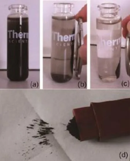
图1显示了水溶液中Fe3O4纳米颗粒对外加磁场的响应变化情况。悬浮在溶液中的Fe3O4纳米颗粒可以在5 s内被吸到永磁体的表面(图1b),15 s内溶液完全澄清透明(图1c)。这说明本研究中制得的Fe3O4纳米材料具有较好的磁响应能力,其粉末在磁性指纹刷刷头具有良好的吸附效果(如图1d)所示)。

图 1 水溶液中的Fe3O4纳米颗粒对外磁场的响应(a~c) 和Fe3O4纳米粉末在磁性指纹刷上的吸附(d)Fig.1 Photographs of magnetic nanoparticles dispersion in a vial (a) without, (b) with magnetic fi eld for 5s, and (c) with magnetic fi eld for 15s. (d) the photograph as prepared Fe3O4magnetic nanopowder was attracted by a magnetic brush.
实验中采用HRTEM对 Fe3O4纳米颗粒的尺寸进行了表征,结果如图2 所示:制备的磁性材料为类球形颗粒,粒径约为80 nm且尺寸分布较为均一。从Fe3O4纳米颗粒的XRD图(图2)中可以看出,制备的磁性材料具有较强的衍射峰,说明产物具有较好的晶体结构,衍射峰分别属于220、311、222、400、422、511和440 等七个不同的晶面[19],且谱图上没有其它衍射峰的存在,说明采用该法制备的材料具有较高的纯度。
2.2 潜指印的显现
潜指印显现中的粉末法主要是利用粉末物质与指纹残留物中的湿润或/和油脂类物质之间的静电吸附实现,该法操作简便,对新鲜指印有较好的显现效果[1]。本研究中亦采用粉末法将制得的Fe3O4纳米粉末用于多种不同性质的物体表面潜指印的显现。

图 2 Fe3O4纳米颗粒的HRTEM 图(左)和XRD 谱图(右)Fig.2 HRTEM image (left) and XRD pattern (right) of the prepared magnetic nanoparticles
在入室盗窃案件中,潜指印往往出现在房屋的进出口,如门把手及窗户玻璃等,因此这些案发现场的重点部位的潜指印显现是实际工作中的关注点。实验中采用Fe3O4磁性粉末对金属(门把手)、玻璃、塑料等非渗透性客体表面的潜指印进行了显现,结果如图3所示。无论在油漆金属物还是玻璃的表面都获得了高质量的指印,纹线清晰、细节特征显著且与背景有很好的对比度,即使是对不完整的指印其显现效果也非常好(图3b、c)。在塑料瓶上以及盆的边缘上,指印显现的对比度稍逊于前面二者,主要因为客体表面的弧度以及磨损造成了轻微的背景着色,但并不影响纹线的观察和细节特征的比对(图3d、e)。传统的磁性粉末不适用于金属表面,而本研究中制备的Fe3O4纳米粉末则成功的显现了金属把手上的潜指印(图3a,由于门把手面积较小且其表面具有一定的弧度,因此显现之后获得的指印面积较小,但其清晰的纹线走向和细节特征仍能在实际工作中为我们提供线索。

图 3 非渗透性表面潜指印的显现效果。(a)金属表面(门把手);(b)油漆金属表面(空调外壳);(c)玻璃表面(载玻片);(d)塑料表面(矿泉水瓶);(e)塑料表面(塑料盆)。Fig.3 Latent fi ngermarks on non-porous surfaces developed with the prepared Fe3O4magnetic nanopowder. (a) on metal surface; (b) onpaint metal surface; (c) on glass; (d) on water bottle; and (e) on plastic basin.

图 4 各种纸张表面潜指印的显现效果。(a)称量纸;(b) 普通文印纸;(c)滤纸。Fig.4 Latent fi ngermarks on porous (various papers) surfaces developed with the prepared Fe3O4magnetic nanopowder. (a) weighing paper;(b) common offi ce paper; and (c) fi lter paper.

图 5 Fe3O4磁性粉末与传统粉末对颗粒板上潜指印的显现效果对比图。从左至右使用的粉末依次为:实验中制备的Fe3O4磁性粉末、金粉、银粉和黑色磁粉。Fig.5 Comparison of prepared Fe3O4magnetic nanopowder with other common powders using freshly deposited fi ngermarks on granular board. From left to right: prepared Fe3O4magnetic nanopowder, copper powder, aluminum powder and black magnetic powder.
纸张是一类常见的渗透性客体,其粗糙程度的不同使其在渗透性方面存在较大的差异。实验中以称量纸、普通文印纸以及滤纸(三者的粗糙程度递增)上的潜指印为分析对象,考察了Fe3O4磁性纳米粉末的显现能力。如图4所示,在称量纸和普通文印纸上获得了清晰的指印,而滤纸则由于较为粗糙产生了严重的背景着色,从而降低了显现的对比度,尽管如此,仍能辨认出其基本纹型及纹线的流向。
传统的磁性粉末是由还原性的铁粉和硒粉或者碳粉构成的混合物,其尺寸大约为300目(约为50 μm),主要适用于光滑表面以及部分塑料表面。本研究中制备的磁性粉末为纯度较高Fe3O4纳米颗粒,其尺寸约为80 nm,且具有较好的均一性和尺寸分布,除了对光滑客体物表面的潜指印具有较好的显现效果外,对部分粗糙表面潜指印的显现具有显著的优势。实验中,以凹凸不平的实验台面为分析对象,使用Fe3O4纳米粉末对其表面的潜指印进行了显现。如图5所示,连续的纹线走向以及分歧、结合等纹线细节特征都得到了近乎完美的显现,且拥有较好的对比度而几乎没有出现背景着色的现象。

图 6 油漆物表面潜指印的显现效果Fig.6 Latent fi ngermark on paint metal surface developed with the prepared Fe3O4magnetic nanopowder. The inserted on the top right is of partial enlargement.
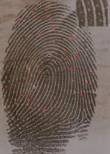
为了比较Fe3O4磁性纳米粉末与传统的常用粉末(金粉、银粉和黑色磁粉)在显现效果和适用范围方面的差异,进行了相应的对比实验。实验中采用刷显的方法依次用金粉、银粉、黑色磁粉和Fe3O4磁性纳米粉末分别对光滑客体(以玻璃、油漆物为代表)和粗糙的客体(以凹凸不平的颗粒板为代表)表面的新鲜潜指印进行了显现。实验结果表明:对于玻璃、光滑的纸张等光滑客体,黑色磁粉和Fe3O4磁性纳米粉末具有相当的显现效果,呈现出清晰的纹线和细节特征,且显现效果均优于金粉和银粉。采用Fe3O4磁性纳米粉末显现后的油漆物表面的指印,纹线细节特征清晰可辨,放大图上甚至清楚的展现了纹线上的汗孔分布(见图6);对于较为粗糙的表面,Fe3O4磁性纳米粉末在显现效果上优势更为明显。如图5所示,金粉、银粉显现的指印纹线难以辨识,黑色磁粉显现的指印纹型和纹线流向基本可辨,而Fe3O4磁性纳米粉末显现的指印纹线清晰,特征辨识度较高。
3 结 论
通过微波辐射辅助的方法高效(仅需60 s)的制备了尺寸约为80 nm的均一、球形磁性Fe3O4纳米颗粒,并采用粉末刷显的方式将其成功的应用于多种非渗透性、渗透性客体表面潜指印的显现中。在玻璃、带漆金属、塑料以及部分纸张(较为光滑的称量纸和普通文印纸)表面获得了高质量的指印,纹线清晰、特征显著,尤其是在粗糙表面上的潜指印显现中,展现出明显的优势,可用于人身认定的实际工作中。Fe3O4磁性纳米粉末对金属把手、滤纸以及粗糙桌面上潜指印的显现,进一步拓展了磁性粉末的使用范围。
参考文献
[1] Sodhi GS, Kaur J. Powder method for detecting latent fi ngerprints: a review. Forensic Sci Int, 2001,120:172-176.
[2] Dilag J, Kobus H, Ellis AV. Cadmium sulfi de quantum dot/chitosan nanocomposites for latent fi ngermark detection. Forensic Sci Int, 2009,187:97-102.
[3] Wang YF, Yang RQ, Wang YJ, et al. Application of CdSe nanoparticle suspension for developing latent fingermarks on the sticky side of adhesives. Forensic Sci Int, 2009,185:96-99.
[4] Liu L, Gill SK, Gao YP, et al. Exploration of the use of novel SiO2nanocomposites doped with fluorescent Eu3+/sensitizer complex for latent fingerprint detection. Forensic Sci Int,2008,176:163-172.
[5] Theaker BJ, Hudson KE, Rowell FJ. Doped hydrophobic silica nano- and micro-particles as novel agents for developing latent fi ngerprints. Forensic Sci Int, 2008,174:26-34.
[6] Liu J, Shi Z, Yu Y, et al. Water-soluble multicolored fl uorescent CdTe quantum dots: Synthesis and application for fi ngerprint developing. J Colloid Interf Sci, 2010,342:278-282.
[7] Islam NUI, Ahmed KF, Sugunan A, et al. Forensic fi ngerprint enhancement using bioadhesive chitosan and gold nanoparticles//Proceedings of the 2nd IEEE International Conference on Nano/Micro Engineered and Molecular Systems. Bangkok,Thailand, 2007:16-19.
[8] Choi MJ, McDonagh AM, Maynard P, et al. Metal-containing nanoparticles and nano-structured particles in fi ngermark detection. Forensic Sci Int, 2008,179:87-97.
[9] Jin YJ, Luo YJ, Li GP, et al. Application of photoluminescent CdS/PAMAM nanocomposites in fi ngerprint detection. Forensic Sci Int, 2008,179:34-38.
[10] Theaker BJ, Hudson KE, Rowell FJ. Doped hydrophobic silica nano- and micro-particles as novel agents for developing latent fi ngerprints. Forensic Sci Int, 2008,174:26-34.
[11] Becue A, Scoundrianos A, Champod C, et al. Fingermark detection based on the in situ growth of luminescent nanoparticles-Towards a new generation of multimetal deposition. Forensic Sci Int,2008,179:39-43.
[12] Becue A, Moret S, Champod C, et al. Use of quantum dots in aqueous solution to detect blood fingermarks on non-porous surfaces. Forensic Sci Int,2009,191:36-41.
[13] Algarra M, Jiménez-Jiménez J, Moreno-Tost R, et al. CdS nanocomposites assembled in porous phosphate heterostructures for fi ngerprint detection. Optl Mater, 2011,33:893-898.
[14] Hussain I, Hussain SZ, Habib ur R, et al. In situ growth of gold nanoparticles on latent fi ngerprints-from forensic applications to inkjet printed nanoparticle patterns. Nanoscale, 2010,2:2575-2578.
[15] Choi MJ, McBean KE, Ng PHR, et al. An evaluation of nanostructured zinc oxide as a fluorescent powder for fingerprint detection. J Mater Sci, 2008,43:732-737.
[16] Choi MJ, Smoother T, Martin AA, et al. Fluorescent TiO2powders prepared using a new perylene diimide dye: Applications in latent fi ngermark detection. Forensic Sci Int, 2007,173:154-160.
[17] Leggett R, Lee-Smith EE, Jickells SM, et al. “Intelligent” fi ngerprinting: Simultaneous identification of drug metabolites and individuals by using antibody-functionalized nanoparticles. Angew Chem Intl Ed, 2007,46:4100-4103.
[18] Algarra M, Radotić K, Kalauzi A, et al. Fingerprint detection and using intercalated CdSe nanoparticles on non-porous surfaces. Anal Chim Acta, 2014,812:228-235.
[19] Zheng B, Zhang M, Xiao D, et al. Fast microwave synthesis of Fe3O4and Fe3O4/Ag magnetic nanoparticles using Fe2+as precursor. Inorg Mater, 2010,46:1106-1111.
重庆市科委科技计划项目自然科学基金(cstcjjA00026);西南政法大学校级项目(2014XZQN-02)
中图分类号:DF794.1
文献标识号:A
文章编号:1008-3650(2016)01-0057-05
收稿日期:2015-04-22
基金项目:国家自然科学基金 (51401174);重庆市教委科技项目 (KJ130109, No.KJ120103);
作者简介:喻彦林,讲师,博士,研究方向为物证技术学。 E-mail: yuyanlin@sina.com
* 通讯作者:颜 磊,讲师,博士,研究方向为物证技术学。 E-mail: yanlei@126.com
Application of Fe3O4Nanopowder in the Development of Latent Fingermarks
YU Yanlin1,2, YAN Lei1, *(1. Forensic Engineering Research Center of Universities in Chongqing, Criminal Investigation College of Southwest University of Political Science and Law, Chongqing 401120, China; 2. Bioengineering College of Chongqing University, Chongqing 400044, China)
ABSTRACT:Objective To prepare novel magnetic Fe3O4nanopowder for developing latent fi ngermarks on various surfaces,including the porous and nonporous, the smooth and rough. Methods Highly pure Fe3O4nanoparticles were synthesized by microwave irradiation, and characterized by high resolution transmission electron microscopy (HRTEM) and X-ray diffraction (XRD). The process of synthesis was fast, simple and of higher yield. It was also an effective and environmentfriendly technique. The prepared magnetic Fe3O4nanopowder was applied to develop latent fi ngermarks on various surfaces,including porous and nonporous, smooth and rough, using the traditional magnetic brushing technique. The nanopowders were compared with some conventional powders, such as copper powder, black magnetic powder and aluminum powder. Results Magnetic Fe3O4nanopowder developed the sharp and clear latent fi ngermarks with good contrast. Either on the painted metal or glass surface, the developed prints showed clear ridges and good contrast, even for the minutiae and pore. Especially for the rough surface, the small, fi ne nano-sized magnetic powder demonstrated great advantages. Conclusions The magnetic Fe3O4nanopowder can be widely used to develop latent fingerprints on different surfaces, such as non-porous surfaces (painted metal, glasses, plastic etc.) and porous surfaces (commonly used papers), ideal for latent fi ngerprints to develop on the rough surface, such as that of fi lter paper and uneven panel. Powdering technique of latent fi ngerprint development with this novel Fe3O4nanoparticle is proven to be relatively simple, inexpensive, and effective.
KEY WORDS:latent fi ngermark detection; magnetic nanopowder; microwave irradiation; Fe3O4nanoparticles

