Reducing oxidative stress and hepatoprotective effect of water extracts from Pu-erh tea on rats with high-fat diet
Jingjing SuXueqing WngWenjun SongXioli BiChngwen Li
a Tianjin Key Laboratory of Food Biotechnology,College of Biotechnology and Food Science,Tianjin University of Commerce,Tianjin 300134,China
b Yunnan Tasly Deepure Biological Tea Group Company Limited,Pu Erh,Yunnan 665100,China
Abstract Reducing oxidative stress and hepatoprotective effect of Pu-erh tea water extracts on rats fed with high-fat diet were researched for explaining health care of Pu-erh tea.Fifty SD rats were divided into five groups.The body weight was measured once a day.The malondialdehyde(MDA)and glucose (Glu) levels and the activities of alanine aminotransferase (ALT), aspartate aminotransferase (AST), nitric oxide synthase (NOS),and pyruvate kinase(PK)in serum were determined.Furthermore,the hepatic glycogen level(HGL)and the activities of hepatic total superoxide dismutase(T-SOD),catalase(CAT),and glutathione peroxidase(GSH-Px)were also measured after continuous administration for 12 weeks.The result demonstrated that Pu-erh extract caused the decreases in body weight,fat index,MDA and NOS levels,and the increases in hepatic T-SOD,CAT and GSH-Px activities,indicating that the extract may be due to inhibiting the increases of body weight and fat index,reducing oxidant stress state and inhibiting lipid peroxidation,thus decreasing the activities of ALT and AST,and protecting the liver in rat.Meanwhile,the extracts could increase the production of hepatic glycogen and the activity of PK,and reduce glucose level,protecting the liver from the diseases associated with type II diabetes.
Keywords: Pu-erh tea extracts;Oxidative stress;Hepatoprotective effect
1.Introduction
During the process of the aerobic metabolism, a certain amount of reactive oxygen species (ROS) inevitably produce along with the generation of energy,including superoxide radical anion,hydroxyl radical,hydrogen peroxide and nitric oxideetc.[1].Under normal physiological conditions,ROS is quickly removed by antioxidant enzymes associated with scavenging ROS or reducing substancesin vivo,thereby not leading to the accumulation of ROS.However,under pathological conditions,the amount of ROS production increases with the decrease of antioxidant enzymes activity.The accumulation of ROS will lead to the oxidative stress status and further cause membrane lipid peroxidation, intracellular protein and enzyme denaturation, DNA oxidative damage and tissue damage [2].It has been reported that oxidative stress is an important pathogenic mechanism of obesity-associated metabolic syndrome [3–8].Therefore, in recent years, increasing interests have been seen in researching and developing drugs for effectively reducing oxidative stress and improving antioxidant effects in the medical profession.
Although synthetic antioxidants such as butylated hydroxy toluene (BHT) and butyl hydroxy anisole (BHA) have exhibited limited therapeutic benefits for improving oxidative stress in pharmacotherapy[9,10],these compounds exist serious risks of side effects and toxicity.Therefore, developing alternatives for existing synthetic antioxidants with high efficiency and low toxicity of novel antioxidants from natural plants is of great significance for the treatment and prevention of diseases associated with oxidative stress.Teas,such as Pu-erh tea,green tea and oolong tea have numerous biological activities including anti-obesity, -oxidant, -allergic, -inflammatory and neuroprotective activities [11–15] and other functions are attributed to the chemical ingredients in teas, including theabrownins, tea polysaccharides, polyphenols, and flavonoids.These ingredients can be used as potentially natural antioxidants for reducing the oxidative stress statusin vivo[16–20].
Pu-erh, a kind of fermented tea, is producedviaa special fermentation process with high temperature and high humidity.During the process, microbes likeAspergillus nigerandSaccharomycetesgrow and convert some substances in Puerh into its unique flavor ingredients including theabrownins(TB), thearubigins, and polyphenols.Recently, TB, one of the main composition of tea extracts,has been demonstrated to play an important role in scavenging 1,1-diphenyl-2-picrylhydrazyl radical 2,2-diphenyl-1-(2,4,6-trinitrophenyl) hydrazyl (DPPH)and antioxidant functions [21].Pu-erh tea has been proven to exhibite a wide range of health benefits including anti-diabetics,-oxidation, -obesity, -mutagenic, and -atherosclerosis but few was reported on the mechanism of inducing oxidative stress of the liver[22–26].
Oxidative stress status occurs when ROS accumulate.Superoxide dismutases(SOD),one of antioxidant enzymes,catalyzes the dismutation of superoxide anion(•O−2)into O2and hydrogen peroxide(H2O2).Subsequently,H2O2is reduced to H2O by glutathione peroxidase(GSH-Px)in the cytosol,or by catalase(CAT) in the peroxisomes.SOD, CAT and GSH-Px are easily induced by oxidative stress.The activity levels of these enzymes have been used to quantify oxidative stress incells[27,28].Nitric oxide synthases(NOS)in a large number of different tissues synthesize nitric oxide(NO).NO is a very small,lipophilic,readily diffusible,chemically unstable molecule with a very short halflife(seconds).No plays a relevant role in signal transduction in physiopathology[29].Pyruvate kinase(PK)is a key enzyme that regulates glycolytic flux in response to the intracellular energy level.Malonic dialdehyde(MDA)is a major reactive aldehyde resulting from the peroxidation of polyunsaturated fatty acid in biological membrane.MDA generally is used as an indicator of the tissue damage involving a series of chain reactions.Excessively activated radicals can react with the unsaturated fatty acid of cytomembrane, causing lipid peroxidation and formingMDA.The liver is an organ with the multi-functions in the body,such as storing glycogen,secreting protein.Aminotransferase(ALT)and aspartate aminotransferase (AST), mainly distributed in hepatocytes,are sensitive indicators of liver injury.In this work,activities of nitric oxide synthase(NOS),pyruvate kinase(PK),alanine aminotransferase(ALT)and aspartate aminotransferase(AST)were determined;malondialdehyde(MDA)and glucose levels in serum, were measured; activities of total superoxide dismutase (T-SOD), catalase enzyme (CAT), and glutathione peroxidase (GSH-Px) in the liver were also determined.Then,the improving oxidative stress status and hepatoprotective effect of Pu-erh tea extracts in normal rats fed with high-fat diet were carried out in order to provide some information on Pu-erh tea’s health benefits.
2.Materials and methods
2.1.Materials and reagents
Pu-erh tea extracts were provided by the Yunnan Tasly Deepure Biological Tea Group Company Limited(Pu’er,Yunnan province, China).The Pu-erh tea was extracted with a ten-fold volume of water (1:10, w/v) at 90°C for 30 min twice.The extract solutions were combined, concentrated,and sprayed under vacuum to obtain Pu-erh tea extract powders.The contents of regular ingredients in Pu-erh tea powder including caffeine (89.6 mg/g), total polyphenols (211 mg/g),polysaccharide (103 mg/g), theaflavins (0.297 mg/g), thearubigins (1.82 mg/g) and theabrownins (48.6 mg/g).The total tea polyphenols were determined according to Folin–Ciocalteu’s test using GA as the standard [27].Polysaccharides were quantitated using the anthrone-sulfuric acid method with modifications as described [29].Three kinds of tea pigments,including TFs, thearubigins (TRs), and theabrownins (TBs),were analyzed using spectrophotometry [30].The flavonoid content was calculated using the aluminum trichloride colorimetric method [31].The compositions of polyphenol in Pu-erh tea powder, which were gallic acid (GA, 1.57%),(−)-gallocatechin (GC, 0.64%), (−)-epigallocatechin (EGC,0.25%),(+)-catechin(C,3.27%),(−)-epicatechin(EC,0.94%),(−)-epigallocatechin −gallate(EGCG,1.75%),(−)-epicatechin gallate (ECG, 1.16%) and (+)-catechin gallic acid ester(CG,0.54%), respectively, were determined by high-performance liquid chromatography (HPLC) analysis using a Waters 2695 system controller.The stationary phase was a Hypersil ODS2-C18 packed column (4.6×250 mm, 5 μm) (Thermo Fisher,Inc., United States), the mobile phase A & B is 2% acetic acid and 100% acetonitrile at a flow rate of 1 mL/min.The tea extract was filtered through a 0.45 μm filter disk,and then,5 μL of extracted solution was directly injected into the HPLC system.The column temperature was 30°C and the detection wavelength was 280 nm with Waters 484 turnable absorbance detector.Gradient elution conditions: 6.5%–8% B, 0–16 min;8%–15% B, 16–20 min; 15%–25% B, 20–25 min; 25%–6.5%B,25–30 min,equilibration time,3 min.All eluents used were HPLC grade.
The commercially available diagnostic kits of GSH-Px, TSOD, CAT, ALT, AST, PK, NOS and MDA were obtained from the Jiancheng Institute of Biotechnology(Nanjing,China).Assay kit for glucose(Glu)was the product of Rongsheng Company of Biotechnology(Shanghai,China).
Basal and high fat chows(license number:SCXK-2002-018),were purchased from Experimental Animal Center, Academy of Military Medical Sciences(Beijing,China)as follows:basal chow(a mixture of 36.9%corn flour,15.5%wheat flour,15.5%bran, 4.5% fish meal, 15.5% bean cake, 2.5% mineral, peanut cake 9.5%and 0.1%vitamin)and high fat chow(a mixture of 59.25% rodent chow, 2% whole milk powder, 20% lard, 10%sucrose,8%egg yolk powder,0.5%yeast powder,0.1%vitamin and 0.15%trace element).
2.2.Animals
Male Sprague-Dawley(SD)rats weighing 160–170 g at the SPF level(license number:SCXK-2012-0004)were purchased from Experimental Animal Center,Academy of Military Medical Sciences (Beijing, China).The rats were housed in an environmentally controlled room(19–26°C,40%–70%relative humidity,12 h light-dark cycle)and allowed free access to tap water and basal chow.The rats were acclimatized for at least 7 days and then randomly divided into the following five groups(10 rats each group)i.e., normal-feed control group, high-fat feed control group,Pu-erh extract-treated groups with three levels of dose (0.45 g/kg BW for low-dose group, 0.90 g/kg BW for middle-dose group and 1.35 g/kg BW for high-dose group).In the normal control group,the rats were fed with basal chow and tap water,and those of other groups were given with high fat chow and tap water.And rats were gavaged the 0.2 mL/10 g volume of Pu-erh tea extract with three doses of 0.45 g/kg BW,0.90 g/kg BW and 1.35 g/kg BW and distilled water for the Puerh extract-treated groups and control groups of basal-,high-fat feed once a day,respectively,and conducted at 9:00 a.m.every day for 12 consecutive weeks.All procedures involving animals were conducted in strict accordance with the Chinese legislation on the use and care of laboratory animals.
2.3.Tissue and blood samples
The rats were fasted strictly after continuous administration of Pu-erh tea extracts for 12 weeks,but had access to tap waterad libitumas usual for 24 h.Finally, all rats were fully anesthetized by the inhalation of ether,weighed,and then sacrificed to obtain blood and livers, pararenal and epididymal fat.The blood samples were centrifuged at 1200×gfor 20 min and the serum was collected and stored at 4°C until use.The isolated livers, pararenal and epididymal fat were weighed after washing with ice-cold physiological saline and frozen at −80°C for further analysis.
Hepatosomatic index (HI) and fat index were calculated according to the following formulas:

2.4.Determination of ALT,AST and PK activities,and glucose and HGLs
Liver function of the rat was assessed by estimating the serum enzyme activities of ALT and AST,and the results were expressed in U/L.The PK activity and glucose level in serum and HGL were performed by using commercially available diagnostic kits,and the results were expressed as U/L,mmol/L,and mg/g tissue,respectively.All the examinations were conducted in triplicates, and the average values were acquired from each individual sample.
2.5.Analysis of antioxidant activities in vitro
0.5 g hepatic tissue of the rat was homogenized in nine-fold(w/v)cold normal saline and centrifuged at 2000×gfor 10 min.The supernatant was collected for the T-SOD,GSH-Px,and CAT assays which reflected as common indexes of antioxidant status of tissues.The protein concentration in homogenates was measured by the method of Coomassie brilliant blue[32].The assay for hepatic T-SOD,GSH-Px and CAT activities were performed with commercially available diagnostic kits,and the results were expressed as U/mg prot, U/g prot and U/g prot, respectively.The MDA level and NOS activity in serum were assessed using commercial kits,and the results were expressed as nmol/mL and U/mL,respectively.
2.6.Statistical analysis
All experiments were performed at least in triplicate and the results were expressed as means±SD(standard deviation).Comparisons between multiple groups were evaluated using one-way analysis of variance(ANOVA)and Duncan’s multiple range tests.p-Values of<0.05 were considered to be statistically significant(SPSS,version 16.0).
3.Results and analysis
3.1.Effects of Pu-erh extract on body weight,liver weight,HI and fat index in rat
Table 1 shows the changes in body weight,liver weight,HI and fat index of different experimental groups.In comparison with the normal control group,the body weight,liver weight and fat index(p<0.01)of high fat control rats were increased significantly as well as HI(p<0.01).Compared to high fat control rats,the body weight,liver weight,fat index and HI of the rats fed with Pu-erh tea extracts showed dose-dependent decreasing.When the rats treated with Pu-erh tea extracts at the high dosages of 1.35 g/kg BW,the data of above all could be below the high fat control group(p<0.01)(Table 1).The results exhibited that Pu-erh tea extracts in the experimental concentrations have the functions of preventing obesity and liver lesions, and these protective effects were dose-dependent.
3.2.Effects of Pu-erh extract on the hepatic GSH-Px activity and MDA level in serum
MDA,as an indicator of the cytomembrane damage causing lipid peroxidation, can enter the aqueous phase of membrane phospholipids, lead to damaging or losing the function of cytomembrane by hardening,reducing the fluidity and increasing the permeability of cytomembrane, thus making the cell swelling and necrotic.However, GSH-Px is an important enzyme of decomposing peroxidein vivo.The physiologicalfunction of GSH-Px is mainly involved in the catalytic effect of glutathione peroxidatic reaction, removing peroxide and hydroxyl radicals generated in the metabolic process of cellular respiration,thereby reducing the polyunsaturated fatty acid peroxidation in the cell membrane[33,34].The GSH-Px activity is proportional to antioxidant capacity.Thus,increasing the GSHPx activity and reducing the MDA level can effectively protect the liver.

Table 1 Effects of Pu-erh extract on body weight,liver weight,HI and fat index in rat.
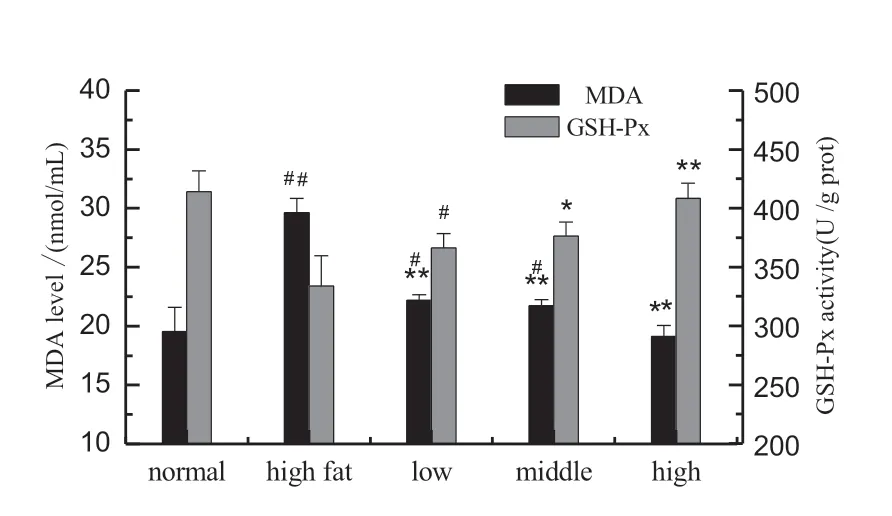
Fig 1.Effects of Pu-erh extract on hepatic GSH-Px activity and serum MDA level in rats fed with high-fat diet.Results are presented as the mean±SD(n=10).normal control group;high fat control group;low-,middle-and highconcentration groups administrated with Pu-erh tea extract at doses of 0.45 g/kg BW,0.9 g/kg BW,1.35 g/kg BW treatment groups;notes:#p<0.05,##p<0.01,significant differences from the normal control group; *p<0.05, **p<0.01,significant differences from the high fat control group.
The level of MDA and the activity of GSH-Px in rats fed with high-fat diet were determined.It was shown that the MDA level in high fat control group caused a significant increase compared to the normal control(p<0.01),and the MDA level increased from 19.55±2.05 nmol/L(the normal control group)to 29.62±1.22 nmol/L (the high fat control group) (Fig.1).However, the concentrations of MDA in Pu-erh tea extract groups with low-,middle-,and high-dose groups were decreased by 25.11%,26.62%,and 35.50%in comparison with the high fat control group,respectively(p<0.01).Meanwhile,the GSH-Px activity in Pu-erh extract groups significantly increased compared to high fat control group(p<0.05).Therefore, Pu-erh tea extracts could protect liver cells from free radical damage through significantly elevating GSH-Px activity,effectively inhibiting lipid peroxidation,reducing MDA level.
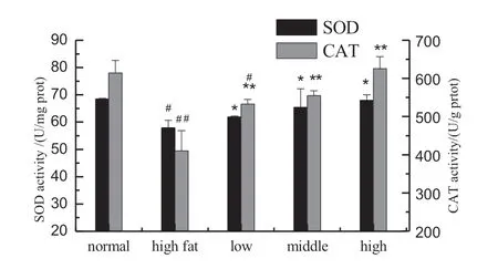
Fig.2.Effects of Pu-erh tea extract on hepatic T-SOD and CAT activities in rats fed with high-fat diet.Rats were administrated intragastrically with Pu-erh extract(0.45,0.9 and 1.45 g/kg BW)once daily for 12 consecutive weeks.Values are expressed as means±SD of 10 rats in each group.#p<0.05,and##p<0.01,vs the normal control group.*p<0.05,and**p<0.01,compared to the high fat control group.
3.3.Effects of Pu-erh tea extract on the activities of hepatic T-SOD and CAT
The activities of SOD and CAT as two of the most important defense enzymes in the liver were further investigated for better understanding the hepatoprotective mechanisms of Pu-erh tea extracts in rat.SOD can catalyze the dismutation of superoxide anions into hydrogen peroxide (H2O2) and remove excess radicals [35].CAT catalyzes the decomposition of H2O2into O2and H2O.Therefore,SOD and CAT could remove harmful superoxide radicals and H2O2in vivoand protect tissue from oxidative damage.
Effects of Pu-erh tea extracts on the activities of hepatic T-SOD and CAT are shown in Fig.2.Compared to the normal control group, the T-SOD activity in the high fat control group was remarkably decreased.However,the T-SOD activity in the Pu-erh tea extract groups with low-, middle- and highdose groups were increased by 6.87%, 12.89% and 17.44%,respectively,in comparison with the high fat control group,and the middle-,and high-dose groups were increased significantly(p<0.05).Meanwhile,the CAT activity in the high fat control group was lower than the normal control group significantly(p<0.01).And contrast to the high fat control group,the CAT activity in Pu-erh tea extracts with three doses were elevated by 29.86%, 35.28%, and 52.43%, respectively (p<0.01) at a dose-dependent manner.

Fig.3.Effect of Pu-erh tea extract on the serum NOS activity in rats fed with high-fat diet.Normal,normal control group;high fat,high fat control group;low,administrated with Pu-erh tea extract at a dose of 0.45 g/kg BW;middle,administrated with Pu-erh tea extract at a dose of 0.90 g/kg BW;high,administrated with Pu-erh tea extract at a dose of 1.35 g/kg BW;notes:#p<0.05,##p<0.01,significant differences from the normal control group; *p<0.05, **p<0.01,significant differences from the high fat control group.
3.4.Effect of Pu-erh tea extract on the NOS activity in serum of rats
Low-concentration NO plays an important role in maintaining the normal physiological function.However,under the pathological conditions, large amounts of NO combined with ROS, can generate highly toxic peroxynitrite anions, inducing thiol oxidation and lipid peroxidation.Furthermore, NO can inhibit the synthesis of protein in the hepatocytes, damage the mitochondrial structure, inhibit the activity of ribonucleotide reductase and damage DNA double helix structure, thereby inducing hepatocyte apoptosis and necrosis.Effect of Pu-erh tea extracts on the serum NOS activity was assayed in Fig.3.Compared to the normal control group,the NOS activity in high fat control group was increased from 22.69877±0.49719 U/mL to 27.84257±7.55303 U/mL.As expected,the NOS activity was effectively attenuated by pretreatment with Pu-erh tea extracts at the tested dosages of 0.45,0.90 and 1.35 g/kg BW(p<0.05)in a dose-dependent manner.Based on the biochemical analysis,it is suggested that Pu-erh tea extracts may prevent liver-damagedviainhibiting lipid peroxidationin vivo.
3.5.Effects of Pu-erh tea extract on ALT and AST activities in serum
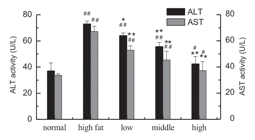
Fig.4.Effects of Pu-erh extract on hepatic ALT and AST activities in serum of rats fed with high-fat diet.Results are presented as the mean±SD (n=10 rats in each group).Normal control group;high fat control group;low-,middleand high-concentration groups administrated with Pu-erh tea extract at doses of 0.45 g/kg BW,0.9 g/kg BW,1.35 g/kg BW treatment groups;Notes:#p<0.05,##p<0.01, significant differences from the normal control group; *p<0.05,**p<0.01,significant differences from the high fat control group.
It is well known that ALT and AST enzymatic activities in serum have been used as a representative marker of hepatic injury[36]while the hepatocytes alters their transport function.Mild to asymptomatic elevation of the serum aminotransferases (ALT,AST)is the most common and often the only laboratory abnormality found in patients with nonalcoholic fatty liver disease(NAFLD)[37].The ALT and AST activities in NAFLD patients are 2–3 fold-times higher than those in the normal people.Furthermore,the AST/ALT ratio of the majority NAFLD patients is less than one,and nearly 20%of NAFLD patients can develop steatohepatitis.Effects of Pu-erh tea extracts on high-fat induced liver damage rats were performed and the ALT and AST activities in serum are shown in Fig.4.Compared to the normal control group,a notable elevation of ALT and AST enzymes activities was found in the high fat control group, indicating the livers of rats had formed hepatic lesion (p<0.01).In contrast to the high fat control,the ALT activity with three doses was significantly lowered by 12.27%, 23.74%, and 44.94%, respectively,and the middle-,high-dose groups were decreased significantly(p<0.01).The AST activity in Pu-erh tea extracts with three doses were reduced to 21.33%, 32.38% and 44.49%, respectively(p<0.01).Hence,Pu-erh tea extracts showed significantly hepatoprotective activity.
3.6.Effects of Pu-erh tea extract on the serum PK activity and HGL in rats
Many enzymes are involved in glucose metabolism,in which the key enzyme,such as PK,plays a particularly important role.When PK is activated once, the decomposition of glucose is accelerated,whereas glucoseviaglycolysis is reduced.Therefore,PK activity influences glucose metabolism and the blood glucose level.Meanwhile,hepatic glycogen is an energy storage substance,which can efficiently balance the level of blood glucose.When the blood glucose level in serum is full enough for metabolism, hepatic glycogen will not be decomposed.However, when the blood glucose level is higher than the normal,insulin will promote glucose in the blood into the liver,forming hepatic glycogen and lowering blood glucose level.In addition,hepatic glycogen has the function of repairing hepatocytes.Therefore,HGL reflects the balance condition of glucose in the blood and the status of liver.
The HGL and PK activity were determined (Fig.5).The rats in high fat control group caused a significant decrease(p<0.01)in HGL,ranging from 5.14±0.79 mg/g tissue for the normal control to 4.98±0.11 mg/g tissue.However,the HGLs treated Pu-erh tea extracts with low-, middle-, and high-dose were increased by 23.86%, 28.79%, and 37.53%, respectively(p<0.01).Meanwhile, the PK activity in the high fat control group decreased significantly(p<0.01)in comparison with the normal control group, and increased by 36.73%,41.23%, and 44.31%,respectively(p<0.01),in the Pu-erh tea extract groups for low-,middle-,and high-concentrations.The results indicated Pu-erh tea extracts could significantly elevate the PK activity and HGL,accelerating the decomposition and synthesis of glucose,thereby effectively regulating the blood glucose level.

Fig.5.Effects of Pu-erh tea extract on hepatic glycogen level and PK activity in rats.Rats fed with high-fat diet were administrated intragastrically with Puerh extract(0.45,0.9 and 1.45 g/kg BW)once daily for 12 consecutive weeks.Values are expressed as means±SD of 10 rats in each group.#p<0.05, and##p<0.01,vs the normal control group.*p<0.05,and**p<0.01,compared to the high fat control group.
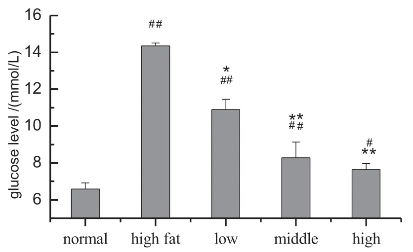
Fig.6.Effect of Pu-erh tea extract on the glucose level in rats.Normal,normal control group;high fat,high fat control group;low,administrated with Pu-erh tea extract at a dose of 0.45 g/kg BW;middle,administrated with Pu-erh tea extract at a dose of 0.90 g/kg BW;high,administrated with Pu-erh tea extract at a dose of 1.35 g/kg BW;Notes:#p<0.05,##p<0.01,significant differences from the normal control group;*p<0.05,**p<0.01,significant differences from the high fat control group.
3.7.Effects of Pu-erh tea extract on the glucose level
Effects of Pu-erh tea extracts on the level of glucose in rats are shown in Fig.6.The glucose level in high fat control group was notably increased as compared to those in normal control group(p<0.01).In comparison with the high fat control group,the level of glucose in low-,middle-and high-concentration of Pu-erh extract groups were decreased significantly, especially in the middle-and high-dose groups,which were decreased by 42.33%and 46.75%(p<0.01),respectively,indicating that Puerh tea extracts have the significant effect on regulating glucose level.
4.Discussion
ROS induced by obesity involves in multi-mechanisms,including antioxidant defense system reducedin vivoand the activities of SOD,GSH-Px and catalase decreased,which leads to the oxidative stress status [38–41].The reducing the body mass index (BMI) can markedly improve the oxidative stress state[42].The experimental results showed that the body weight of rats in low-, middle-, and high- concentration groups were significantly lower than those in the high fat control group by gavaging with Pu-erh tea extracts for twelve weeks, confirming that Pu-erh tea extracts had the function of preventing obesity (Table 1).Meanwhile, by comparison with the normal control group, the T-SOD, GSH-Px and CAT activities were significantly reduced, and MDA level and NOS activity were significantly increased in the high fat control group,indicating ROS in the rats fed with high-fat diet was excessively produced,and the innate antioxidant defense systemin vivowas damaged,thereby leading to the oxidative stress state and lipid peroxidation (Figs.1–3).However, the SOD, GSH-PX, CAT, NOS activities and the MDA level in the high-dose treatment group were roughly equivalent to that of the normal control group,demonstrating that excessive ROS in the rats fed with high-fat diet was effectively removed and remained ROS at a lower levelviathe intervention of high-dose tea pigments.Therefore, this study substantiated that Pu-erh tea extracts can efficiently reduce oxidative stress status and inhibit lipid peroxidation.This may be due to the mechanisms of preventing the weight of rats fed with high-fat diet from increasing,thus restoring the innate antioxidant defense system and effectively dampening the generation of free radicals, reducing oxidative stress status and inhibiting lipid peroxidation, and preventing the occurrence of diseases associated with oxidative stress.It has been reported that the tea water extracts can significantly decrease body weight in hyperlipidemia-rats, increasing the SOD and GSH-PX activities,decreasing MDA level[24],and this is consistent with the experimental results.
The essence of obesity is that the energy intake is larger than that consumption.When the amount of glucose used as direct energy material excesses that of daily requirement in the body,the extra glucose is transferred to the cells of liver or skeletal muscle and is transformed into liver-or muscle-glycogen to provide energy of the body during times of energy deficit.Because of the limited storage capacity of glycogen in the body,the extra part on the one hand is turned into fat by the liver cells and stored in them; on the other hand, the fat by fat mobilization or gluconeogenesis is removed.When the amount of fat storage is greater than the clearance, the fat accumulation in the liver cells occurs and fatty liver is formed.Therefore,obesity or fatty liver often accompanied by the increased free fatty acids and blood glucose[43].Free fatty acid or glucose accumulation in obesity can enter the mitochondria to undergo β-oxidation or tricarboxylic acid cycle (TCA).During the process of oxidation,ROS are generatedviathe mitochondrial respiratory chain,and elevated ROS also contributes to proinflammatory cytokine production,such as NO,TNF-α,and cellular apoptosis,meanwhile,increased ROS can result in increased lipid peroxidation,causing injury of liver cell membrane and organelles[44].In our study,the activities of ALT,AST and liver index in high-fat diet rats increased significantly,relative to the normal control group,indicating that the liver of rats fed with high-fat diet appeared lesions (Fig.4).However, after the intervention of Pu-erh tea extracts, the ALT and AST activities and liver index in low-,middle- and high-concentrations groups reduced significantly and these results showed Pu-erh tea extracts have hepatoprotective effect in the rats fed with high-fat diet.The mechanism may be that Pu-erh tea extracts can increase the SOD,GSH-PX and CAT activities in liver, reduce the MDA level and NOS activity by restraining the growth of body weight, thus enhancing the innate antioxidant defense system in the liver.By reducing oxidative stress and inhibiting liver lipid peroxidation, Pu-erh tea extracts can protect liver tissue from ROS damaging,thereby reducing the ALT and AST activities in serum.Weight loss can lead to the reduction in ALT and AST activities and MDA level,and improve oxidative stress status and restrain lipid peroxidation so as to protect liver from damaging, and this result is consistent with our the experiment.
Obesity,especially abdominal obesity,have enlarged fat cells in common.The sensitivity of fat cells to insulin is greatly reduced and thereby causes insulin resistance.Obesity can lead to the increase of blood glucose level,and further stimulate the production of ROS.ROS can damage islet cells lead to impaired glucose tolerance in body,and eventually cause diabetes.In this study, the levels of body weight and abdominal fat in high fat control group were significantly higher than those of the normal control group (Table 1).And the synthesis ability of glycogen and the PK activity in the high fat control group were significantly reduced, and the glucose level significantly increased(Figs.5 and 6).However, under the Pu-erh tea extract intervention, the rising trends of weight and fat had been under control, and the HGL and PK activity in Pu-erh tea extract treatment groups were significantly increased,and the glucose level reduced significantly.In the previous experiment,we have proved that Pu-er tea extracts can effectively improve insulin resistance in mice fed with high fat diet by reducing body weight and abdominal fat mass,keeping the glucose level at appropriate level[45].Therefore,the mechanism of Pu-erh tea extracts on regulating glucose may be attributed to reducing the weight and visceral fat of rats, and these effects can improve insulin resistance, increase the sensitivity of insulin, and promote the synthesis and decomposition of glucose,thus regulating the glucose level and preventing the occurrence of type II diabetes and fatty liver.
5.Conclusion
Pu-erh extracts showed a strong protection effect against the hepatic damage by oxidative stress.The protection mechanism of Pu-erh extracts is confirmed in the studyviathe activation of the hepatic antioxidant system and the reversal of lipid peroxidation.In addition,Pu-erh tea extracts have important effect on regulating glucose level by enhancing the glycogen synthesis and the PK activity,and thus prevent people from liver disease.Therefore, Pu-erh tea can be used as a potential healthy drink for prevention and/or treatment of fatty liver disease and many diseases associated with oxidative stress.
Conflict of interest
The authors have declared that no conflict of interest exists.
Acknowledgments
The financial supports from the National Natural Science Fund of China (31371773), Tianjin Higher School Educational Projects of Science and Technology Development Fund(20120603)and the Yunnan Tasly Deepure Biological Tea Group Company Limited(20100908)are gratefully acknowledged.
- 食品科学与人类健康(英文)的其它文章
- About the Beijing Academy of Food Sciences
- Effect of Roselle calyces extract on the chemical and sensory properties of functional cupcakes
- Thermo-mechanical and micro-structural properties of xylanase containing whole wheat bread
- Biological activities of silver nanoparticles from Nothapodytes nimmoniana(Graham)Mabb.fruit extracts
- Processed beetroot(Beta vulgaris L.)as a natural antioxidant in mayonnaise:Effects on physical stability,texture and sensory attributes
- Coagulase gene polymorphism of Staphylococcus aureus isolates:A study on dairy food products and other foods in Tehran,Iran

