小分子化合物诱导干细胞分化为胰岛素分泌细胞研究进展
廖菁菁,郝好杰,刘建萍井冈山大学附属医院 内分泌科,江西吉安 4000;解放军总医院,北京 0085;南昌大学第二附属医院 内分泌科,江西南昌 0006
根据国际糖尿病协会(IDF)调查,2011年全球糖尿病患者为3.66亿,到2030年预计将达到5.22亿,即患病率将达到7.7%[1]。而国内最新大样本流行病学调查显示,中国成人糖尿病患病率为11.6%[2]。糖尿病已经成为一种非常普遍的疾病,目前主要是通过口服降糖药及注射外源性胰岛素进行治疗,但长期注射胰岛素给患者带来很多不便,也不能阻止各种糖尿病慢性并发症的发生[3],因此迫切需要一些新的治疗方法。糖尿病是由于胰岛B细胞受损,导致胰岛素分泌缺陷和(或)作用缺陷,从而使患者血液中葡萄糖水平增高。如能重建患者体内有功能的B细胞,就有可能治愈糖尿病。近年来,细胞治疗成为非常有前景的治疗方法。目前,将各种来源的细胞诱导分化或转分化为胰岛素分泌细胞(insulin-producing cells,IPCs)[4-6],为糖尿病细胞治疗开辟了一条新道路。关于IPCs再生来源的干细胞已有综述报道[7],但有关IPCs的诱导方法及小分子化合物在干细胞向IPCs诱导分化过程中的作用的文献报道较少。本文就各种来源干细胞向IPCs分化的诱导方法及相关小分子化合物在诱导过程中所起的作用进行综述。
1 胰岛素分泌细胞的来源
现阶段研究较多的用于治疗糖尿病的干细胞可分为3类:胚胎干细胞、诱导性多能干细胞(induced pluripotent stem cells,iPs)及成体干细胞。胚胎干细胞是一种全能干细胞,具有多向分化潜能及自我更新能力,在适当的条件下可分化为3个胚层细胞,包括IPCs[8-9]。最初诱导效率较低,且生成的IPCs对葡萄糖刺激不敏感,随着不断的研究,尤其是利用小分子化合物分阶段诱导后,诱导效率明显提高,诱导出的IPCs对葡萄糖的敏感性也大大提高了[7]。
目前,用确定的转录因子将体细胞重编程为诱导性多能干细胞的方法已经建立[10-11],这些iPS细胞与胚胎干细胞有相似的特性,具有多向分化及自我更新能力。同时自体细胞来源的多能干细胞避免了免疫排斥问题,也避开了伦理及法律上的争议。不同来源的iPS细胞在动物模型中可治疗各种疾病,包括将其诱导分化为IPCs治疗糖尿病[12-14]。
成体干细胞是胚胎干细胞分化形成的各特定类型细胞的干细胞或前体细胞,在适当的条件下可以转分化为其他类型的细胞,包括转分化为IPCs[15]。这种转分化过程包括几种可能的途径:1)从一种已分化细胞转分化为另一种分化细胞;2)已分化的细胞去分化为一种共同的前体细胞后再分化为另一种细胞;3)将成体组织中存在的多能细胞诱导分化为另一种细胞;4)与另一已分化的细胞融合为多能细胞。成体干细胞相对胚胎干细胞来说,取材更方便,成瘤风险更低,且无伦理上的争议[16]。
2 小分子化合物诱导IPCs生成
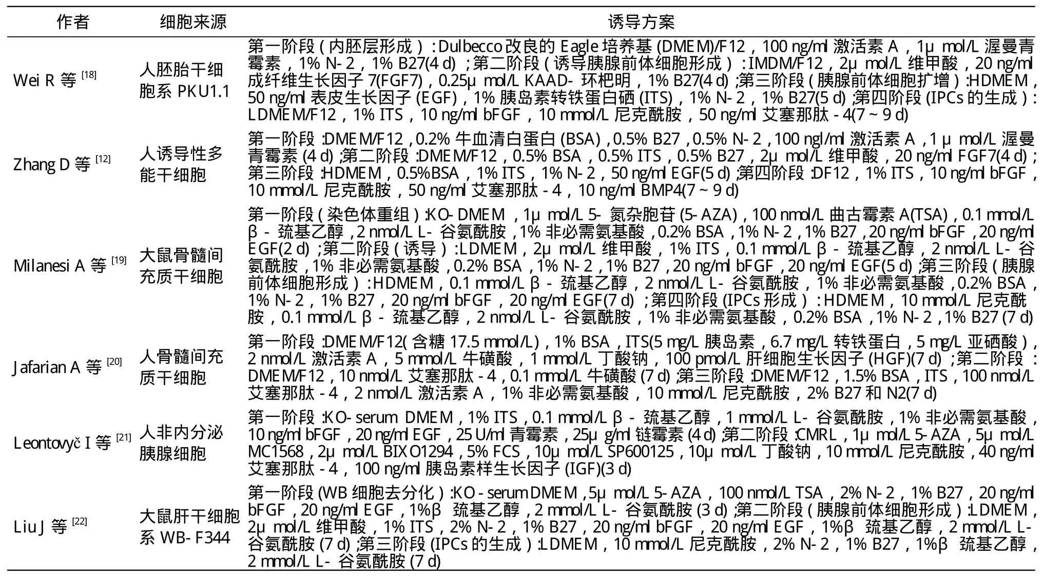
Hou等[17]用7种小分子化合物将小鼠成体细胞诱导为多能细胞,这种应用小分子化合物进行重编程的非转基因方法较转基因方法对临床意义更大,因为小分子化合物无免疫原性、可渗入细胞、操作方便、容易合成及保存,且其对特异蛋白的抑制或激活功能多是可逆的,应用时浓度可以准确控制[17]。目前,已有很多实验室通过不同的生长因子与小分子化合物的组合,直接将胚胎干细胞、诱导性多能干细胞及成体干细胞分阶段诱导分化为IPCs,表1中归纳了部分作者的诱导方案。
3 小分子化合物在IPCs诱导分化过程中的作用
从表1中可以看到不同的细胞诱导分化为IPCs需要不同的培养基、生长因子及小分子化合物,但它们也有一些共同的特点,如都是分阶段进行诱导,诱导分化过程中使用的生长因子及小分子化合物部分是一样的。这些生长因子及小分子化合物可促进B细胞的增殖和分化、增加胰岛素含量。如表皮生长因子(epidermal growth factor,EGF)是EGF蛋白家族中的一员,对细胞的存活、增殖及分化起重要作用[23],已有研究发现其可以促进胰十二指肠同源框蛋白1阳性细胞增殖[12]。碱性成纤维生长因子与EGF联合使用或单独使用,有利于IPCs分化[24-25]。激活素A是转化生长因子-β超家族的成员,可调节细胞的增殖和分化,诱导胚胎干细胞向终末内胚层分化,与其他细胞因子联合作用可进一步诱导分化为IPCs[24,26]。5-氮杂胞苷,一种DNA甲基化酶抑制剂,可逆转DNA的甲基化过程,刺激分化过程中胰腺内分泌细胞相关转录因子(Ngn3、Pdx1)的表达。Pennarossa等[27]利用5-AZA使人成纤维细胞经过一个短暂的去甲基化状态后,成功转分化为IPCs,将重编程所得IPCs移植入糖尿病小鼠体内后可逆转其高血糖状态。曲古霉素A,一种非特异组蛋白去乙酰化酶抑制剂,可调节染色体重构,使干细胞去分化,并进一步向胰腺内分泌细胞分化,增加内分泌细胞的数量,与5-AZA联合应用可促进骨髓间充质干细胞、肝干细胞向IPCs分化[19-22]。维甲酸可诱导人胚胎干细胞表达胰腺发育过程中重要的转录因子Ngn3和神经源性分化因子,并进一步促进Pdx1、成对盒基因4(Pax4)及同源盒基因6.1(Nkx6.1)表达,诱导生成IPCs[28-29]。尼克酰胺是各种诱导方案中常用的一种小分子化合物,早在20世纪90年代,Otonkoski等就利用尼克酰胺诱导人胎胰向内分泌细胞分化,其研究表明,用10 nmol/L尼克酰胺作用于未分化的上皮细胞簇,可使发育过程中的胰岛B细胞DNA含量增加2倍,胰岛素含量增加3倍,同时,胰岛素、胰高血糖素及生长抑素的基因表达量也增加了[16]。艾塞那肽-4是胰高血糖素样肽1(GLP-1)激动剂,可通过刺激B细胞的复制及再生增加B细胞团的数量,并可增加胰岛素基因表达及胰岛素原的合成,与利索茶碱共作用时可逆转受损的糖尿病鼠的B细胞[30]。也有报道体外高浓度的艾塞那肽-4可增加小鼠胚胎干细胞来源的IPCs胰岛素的分泌量[31]。艾塞那肽-4与激活素A联合作用,可上调Pdx1、Ngn3,胰岛素、胰高血糖素、生长抑素、胰多肽及葡萄糖转运子2(Glut2)的表达,这些基因可促进有功能的B样细胞的生成[20]。

表1 小分子化合物诱导干细胞分化为胰岛素分泌细胞Tab. 1 Differentiation of stem cells into insulin-producing cells using small molecule compounds
4 展望
近年来的研究表明,不同来源的细胞通过转基因[26,32]及非转基因[12,18,22]的方法都可以诱导分化或转分化为IPCs,因此选择一个最佳的细胞类型及诱导方案也是目前一个比较棘手的问题。胚胎发育过程中,肝与胰腺共同来源于内胚层,发育上的同源性提示肝和胰腺在细胞增殖及分化调控等方面有很高的相似性[33],两者之间的转化涉及相对较少的表观基因改变,且肝具有强大的修复与再生能力,能为胰岛素分泌细胞的重编程提供大量的细胞来源。近来的研究发现,miR-302可促进Pdx1、Ngn3、V-MAF肌肉腱膜纤维肉瘤癌基因同源A(MafA)将人成体肝细胞重编程为胰岛素分泌细胞,过表达miR-302a可增加胰腺发育相关基因Pdx1的表达[34]。因此,将肝细胞重编程为胰岛素分泌细胞在糖尿病细胞治疗方面具有很大潜力。非内分泌的胰腺细胞及胰岛细胞与胰岛B细胞来源同一胚层,或许会是一个比较好的选择[35]。转基因方法存在转染基因整合及诱变的风险[8],因此通过小分子化合物这种非转基因的方法诱导分化或重编程出IPCs的策略越来越受到人们的关注。小分子化合物更像是一种药物,通过小分子化合物抑制或激活组织细胞发育过程中相关基因及信号通路将细胞进行诱导分化及转分化。但是,目前我们对于各种小分子化合物的作用机制及其与胰腺发育过程中各信号通路是如何相互影响的并不清楚。想要通过小分子化合物诱导出我们需要的产胰岛素细胞,就需要了解各组织自然发育过程中基因表达及小分子化合物是如何调节这种复杂的基因网络系统的。这需要我们进行深入研究,这是一个很艰辛的过程,是一个挑战,但又吸引我们不断探索,因为我们了解的越多,解析的越清楚,离治愈糖尿病的日子就越近。
1 Canivell S, Gomis R. Diagnosis and classification of autoimmune diabetes mellitus[J]. Autoimmun Rev, 2014, 13(4/5): 403-407.
2 Xu Y, Wang LM, He J, et al. Prevalence and control of diabetes in Chinese adults[J]. JAMA, 2013, 310(9): 948-958.
3 Muir KR, Lima MJ, Docherty HM, et al. Cell therapy for type 1 diabetes[J]. QJM, 2014, 107(4):253-259.
4 Segev H, Fishman B, Ziskind A, et al. Differentiation of human embryonic stem cells into insulin-producing clusters[J]. Stem Cells, 2004, 22(3): 265-274.
5 Alipio Z, Liao WB, Roemer EJ, et al. Reversal of hyperglycemia in diabetic mouse models using induced-pluripotent stem (iPS)-derived pancreatic beta-like cells[J]. Proc Natl Acad Sci U S A, 2010,107(30): 13426-13431.
6 Nagaya M, Katsuta H, Kaneto H, et al. Adult mouse intrahepatic biliary epithelial cells induced in vitro to become insulin-producing cells[J]. J Endocrinol, 2009, 201(1): 37-47.
7 Shen J, Cheng Y, Han Q, et al. Generating insulin-producing cells for diabetic therapy: existing strategies and new development[J].Ageing Res Rev, 2013, 12(2): 469-478.
8 Kroon E, Martinson LA, Kadoya K, et al. Pancreatic endoderm derived from human embryonic stem cells generates glucoseresponsive insulin-secreting cells in vivo[J]. Nat Biotechnol,2008, 26(4):443-452.
9 Rezania A, Bruin JE, Arora P, et al. Reversal of diabetes with insulin-producing cells derived in vitro from human pluripotent stem cells[J]. Nat Biotechnol, 2014, 32(11): 1121-1133.
10 Park IH, Zhao R, West JA, et al. Reprogramming of human somatic cells to pluripotency with defined factors[J]. Nature, 2008, 451(7175): 141-146.
11 Lewitzky M, Yamanaka S. Reprogramming somatic cells towards pluripotency by defined factors[J]. Curr Opin Biotechnol, 2007,18(5): 467-473.
12 Zhang D, Jiang W, Liu M, et al. Highly efficient differentiation of human ES cells and iPS cells into mature pancreatic insulinproducing cells[J]. Cell Res, 2009, 19(4): 429-438.
13 Thatava T, Kudva YC, Edukulla R, et al. Intrapatient variations in type 1 diabetes-specific iPS cell differentiation into insulin-producing cells[J]. Mol Ther, 2013, 21(1):228-239.
14 Raikwar SP, Kim EM, Sivitz WI, et al. Human iPS cell-derived insulin producing cells form vascularized organoids under the kidney capsules of diabetic mice[J]. PLoS One, 2015, 10(1):e0116582.
15 Wu Y, Li J, Saleem S, et al. c-Kit and stem cell factor regulate PANC-1 cell differentiation into insulin- and glucagon-producing cells[J]. Lab Invest, 2010, 90(9):1373-1384.
16 Wong RS. Extrinsic factors involved in the differentiation of stem cells into insulin-producing cells: an overview[J]. http://www.hindawi.com/journals/jdr/2011/406182.
17 Hou P, Li Y, Zhang X, et al. Pluripotent stem cells induced from mouse somatic cells by small-molecule compounds[J/OL].Science, 2013, 341(6146):651-654.
18 Wei R, Yang J, Liu GQ, et al. Dynamic expression of microRNAs during the differentiation of human embryonic stem cells into insulinproducing cells[J]. Gene, 2013, 518(2): 246-255.
19 Milanesi A, Lee JW, Xu Q, et al. Differentiation of nestin-positive cells derived from bone marrow into pancreatic endocrine and ductal cells in vitro[J]. J Endocrinol, 2011, 209(2): 193-201.
20 Jafarian A, Taghikhani M, Abroun S, et al. Generation of high-yield insulin producing cells from human bone marrow mesenchymal stem cells[J]. Mol Biol Rep, 2014, 41(7):4783-4794.
21 Leontovyč I, Koblas T, Pektorova L, et al. The effect of epigenetic factors on differentiation of pancreatic progenitor cells into insulinproducing cells[J]. Transplant Proc, 2011, 43(9): 3212-3216.
22 Liu J, Liu Y, Wang H, et al. Direct differentiation of hepatic stemlike WB cells into insulin-producing cells using small molecules[J].Sci Rep, 2013, 3:1185.
23 Herbst RS. Review of epidermal growth factor receptor biology[J].Int J Radiat Oncol Biol Phys, 2004, 59(2 Suppl): 21-26.
24 Sun Y, Chen L, Hou XG, et al. Differentiation of bone marrowderived mesenchymal stem cells from diabetic patients into insulinproducing cells in vitro[J]. Chin Med J, 2007, 120(9): 771-776.
25 Lumelsky N, Blondel O, Laeng P, et al. Differentiation of embryonic stem cells to insulin-secreting structures similar to pancreatic islets[J]. Science, 2001, 292(5520): 1389-1394.
26 Tang DQ, Lu S, Sun YP, et al. Reprogramming liver-stem WB cells into functional insulin-producing cells by persistent expression of Pdx1- and Pdx1-VP16 mediated by lentiviral vectors[J]. Lab Invest, 2006, 86(1):83-93.
27 Pennarossa G, Maffei S, Campagnol M, et al. Brief demethylation step allows the conversion of adult human skin fibroblasts into insulinsecreting cells[J]. Proc Natl Acad Sci U S A, 2013, 110(22):8948-8953.
28 Cai J, Yu C, Liu Y, et al. Generation of homogeneous PDX1(+)pancreatic progenitors from human ES cell-derived endoderm cells[J]. J Mol Cell Biol, 2010, 2(1): 50-60.
29 Oström M, Loffler KA, Edfalk S, et al. Retinoic acid promotes the Generation of pancreatic endocrine progenitor cells and their further differentiation into beta-cells[J]. PLoS One, 2008, 3(7):e2841.
30 Yang Z, Chen M, Carter JD, et al. Combined treatment with lisofylline and exendin-4 reverses autoimmune diabetes[J].Biochem Biophys Res Commun, 2006, 344(3): 1017-1022.
31 Li H, Lam A, Xu AM, et al. High dosage of Exendin-4 increased early insulin secretion in differentiated beta cells from mouse embryonic stem cells[J]. Acta Pharmacol Sin, 2010, 31(5):570-577.
32 Miyashita K, Miyatsuka T, Matsuoka TA, et al. Sequential introduction and dosage balance of defined transcription factors affect reprogramming efficiency from pancreatic duct cells into insulinproducing cells[J]. Biochem Biophys Res Commun, 2014, 444(4):514-519.
33 Deutsch G, Jung J, Zheng M, et al. A bipotential precursor population for pancreas and liver within the embryonic endoderm[J].Development, 2001, 128(6):871-881.
34 Lu J, Dong H, Lin L, et al. miRNA-302 facilitates reprogramming of human adult hepatocytes into pancreatic islets-like cells in combination with a chemical defined media[J]. Biochem Biophys Res Commun, 2014, 453(3): 405-410.
35 Pandian GN, Taniguchi J, Sugiyama H. Cellular reprogramming for pancreatic β-cell regeneration: clinical potential of small molecule control[J]. Clin Transl Med, 2014, 3(1): 6.

