Lin28调控let-7 microRNA合成机制的研究进展
Lin28调控let-7 microRNA合成机制的研究进展
张亚莉1,曹贵玲1*,储明星2,王桂英1
(1 聊城大学 农学院,山东 聊城252059;2.中国农业科学院 北京畜牧兽医研究所,农业部畜禽遗传资源与种质创新重点实验室 北京 100193)
[摘要]Lin28是在胚胎干细胞中广泛表达的RNA结合蛋白,参与分化、发育、肿瘤发生、葡萄糖代谢等多个生命过程。Lin28执行这些功能主要是通过抑制let-7 miRNA的生物合成,并且主要依赖Lin28氨基端冷休克结构域和羧基端锌指结构域。最近一些结构学研究揭示了Lin28调控let-7合成机制,Lin28结合在pri-let-7和pre-let-7的末端loop上,从而阻断Drosha和Dicer的加工,这些互作的特异性主要由锌指结构域和保守的GGAG模体介导。本文就Lin28如何通过其结构域与let-7miRNA前体特异性结合,特异性结合的结构基础等进行了综述,以期为相关研究提供借鉴。
[关键词]Lin28;let-7 microRNA;调控机制;结构基础
Lin28(Cell lineage abnormal 28)是高等真核细胞中保守的RNA结合蛋白,调控分化、发育、多潜能性、肿瘤发生及葡萄糖代谢等几个重要的细胞功能。Lin28最初是在线虫(Caenorhabditiselegans)中发现的一个异时基因,该基因的突变会导致幼虫发育时间发生紊乱[1-2]。Lin28含有两个RNA结合结构域,一个是氨基端的冷休克结构域(Cold shock domain, CSD),一个是羧基端的两个串联排列的CCHC (Cys-Cys-His-Cys)锌指结合motif (CCHC×2),这两个结构域在动物中高度保守。哺乳动物含有两个Lin28同源基因,即Lin28a和Lin28b,在分子水平二者是let-7 microRNA合成的负调控因子。
Let-7 miRNA 是目前研究最为广泛的microRNA家族之一。哺乳动物let-7 miRNA家族中含有12个成员(let-7a-1、let-7a-2、let-7a-3、let-7b、let-7c、let-7d、let-7e、let-7f-1、let-7f-2、let-7g、let-7i和Mir-98),由8个位点表达[3-4]。let-7 miRNA转录后加工和其他miRNA一样,首先核定位RNA酶III Drosha将长的miRNA初级转录物(pri-miRNA)切割,产生一个60~80 nt的具有发夹结构、3'-端具有2 nt突出的产物,被称为pre-miRNA;pre-miRNA被exportin-5:RanGTP转运到细胞质内,被Dicer切割产生成熟的miRNA。
1Lin28阻断let-7的成熟过程
Lin28和let-7 miRNA表达模式最初在研究线虫幼虫发育中被发现[2,4-6]。在幼虫发育早期,Lin28和pri-let-7都表达,但pre-let-7和成熟的let-7未被检测到,表明其成熟过程有转录后调控[7]。这种调控关系在哺乳动物细胞中也存在,Lin28a/b主要在未分化细胞中表达,而成熟的let-7只是在已分化或组织发育中能检测到[8-10]。另外,pri-let-7在整个分化或发育过程中表达量不变,说明在干细胞或祖细胞中Lin28a/b 对 let-7有负反馈调控[11-13]。
目前研究发现,Lin28阻断let-7的成熟过程主要有两种机制:一种是Lin28a/b与let-7前体结合,阻碍Drosha和Dicer对let-7前体切割位点的识别。Lin28a/b能特异性与pri-let-7、pre-let-7结合并抑制pri-let-7[9,14-15]、pre-let-7[8]的加工。RNA和染色质免疫共沉淀试验表明,Lin28和pri-let-7之间有特异的互作,从而抑制Drosha对pri-let-7的加工。另外,Lin28b主要定位在细胞核内,并在核内结合pri-let-7使其避免与Drosha结合,阻断Drosha的加工[16]。在人胚胎干细胞(Embryonic Stem cell, ESC)和神经元干/祖细胞中,Lin28与内源pri-let-7也有互作[7]。一个高水平表达的RNA-结合蛋白Musashi 1(Msi 1)可能也参与了上述过程,Msi 1选择性招募Lin28到细胞核内,起到协同作用[17]。Lin28通过与Dicer切割位点附近的双链stem和pre-element (preE terminal loop或 hairpin)结合,阻断Dicer对let-7前体的加工[14]。 此外有研究表明,Lin28a/b能诱导pre-let-7 preE的结构变化,使包括Dicer切割位点在内的双链stem打开[18-20],使Dicer不再识别原有切割位点从而阻断Dicer的切割。
Heo等[21-22]提出了另外一种抑制模式,这种加工模式会使pre-let-7降解。Lin28a/b诱导pre-let-7的3'-突出的寡核苷酸发生尿苷化(oligo-uridylation),oligo-尿苷化的pre-let-7可以抵抗Dicer的切割。正常情况下,Dicer通过它的PAZ 结构域识别miRNA前体上2 nt 3'端突出。尿苷化后,Dicer就不能识别延长了的3'-突出,从而不能对pre-let-7进行加工。oligo-尿苷化的RNA会招募3'-5'核酸外切酶,之后被降解[21-23]。Chang等[24]在小鼠ESC中发现,3'-5'核酸外切酶Dis312能催化pre-let-7的降解,Dis312 敲减会使小鼠ESC中尿苷化的pre-let-7聚集。尿苷化的pre-let-7比未发生尿苷化的 pre-let-7更容易降解[21]。Pre-let-7的oligo-尿苷化由非经典的poly(A)聚合酶--末端转移酶4(Terminal uridyly oligo transferase 4,TUT4,又称Zcchc11)和TUT7(又称Zcchc6)催化,这些酶依赖Lin28[22,25-27]。但是,TUT4和 TUT7在没有Lin28时会催化具有1 nt 3'-突出的pre-miRNA的单核苷酸发生尿苷化(mono-uridylation),此单核苷酸尿苷化会促进Dicer的加工[28]。在有Lin28存在时,pre-let-7和其他preE中含有GGAG模体的miRNA更倾向发生oligo-尿苷化,GGAG模体对于RNA结合和oligo-尿苷化是必需的[22]。线虫中也有类似的加工机制,线虫使用的尿苷化酶是poly(U)聚合酶2(Poly(U) polymerase, PUP2)。PUP2以依赖Lin28的方式使pre-let-7发生oligo-尿苷化,从而抑制let-7的成熟[29]。
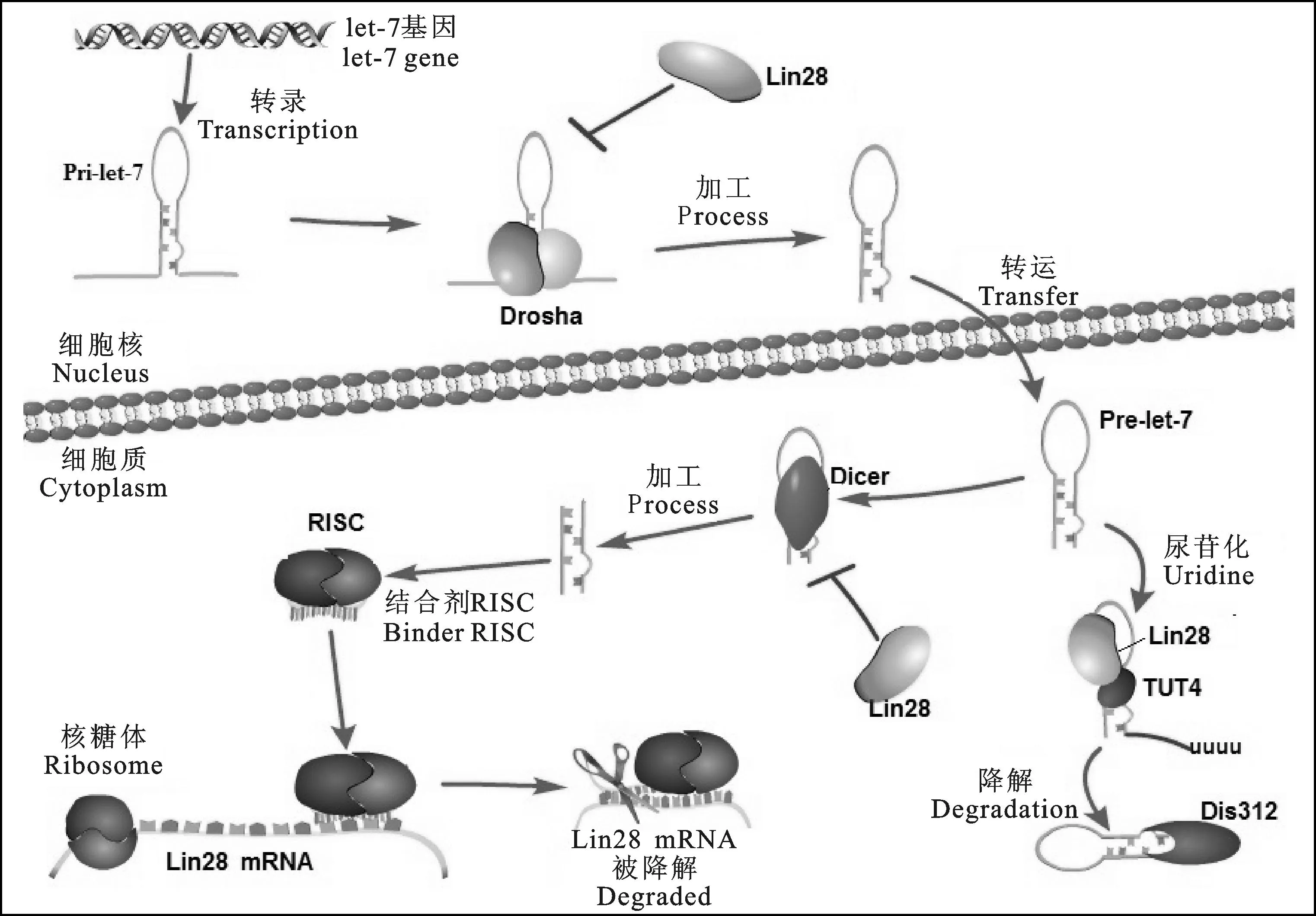
因此,Lin28通过两种机制三种途径调控let-7 miRNA的成熟过程(见图1)。第一,在细胞核内Lin28抑制Drosha对pri-let-7的加工;第二,在细胞质内Lin28抑制Dicer对pre-let-7的加工;第三,Lin28招募TUT4(TUT7)结合到pre-let-7,引起pre-let-7的3'-端发生尿苷化,尿苷化的pre-let-7之后被降解。而成熟的let-7结合在lin28的3'-UTR区域从而对lin28的表达起抑制作用。

图1 Lin28调控let-7 miRNA的加工机制
2Lin28/RNA结合特异性的结构基础
Lin28的CSD和CCHC结构域对pre-let-7的结合都是必须的,删除任何一个都会影响与pre-let-7的高度亲和性,但是删除氨基末端和羧基末端(约30个氨基酸残基)不会影响与pre-let-7的结合[18]。在Lin28和pre-let-7 preE形成的晶体结构中,preE的loop环绕Lin28的CSD,形成一个领带,stem交叉形成领结,并且CCHC锌指与3'-stem的GGAG模体相结合[18]。Desjardins等[30]提出了Lin28与let-7 miRNA前体形成复合体的模型,表明在二者结合过程中CSD和CCHC锌指是相互相互配合、缺一不可的。
2.1 Lin28锌指结构域特异性识别GGAG模体
为了确定Lin28与let-7前体之间特定互作关系,科学家研究了Lin28和let-7前体互作的特异性。用pre-let-7不同序列进行凝胶阻滞分析,结果表明单独的pre-let-7末端loop(也称为pre-element或preE)也能满足与Lin28a的结合[14]。对let-7前体的stem-loop比对发现let-7前体中有一个脊椎动物高度保守的GGAG模体对Lin28的结合非常重要,这个motif发生突变(GGAG--AAAG,GGAG--GUAU)会使Lin28a介导的pri-let-7或pre-let-7的加工阻断解除,且TUT4介导pre-let-7 oligo-尿苷化受损[15,22]。将GGAG模体引入不相关的miRNA(如pre-mir-16-1)pre E中,Lin28a会与这个嵌合pre-miRNA结合,并且TUT4介导的尿苷化也会发生[22]。
Lin28的ZKD发生突变会破坏其与pre-let-7的特异性结合,也会破坏单独ZKD与含GGAG模体 的RNA结合[18,20,22,31-32]。小鼠Lin28a与pre-let-7来源的含GGAG 的寡核苷酸之间的共晶体结构和人Lin28a ZKD结合AGGAGAU的核磁共振(Nuclear magnetic resonance, NMR)溶液结构显示Lin28的ZKD与GGAG模体之间发生了特异性结合[18,32]。两个CCHC锌指直接与Let-7 GGAG模体结合并在RNA骨架中形成一个特征性的扭结。在这个扭结构象中,Lin28的Y140非常关键,它位于AG之间,形成夹心状,并且与H162互作,形成扭结构象。碱基A没有与Lin28形成很多极性互作,但它压紧了第一个G,并与之形成氢键,辅助形成扭结构象。这种特异性结合主要通过氢键完成,CCHC锌指结合GGAG的G1和G4,氢键由锌指刚性部分的氨基酸残基的羰基和氨酰基骨架介导。除了特异性互作,G1和G4插在由第一个锌指中的酪氨酸和组氨酸与第二个锌指中的组氨酸和蛋氨酸形成的一个疏水口袋中。在小鼠的Lin28a:GGAG复合体中,G2也通过A3的羰基骨架和N1氨基形成氢键从而产生序列特异性结合。A3也参与了RNA骨架中扭结的形成,与G1相互作用[18]。NMR溶液结构显示第二个CCHC锌指在与RNA结合时发生了较大的结构变化,中心的锌离子移动了大约25 Å,是由富含脯氨酸的linker区的Pro158导致的这样一个大的构象变化[32]。在人Lin28a:AGGAGAG结构的RNA骨架中未发现如此强的扭结弯曲,RNA与蛋白结构变化可能导致相邻的双链pre-let-7 stem保持开链状态,从而掩盖了Dicer的切割位点[18,20,33]。有趣的是,TUT4和TUT7 也含有CCHC 锌指[27],这些锌指之间的距离比Lin28锌指之间的距离大些,可能表明他们发挥作用不依赖于对方。
2.2 Lin28 CSD决定结合的亲和力,重塑RNA内部结构
尽管Lin28锌指结构域可以特异结合GGAG模体,但单独的Lin28 ZKD自身不能与let-7结合来阻断其加工,需要CSD的参与[18,20]。CSD区和preE的loop区结合在一起,loop区环绕在CSD 结构域,形成一个领带状,stem区交叉形成领结,当loop环绕在Lin28 CSD上时,碱基伸出与芳香侧链形成一些Ψ-堆积作用,疏水作用力、氢键和立体排阻决定了核苷酸的优先次序并增强了特异性[18]。
系统结合分析表明,爪蟾Lin28b CSD具有广泛的序列特异性,与5'-末端至少有一个尿苷的富含嘧啶的RNA八核苷酸具有最高的亲和力[20]。这一现象之后被genome-wild PAR-CLIP实验证实,Lin28a/b结合位点一般富含U,侧翼有一个或多个尿苷,并且这些位点都是位于相应的ZKD结合位点的上游[34-35]。Lin28 CSD与preE-Let-7的共晶体结构提供了有价值的信息,Lin28 CSD通过一个保守的核酸结合平台结合单链核酸。与ZKD不同的是,这个结合平台在结合核酸之前已经形成,核酸结合后只发生一些细微变化[36]。Lin28 CSD结合8个核苷酸,这些核苷酸以确定的方向沿着一个弯曲的单链排列。在小鼠Lin28a:preE-let-7结构中,另外第九个核苷酸可见,并且它与第一个碱基形成氢键,把preE stem-loop 闭合,序列特异性结合主要发生在第6个核苷酸上。
除了影响结合亲和力和特异性,Lin28 CSD还影响和重塑靶RNA内部的二级结构。与Lin28a结合后,preE的某些区域和pre-let-7的一部分双链stem变得更易被单链特异性核酸酶切割,可能是Lin28能够打开pre-let-7 的双链stem,然后封闭Dicer切割位点[19]。Nam等[18]证明,Lin28a CSD能够使双链stem-loop发生部分解链,从而产生一个最合适的结合界面。对爪蟾Lin28b的研究发现Lin28 CSD结合pre-let-7, 诱导其结构变化[20],保守的组氨酸(爪蟾Lin28b结合位点4中的组氨酸618)和苯丙氨酸残基(苯丙氨酸77-结合亚位点1、2)对于重塑反应起关键作用。CSD诱导的结构重塑可能对Lin28 ZKD识别GGAG模体很重要,有利于ZKD的结合。此外,ZKD可能引起靶RNA结构改变从而促进下游过程如pre-let-7的oligo-尿苷化;CSD的广泛特异性能使Lin28识别let-7家族各成员前体。
Lin28的分子伴侣样作用影响下游RBPs与RNPs的结合或解离,从而影响他们的加工。 研究表明Dis312核糖核酸外切酶降解oligo-尿苷化的pre-let-7。Dis312是由一个核糖核酸酶II结构域、2个CSD和一个CSD样S1结构域组成,CSD和S1结构域对于Dis312结合并降解oligo-尿苷化的pre-let-7都非常重要,假如Lin28 CSD具有U-rich 结合位点的优先性,那么CSD可能识别oligo(U)尾巴,此外,这可能通过打开miRNA的双链stem推动核糖核酸外切酶对oligo(U)-pre-let-7的降解[24]。
2.3 Linker区决定Lin28与let-7各成员前体结合的灵活性
Lin28CSD和CCHC锌指之间的Linker区长度约15个氨基酸,带正电。具有灵活性,可以适应与具有不同的preE序列和长度的let-7家族成员相结合。Linker区对Lin28与let-7前体结合的特异性或亲和性影响较少,删除9个氨基酸残基不影响与pre-let-7的结合[18]。
3问题与展望
Lin28调控let-7miRNA的成熟过程,而同时又是let-7的靶基因,二者相互调控,在正常组织中处于平衡状态,一旦平衡打破,就会引起异常,如肿瘤发生。Let-7在许多恶性肿瘤中表达下降,被认为是肿瘤抑制因子,而Lin28在这肿瘤中高水平表达,Lin28可能是通过作用于let-7而促进转化。目前发现的与Lin28有关恶性肿瘤有肝癌[37]、乳腺癌[38]、结肠癌[39]、卵巢癌[40]、肺癌[41]、恶性生殖细胞肿瘤[42]等。这些肿瘤中,Lin28的相关活性都与let-7的表达抑制及let-7靶基因紧密相关。
目前结构和生物化学方面研究已经揭示了Lin28如何识别靶RNA从而完成与miRNA调控相关的多个功能。在let-7的合成中,Lin28 ZKD特异性的识别preE-Let-7的GGAG模体并且诱导RNA骨架发生弯曲扭结,使临近的Dicer 切割位点保持打开,不再被Dicer识别,从而不能再继续加工。CSD对序列没有明显的特异性,只是对侧翼有一个鸟苷酸的富含嘧啶序列表现优先结合。
尽管目前的研究揭示了Lin28/let-7调控的分子机制,但还有一些问题尚未解决,如Lin28在细胞核中抑制pri-let-7的机制是什么,Lin28如何促进TUT4/TUT7介导的pre-let-7的oligo尿苷化,这是否与TUT4、TUT7中的CCHC锌指有关等,理解这些机制有助于开发Lin28的功能,找到一些能够促进组织重编程的途径或者找到一些治疗代谢病或癌症的新的治疗方法。
参考文献:
[1]Ambros V and Horvitz H. Heterochronic mutants of the nematode Caenorhabditis elegans[J]. Science,1984,226(4673): 409-416.
[2]Moss EG, Lee RC and Ambros V. The cold shock domain protein LIN-28 controls developmental timing in C. elegans and is regulated by the lin-4 RNA[J]. Cell, 1997, 88(5): 637-646.
[3]Johnson SM, Grosshans H, Shingara J, et al. RAS is regulated by the let-7 microRNA family[J]. Cell, 2005, 120(5): 635-647.
[4]Pasquinelli AE, Reinhart BJ, Slack F, et al. Conservation of the sequence and temporal expression of let-7 heterochronic regulatory RNA[J]. Nature, 2000, 408(6808): 86-89.
[5]Moss EG and Tang L. Conservation of the heterochronic regulator Lin-28, its developmental expression and microRNA complementary sites[J]. Developmental Biology, 2003, 258(2): 432-442.
[6]Reinhart BJ, Slack FJ, Basson M, et al. The 21-nucleotide let-7 RNA regulates developmental timing in Caenorhabditis elegans[J]. Nature, 2000, 403(6772): 901-906.
[7]Van Wynsberghe PM, Kai ZS, Massirer KB, et al. LIN-28 co-transcriptionally binds primary let-7 to regulate miRNA maturation in Caenorhabditis elegans[J]. Nature structural & molecular biology, 2011, 18(3): 302-308.
[8]Rybak A, Fuchs H, Smirnova L, et al. A feedback loop comprising lin-28 and let-7 controls pre-let-7 maturation during neural stem-cell commitment[J]. Nature Cell Biology, 2008, 10(8): 987-993.
[9]Viswanathan SR, Daley GQ and Gregory RI. Selective blockade of microRNA processing by Lin28[J]. Science, 2008, 320: 97-100.
[10]Darr H and Benvenisty N. Genetic analysis of the role of the reprogramming gene LIN-28 in human embryonic stem cells[J]. Stem Cells, 2009, 27(2): 352-362.
[11]Yang DH and Moss EG. Temporally regulated expression of Lin-28 in diverse tissues of the developing mouse[J]. Gene Expression Patterns, 2003,3(6): 719-726.
[12]Thomson JM, Newman M, Parker JS, et al. Extensive post-transcriptional regulation of microRNAs and its implications for cancer[J]. Genes &Development, 2006, 20(16): 2 202-2 207.
[13]Wulczyn F, Smirnova L, Rybak A, et al. Post-transcriptional regulation of the let-7 microRNA during neural cell specification[J]. FASEB J, 2007, 21(2): 415-426.
[14]Piskounova E, Viswanathan SR, Janas M, et al. Determinants of microRNA processing inhibition by the developmentally regulated RNA-binding protein Lin28[J]. The Journal of Biological Chemistry, 2008, 283(31): 21 310-21 314.
[15]Newman MA, Thomson JM and Hammond SM. Lin-28 interaction with the Let-7 precursor loop mediates regulated microRNA processing[J]. RNA, 2008, 14: 1 539-1 549.
[16]Piskounova E, Polytarchou C, Thornton JE, et al. Lin28A and Lin28B inhibit let-7 microRNA biogenesis by distinct mechanisms[J]. Cell, 2011, 147(5): 1 066-1 079.
[17]Kawahara H, Okada Y, Imai T, et al. Musashi1 cooperates in abnormal cell lineage protein 28 (Lin28)-mediated let-7 family microRNA biogenesis in early neural differentiation[J]. The Journal of Biological Chemistry, 2011, 286(18): 16 121-16 130.
[18]Nam Y, Chen C. Gregory RI, et al. Molecular basis for interaction of let-7 microRNAs with Lin28[J]. Cell, 2011, 147(5):1 080-1 091.
[19]Lightfoot HL, Bugaut A, Armisen J, et al. LIN28-dependent structural change in pre-let-7g directly inhibits dicer processing[J]. Biochemistry, 2011, 50(35): 7 514-7 521.
[20]Mayr F, Schütz A, D?ge N, et al. The Lin28 cold-shock domain remodels pre-let-7 microRNA[J]. Nucleic Acids Reseach, 2012, 40(15): 7 492-7 506.
[21]Heo I, Joo C, Cho J, et al. Lin28 mediates the terminal uridylation of let-7 precursor MicroRNA[J]. Molecular Cell, 2008, 32(2): 276-284.
[22]Heo I, Joo C, Kim YK, et al. TUT4 in concert with Lin28 suppresses microrna biogenesis through pre-microrna uridylation[J]. Cell, 2009, 138(4): 696-708.
[23]Shen B and Goodman HM. Uridine addition after microRNA-directed cleavage[J]. Science, 2004, 306(5698): 997.
[24]Chang HM, Triboulet R, Thornton JE, et al. A role for the Perlman syndrome exonuclease Dis3l2 in the Lin28-let-7 pathway[J]. Nature, 2013, 497(7448): 244-248.
[25]Hagan JP, Piskounova E and Gregory RI. Lin28 recruits the TUTase Zcchc11 to inhibit let-7 maturation in mouse embryonic stem cells[J]. Nature structural & Molecular Biology, 2009, 16(10): 1 021-1 025.
[26]Yeom KH, Heo I, Lee J, et al. Single-molecule approach to immunoprecipitated protein complexes: insights into miRNA uridylation[J]. European Molecular Biology Orgnization reports, 2011, 12(7): 690-696.
[27]Thornton JE, Chang HM, Piskounova E, et al. Lin28-mediated control of let-7 microRNA expression by alternative TUTases Zcchc11 (TUT4) and Zcchc6 (TUT7) [J]. RNA, 2012, 18(10): 1 875-1 885.
[28]Heo I, Ha M, Lim J, et al. Mono-uridylation of pre-microRNA as a key step in the biogenesis of group II let-7 microRNAs[J]. Cell, 2012, 151(3): 521-532.
[29]Lehrbach NJ, Armisen J, Lightfoot HL, et al. LIN-28 and the poly (U) polymerase PUP-2 regulate let-7 microRNA processing in Caenorhabditis elegans[J]. Nature Structural & Molecular Biology, 2009, 16(10): 1 016-1 020.
[30]Desjardins A, Yang A, Bouvette J, et al. Importance of the NCp7-like domain in the recognition of pre-let-7g by the pluripotency factor Lin28[J]. Nucleic Acids Research, 2012, 40(4): 1 767-1 777.
[31]Desjardins A, Bouvette J and Legault P. Stepwise assembly of multiple Lin28 proteins on the terminal loop of let-7 miRNA precursors[J]. Nucleic acids research, 2014, 42(7): 4 615-4 628.
[32]Loughlin FE, Gebert LF, Towbin H,et al. Structural basis of pre-let-7 miRNA recognition by the zinc knuckles of pluripotency factor Lin28[J]. Nature Structural & Molecular Biology, 2012, 19(1): 84-89.
[33]Ali PS, Ghoshdastider U, Hoffmann J, et al. Recognition of the let-7g miRNA precursor by human Lin28B[J]. FEBS Letters, 2012, 586(22): 3 986-3 990.
[34]Hafner M, Max KE, Bandaru P, et al. Identification of mRNAs bound and regulated by human LIN28 proteins and molecular requirements for RNA recognition[J]. RNA, 2013, 19(5): 613-626.
[35]Graf R, Munschauer M, Mastrobuoni G, et al. Identification of LIN28-bound mRNAs reveals features of target recognition and regulation[J]. RNA Biology, 2013, 10(7): 1 146-1 159.
[36]Sachs R, Max KE, Heinemann U, et al. RNA single strands bind to a conserved surface of the major cold shock protein in crystals and solution[J]. RNA, 2012, 18(1): 65-76.
[37]Tian N, Han Z, Li Z, et al. Lin28/let-7/Bcl-xL pathway: the underlying mechanism of drug resistance in Hep3B cells[J]. Oncol Rep, 2014, 32(3): 1 050-1 056.
[38]Sakurai M, Miki Y, Masuda M, et al. LIN28: A regulator of tumor-suppressing activity of let-7 microRNA in human breast cancer[J]. J Steroid Biochem Mol Biol, 2012, 131(3/4/5): 101-106.
[39]Paz EA, LaFleur B, Gerner EW. Polyamines are oncometabolites that regulate the LIN28/let-7 pathway in colorectal cancer cells[J]. Mol Carcinog, 2014, 53 Suppl 1: e96-106.
[40]Tan P, Helland Å, Anglesio MS, et al. Deregulation of MYCN, LIN28B and LET7 in a molecular subtype of aggressive high-grade serous ovarian cancers[J]. PLoS One, 2011, 6(4): e18064
[41]Pan L, Gong Z, Zhong Z, et al. Lin-28 reactivation is required for let-7 repression and proliferation in human small cell lung cancer cells[J]. Mol Cell Biochem, 2011, 355(1/2): 257-263.
[42]Murray MJ, Saini HK, Siegler CA, et al. LIN28 expression in malignant germ cell tumors downregulates let-7 and increases oncogene levels[J]. Cancer Res, 2013, 73(15): 4 872-4 884.
Research Progress in Molecular Mechanism of Lin28 Mediated let-7 miRNA Regulation
ZHANG Ya-li1, CAO Gui-ling1*, CHU Ming-xing2, WANG Gui-ying
(1.CollageofAgriculture,LiaochengUniversity,Liaocheng,Shandong252059,China;
2.KeyLaboratoryofFarmAnimalGeneticResourcesandGermplasmInnovationofMinistryofAgriculture,InstituteofAnimalScience,
ChineseAcademyofAgriculturalSciences,Beijing100193,China)
Abstract:Lin28 is an essential RNA-binding protein that is ubiquitously expressed in embryonic stem cells. Its physiological function has been linked to the regulation of differentiation, development, oncogenesis, and glucose metabolism. Lin28 mediates these functions by inhibiting let-7 miRNA biogenesis and this activity depends on Lin28's amino terminal carboxyl terminal cold shock domain (CSD) and zink knuckle domain (ZKD). Recently, biochemical and structural studies revealed the mechanisms of how Lin28 control let-7 biogenesis. Lin28 binds to the terminal loop of pri- and pre-let-7 miRNA and represses their processing by Drosha and Dicer. The specificity of this interaction is mainly mediated by the ZKD with a conserved GGAG. In this paper, we reviewed the specific interaction and structural basis between Lin28 and precursors of let-7.
Keywords:Lin28; let-7 miRNA; regulation mechanism; structural basis
[中图分类号]S811.6
[文献标识码]A
[文章编号]1005-5228(2015)11-0006-06
*[通讯作者]曹贵玲(1982-),女,山东东明县人,博士,讲师,主要从事羊遗传育种与生产研究。E-mail:cgLLing@126.com
[作者简介]张亚莉(1988-),女,山东聊城人,硕士研究生,研究方向:动物学。
[基金项目]国家自然科学基金(31201778);中国农业科学院科技创新工程(ASTIP-IAS13);山东省农业良种工程项目(2012-2015)
*[收稿日期]2015-03-18修回日期:2015-05-29

