CD44在胃癌发生中的作用
李 渊(综述),林三仁,周丽雅(审校)
(北京大学第三医院消化科,北京 100191)
胃癌是消化系统最常见的肿瘤之一,其与幽门螺杆菌的感染密切相关。幽门螺杆菌形成的炎性因子、遗传及环境因素的共同作用使胃黏膜发生癌变。目前针对胃癌的诊治还存在以下问题:肿瘤的标志物检测很难发现早期胃癌,因此患者确诊时多处于进展期;一旦胃黏膜发生萎缩,根除幽门螺杆菌虽然可以减少胃癌发生概率,但不能完全预防胃癌的发生。针对胃癌诊断和治疗中的这些棘手问题,CD44可能起到关键作用。
1 CD44的生理和病理作用
透明质酸是细胞外基质(extracellular matrices,ECM)的主要成分,而CD44是透明质酸主要的细胞表面受体。CD44是一种跨膜糖蛋白,属于黏附分子家族。早期研究发现,CD44介导外周淋巴细胞的返家效应[1]。当前研究显示,CD44介导细胞与基质的相互作用,这种作用使“信号从细胞外向内”传导,CD44在ECM的运动、基质的降解以及细胞的生存中起重要作用;在特定条件下,CD44能介导肿瘤转移[2]。
CD44包括了一组由单一基因编码的糖蛋白家族[1]。其中,CD44H (hematopoetic,造血型),也称为CD44s(standard,标准型),是CD44的主要形式,它是由ECM(外显子1~5),跨膜区域以及细胞质区域组成;CD44v(variant,变异型)的ECM是由可变的剪切变体,即变异的外显子v1~v10构成。CD44s在全身广泛分布,而CD44v主要分布在特定组织类型的上皮表面,二者功能也不相同[3]。
2 CD44与肿瘤关系的研究进展
CD44与透明质酸结合后能介导细胞-基质的黏附、细胞迁移和淋巴细胞激活[2]。两者结合的紧密程度对细胞在含有透明质酸的基质上的迁移至关重要。CD44s和CD44v与透明质酸的亲和力不同。因此,CD44不同的变异型能产生不同甚至相反的生物学功能。CD44能参与细胞骨架的锚定[4]。肿瘤细胞表达的CD44或CD44v与正常细胞的不同,其能通过CD44-透明质酸的连接从原发部位浸润到ECM。肿瘤的浸润能力越强,肿瘤细胞的CD44-透明质酸结合越紧密[5]。这是肿瘤浸润的重要步骤。
肿瘤细胞能通过淋巴管扩散。在淋巴转移过程中,淋巴细胞和转移的肿瘤细胞需要从脉管中溢出,即接近并穿过脉管的内皮细胞。这一过程部分是由淋巴细胞或肿瘤细胞的CD44分子与内皮细胞的透明质酸结合介导完成的[6]。
CD44参与部分肿瘤信号转导通路。CD44v6能激活酪氨酸蛋白激酶的受体,它是一种转移的肿瘤细胞的受体[7]。CD44能减少死亡受体Fas介导的肿瘤细胞杀伤作用,还能减少Fas的表达和Fas介导的细胞凋亡作用,导致肿瘤细胞从死亡受体-配体(Fas-FasL途径)介导的细胞毒性作用中发生免疫逃逸[8]。
3 CD44在不同胃组织中的表达
正常胃黏膜上皮缺乏炎性细胞,幽门螺杆菌感染后,胃黏膜上皮经历从浅表性胃炎至萎缩性胃炎和肠化的过程。不同的炎性细胞成分以及上皮细胞成分表达不同的CD44或CD44v。但是由于不同研究者采用的方法不同,而且有些研究并未区分幽门螺杆菌感染状态,因此难以归纳总结。并且,许多研究使用针对CD44的广谱抗体进行研究,未能进一步区分CD44和CD44v,因此造成总结时困难。在以下论述中,将尽可能区分CD44和不同的CD44v,而当确实无法区分时,将其定义为CD44x。
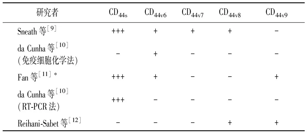
3.1CD44在正常胃组织及幽门螺杆菌感染的胃黏膜中的表达情况 一些研究使用免疫学法或逆转录酶多聚酶链反应(reverse transcript polymerase chain reaction,RT-PCR)来检测CD44在正常胃黏膜中的表达[9-12]。正常胃黏膜上皮强烈表达CD44s,微弱表达或不表达不同的CD44v。一项研究使用巢式RT-PCR法,比较了感染和未感染幽门螺杆菌的胃黏膜中CD44变异型v2~v10的表达以及与炎性反应的关系[12],但该研究并未对71%的幽门螺杆菌感染阴性的慢性炎性患者进一步描述,其中部分患者有可能以前感染过幽门螺杆菌;而且,该研究未使用免疫组织化学法来进一步证实蛋白水平的表达,并未定位表达阳性细胞的类型。尽管存在以上缺陷,该研究仍发现CD44v8~v10在幽门螺杆菌相关性胃炎中的表达为40%,在正常组中表达率为15%~20%,在幽门螺杆菌阴性的慢性胃炎组中的表达率为18%~24%,CD44v2~v7无表达。该研究认为,CDv8~v10在正常黏膜和幽门螺杆菌感染的黏膜中均有表达,炎症的存在并不能显著改变其表达率。Fan等[11]使用CD44s、CD44v6和CD44v9抗体研究幽门螺杆菌感染和非感染个体的胃上皮细胞和上皮内淋巴细胞的表达情况:正常胃上皮细胞和上皮内淋巴细胞均表达CD44s和CD44v6;CD44v9在非感染个体的胃上皮细胞中无表达,但在感染的上皮细胞中表达(表1);感染幽门螺杆菌后CD44在胃黏膜上皮细胞中的表达增加,而在上皮内淋巴细胞中则无此现象。Yasui等[13]使用CD44v9特异性抗体研究幽门螺杆菌感染的胃黏膜发现,在幽门螺杆菌感染的幽门腺体、胃腺瘤及胃腺癌基膜外侧有CD44v9表达。

表1 CD44在正常胃黏膜上皮中的表达
*:幽门螺杆菌感染阳性; -:未做
幽门螺杆菌感染还可以增加CD44v6的表达(幽门螺杆菌感染者为65%,非感染者为45%)[14]。在感染幽门螺杆菌的胃黏膜中,da Cunha等[10]报道在胃的腺颈部有非常弱的CD44v6表达,主要位于胃窦部的腺管中。
3.2CD44与胃癌 从正常胃黏膜、肠化生、临近胃癌的未受累组织、邻近胃癌的肠化组织、直至胃癌组织,CD44v6的表达逐渐增加,认为CD44v6表达增加是从正常黏膜进展到胃癌的重要事件[15]。da Cunha等[10]进一步证实了CD44v6很少在正常胃黏膜中表达,但在胃增生性息肉、不完全和完全肠上皮化生中表达增加;在轻度和重度不典型增生和癌变组织中表达显著增加。在癌前病变以及散发和遗传性胃癌中出现CD44v6原位表达现象,提示CD44v6表达是胃黏膜恶性转化早期潜在的标志物。
研究显示,肠型胃癌较弥漫性胃癌更易出现CD44x的过表达[16-18]。多数印戒细胞癌表达CD44v5,而肠型胃癌通常同时表达CD44v5和CD44v6。肠上皮化生是一种癌前病变,它与肠型胃癌相似,同时表达CD44v5和CD44v6。Heider等[19]研究显示,依据肠型胃癌和弥漫性胃癌在CD44变异型上表达的差异,有可能从分子水平将两者区分。肠化黏膜中CD44v6的表达使其更容易与正常黏膜区分。
4 CD44和胃癌转移的关系
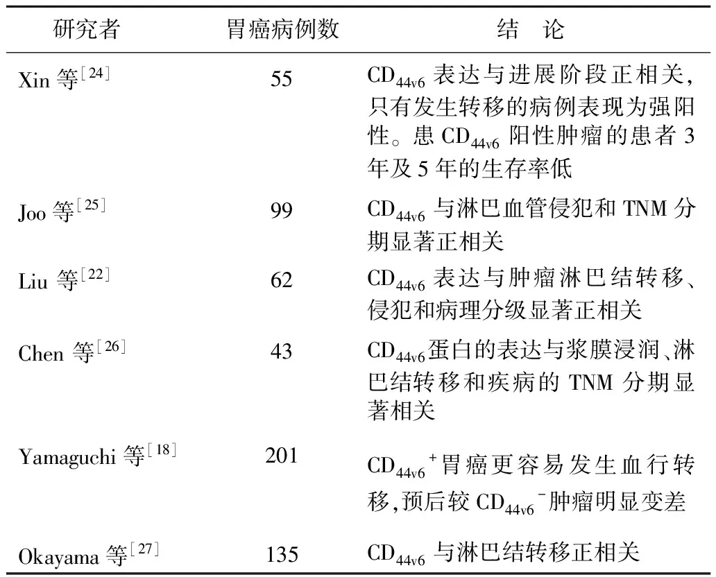
Günthert等[20]将表达CD44s或CD44剪切变异型的质粒转染到未发生转移的大鼠胰腺癌细胞中,发现CD44v6表达与淋巴结转移、淋巴浸润、浸润深度和肿瘤分级有关。该研究为细胞黏附分子在转移过程中起重要作用这一理论提供了第一个例证,该实验推动了CD44v6在人类癌症中的研究。在胃癌中,CD44v6表达与淋巴结转移、血行转移、浸润以及肿瘤病理分级相关(表2和表3)[16,18,21-27]。在一项关于黏膜下胃癌的研究中,学者发现CD44v6是显示淋巴结转移的唯一指标[27]。在胃微乳头状腺癌中,CD44v6的表达与更高的肿瘤分期、淋巴结转移以及淋巴结、血管浸润有关。使用RT-PCR法检测CD44v6mRNA是诊断外周血和骨髓微转移的敏感和特异的方法,有可能作为一个显示肿瘤负荷和治疗效应的指征[23]。另一项研究显示,CD44v5在分化差的胃癌和转移的淋巴结中表达增加[28]。
5 CD44+细胞与胃癌干细胞
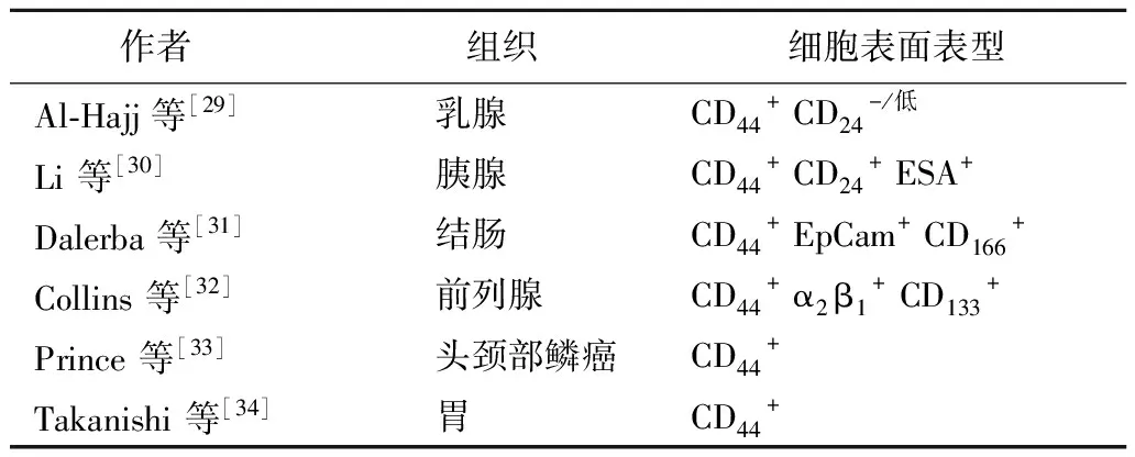
肿瘤干细胞是一种具有自我更新和不定分化潜能、并驱使肿瘤形成的细胞,它们可启动肿瘤生长并保持肿瘤自我更新的能力。目前多数研究认为,实体瘤如乳腺肿瘤、胰腺肿瘤、结肠肿瘤、前列腺癌以及头颈部鳞癌(head and neck squamous cell carcinoma,HNSCC)[29-33]与白血病相似,具有肿瘤干细胞(表4)。肿瘤对放疗和化疗的抵抗力以及肿瘤复发可能都是由肿瘤干细胞引起。2009年,Takaishi等[34]发现三种胃癌细胞系中均有一定体积的CD44+的细胞亚群,当把这些细胞注射到严重联合免疫缺陷(severe combined immunodeficient,SCID)鼠的胃和皮肤中时,CD44+细胞显示出致癌能力和干细胞潜能,而CD44-细胞则不具备该能力;注射CD44+细胞的SCID鼠在8~12周产生肿瘤;使用短发夹RNA(short hairpin RNA,shRNA)敲除CD44后,鼠体内产生肿瘤的能力下降;CD44+胃癌细胞对放疗和化疗有显著的抵抗能力。由于研究者使用的是CD44的广谱抗体,所以无法认定是何种变异型在胃癌的干细胞中起关键作用。
目前研究显示,CD44v9最有可能成为胃癌干细胞的表面标志物[13,35-36]。CD44v9的表达与增殖细胞核抗原的表达及增殖活力显著相关,并与其共表达,显示CD44v9在胃癌的产生和发展中的作用[13]。CD44v6虽然能增加p53表达,但它在除肿瘤细胞外的其他细胞也有表达,在结直肠癌中,CD44v6能够增加p53基因突变,并增加腺瘤癌变概率[21]。肿瘤干细胞的其他特征还包括对放疗、化疗及活性氧应激的不敏感。CD44v9能调节胃癌细胞的氧化还原状态,使其对活性氧所导致的应激不敏感[35]。Ishimoto等[36]的研究显示,鼠的胃癌干细胞表达CD44v9。在食管鳞状上皮与胃柱状上皮交界处可能存在组织干细胞,其也为CD44v9阳性,但增殖细胞核抗原阴性。因此,根据增殖细胞核抗原表达与否,可以将组织干细胞和胃癌干细胞加以区分。
另外,幽门螺杆菌可显著诱发肠化黏膜中干细胞的增生,促使CD44异常转录、p53突变以及DNA的高甲基化[37]。

表2 胃癌中 CD44和CD44变异型的表达

表3 CD44v6的表达及胃癌的临床病例特征

表4 不同器官肿瘤干细胞的细胞表面标志
6 CD44的基因多态性与胃癌的预后
Winder等[38]初步研究显示,CD44等位基因的多态性在基因位点(CD44+4883G→A)(rs187116)出现1个以上的G(GG;AG)时,与局部胃腺癌患者的临床预后相关,并显著影响患者的复发时间和整体生存率;具有CD44T-A的肿瘤患者复发率最低,生存率最高,CD44基因多态性与胃癌的复发相关。
7 CD44在肿瘤治疗中的作用
研究显示,针对CD44和CD44v,尤其是针对CD44v9和v6的治疗有可能产生治疗胃癌的新方法。使用载体shRNA可以使CD44基因沉默[34];使用针对CD44的小干扰RNA可以达到基因敲除的目的[39]。目前这些研究仅在离体实验中进行,在人体胃癌治疗中的意义尚不明了,但在其他系统肿瘤中的研究工作已经有了进展。Misra等[40]用纳米材料包被了针对CD44v6的shRNA,使CD44v6~v9mRNA沉默,从而治疗小鼠的小肠腺癌,其可以减少肿瘤细胞的数量,且对正常细胞的损伤较小。
使用RT-PCR等方法甄别不同CD44剪切变异型的表达,可能会使治疗变得更有针对性和个体化。
8 结 论
CD44和CD44v的表达与胃癌的发生和发展相关,它们在胃癌的诊断、治疗和预后中有可能起到重要作用。将来CD44和CD44v可能研究方向如下:①在幽门螺杆菌感染后,特别是幽门螺杆菌相关的上消化道疾病中,对CD44和CD44v的分布特点和作用的深入研究;②获得可靠的胃癌特别是早期胃癌的检测方法以及预后评估的指标;③ 精确识别胃癌干细胞表面标志物,针对胃癌干细胞进行靶向治疗。随着这些研究的开展,将有希望引起胃癌及其他肿瘤治疗的重大改变。
[1] Screaton GR,Bell MV,Bell JI,etal.The identification of a new alternative exon with highly restricted tissue expression in transcripts encoding the mouse Pgp-1 (CD44) homing receptor.Comparison of all 10 variable exons between mouse,human,and rat[J].J Biol Chem,1993,268(17):12235-12238.
[2] Marhaba R,Zöller M.CD44in cancer progression:adhesion,migration and growth regulation[J].J Mol Histol,2004,35(3):211-231.
[3] Naor D,Sionov RV,Ish-Shalom D.CD44:structure,function,and association with the malignant process[J].Adv Cancer Res,1997,71:241-319.
[4] Seveau S,Eddy RJ,Maxfield FR,etal.Cytoskeleton-dependent membrane domain segregation during neutrophil polarization[J].Mol Biol Cell,2001,12(11):3550-3562.
[5] Nemec RE,Toole BP,Knudson W.The cell surface hyaluronate binding sites of invasive human bladder carcinoma cells[J].Biochem Biophys Res Commun,1987,149(1):249-257.
[6] DeGrendele HC,Estess P,Picker LJ,etal.CD44and its ligand hyaluronate mediate rolling under physiologic flow:A novel lymphocyte-endothelial cell primary adhesion pathway[J].J Exp Med,1996,183(3):1119-1130.
[7] Orian-Rousseau V,Chen L,Sleeman JP,etal.CD44is required for two consecutive steps in HGF/c-Met signaling[J].Genes Dev,2002,16(23):3074-3086.
[8] Yasuda M,Nakano K,Yasumoto K,etal.CD44:functional relevante to inflammation and malignancy[J].Histol Histopathol,2002,17(3):945-950.
[9] Sneath RJ,Mangham DC.The normal structure and function of CD44and its role in neoplasia[J].Mol Pathol,1998,51(4):191-200.
[10] da Cunha CB,Oliveira C,Wen X,etal.De novo expression of CD44variants in sporadic and hereditary gastric cancer[J].Lab Invest,2010,90(11):1604-1614.
[11] Fan X,Long A,Goggins M,etal.Expression of CD44and its variants on gastric epithelial cells of patients with Helicobacter pylori Colonization[J].Gut.1996,38(4):507-512.
[12] Reihani-Sabet F,Eskandarpour M,Khanipour-Roshan M,etal.Effects of inflammation and H.pylori infection on expression of CD44variant exons in gastric tissue[J].J Sci Islam Repub Iran,2003,14(1):11-16.
[13] Yasui W,Kudo Y,Naka K,etal.Expression of CD44containing variant exon 9 (CD44v9) in gastric adenomas and adenocarcinomas:relation to the proliferation and progression[J].Int J Oncol,1998,12(6):1253-1258.
[14] Peng AB,Shi W,Hu SH,etal.Expression of CD44v6in gastric cancer and its correlation with Helicobacter pylori infection[J].Ai Zheng,2003,22(11):1184-1187.
[15] Gulmann C,Grace A,Leader M,etal.CD44v6:a potential marker of malignant transformation in intestinal metaplasia of the stomach? An immunohistochemical study using tissue microarrays[J].Eur J Gastroenterol Hepatol,2003,15(9):981-986.
[16] Ghaffarzadehgan K,Jafarzadeh M,Raziee HR,etal.Expression of cell adhesion molecule CD44in gastric adenocarcinoma and its prognostic importance[J].World J Gastroenterol,2008,14(41):6376-6381.
[17] Dämmrich J,Vollmers HP,Heider KH,etal.Importance of different CD44v6expression in human gastric intestinal and diffuse type cancers for metastatic lymphogenic spreading[J].J Mol Med(Berl),1995,73(8):395-401.
[18] Yamaguchi A,Goi T,Yu J,etal.Expression of CD44v6in advanced gastric cancer and its relationship to hematogenous metastasis and long-term prognosis[J].J Surg Oncol,2002,79(4):230-235.
[19] Heider KH,Dämmrich J,Skroch-Angel P,etal.Differential expression of CD44splice variants in intestinal-and diffuse-type human gastric carcinomas and normal gastric mucosa[J].Cancer Res,1993,53(18):4197-4203.
[20] Günthert U,Hofmann M,Rudy W,etal.A new variant of glycoprotein CD44confers metastatic potential to rat carcinoma cells[J].Cell,1991,65(1):13-24.
[21] Kim MA,Lee HS,Yang HK,etal.Clinicopathologic and protein expression differences between cardia carcinoma and noncardia carcinoma of the stomach[J].Cancer,2005,103(7):1439-1446.
[22] Liu YJ,Yan PS,Li J,etal.Expression and significance of CD44s,CD44v6,and nm23 mRNA in human cancer[J].World J Gastroenterol,2005,11(42):6601-6606.
[23] Wang DR,Chen GY,Liu XL,etal.CD44v6in peripheral blood and bone marrow of patients with gastric cancer as micro-metastasis[J].World J Gastroenterol,2006,12(1):36-42.
[24] Xin Y,Grace A,Gallagher MM,etal.CD44v6in gastric carcinoma:a marker of tumor progression[J].Appl Immunohistochem Mol Morphol,2001,9(2):138-142.
[25] Joo M,Lee HK,Kang YK.Expression of E-cadherin,beta-catenin,CD44sand CD44v6in gastric adenocarcinoma:relationship with lymph node metastasis[J].Anticancer Res,2003,3(2B):1581-1588.
[26] Chen JQ,Zhan WH,He YL,etal.Expression of heparanase gene,CD44v6,MMP-7 and nm23 protein and their relationship with the invasion and metastasis of gastric carcinomas[J].World J Gastroenterol,2004,10(6):776-782.
[27] Okayama H,Kumamoto K,Saitou K,etal.CD44v6,MMP-7 and nuclear Cdx2 are significant biomarkers for prediction of lymph node metastasis in primary gastric cancer[J].Oncol Rep,2009,22(4):745-755.
[28] Hsieh HF,Yu JC,Ho LI,etal.Molecular studies into the role of CD44variants in metastasis in gastric cancer[J].Mol Pathol,1999,52(1):25-28.
[29] Al-Hajj M,Wicha MS,Benito-Hernandez A,etal.Prospective identification of tumorigenic breast cancer cells[J].Proc Natl Acad Sci U S A,2003,100(7):3983-3988.
[30] Li C,Lee CJ,Simeone DM.Identification of human pancreatic cancer stem cells[J].Methods Mol Biol,2009,568:161-173.
[31] Dalerba P,Dylla SJ,Park IK,etal.Phenotypic characterization of human colorectal cancer stem cells[J].Proc Natl Acad Sci U S A,2007,104(24):10158-10163.
[32] Collins AT,Berry PA,Hyde C,etal.Prospective identification of tumorigenic prostate cancer stem cells[J].Cancer Res,2005,65(23):10946-10951.
[33] Prince ME,Sivanandan R,Kaczorowski A,etal.Identification of a subpopulation of cells with cancer stem cell properties in head and neck squamous cell carcinoma[J].Proc Natl Acad Sci U S A,2007,104(3):973-978.
[34] Takaishi S,Okumura T,Tu S,etal.Identification of gastric cancer stem cells using the cell surface marker CD44[J].Stem Cells,2009,27(5):1006-1020.
[35] Ishimoto T,Nagano O,Yae T,etal.CD44variant regulates redox status in cancer cells by stabilizing the xCT subunit of system xc(-)and thereby promotes tumor growth[J].Cancer Cell,2011,19(3):387-400.
[36] Ishimoto T,Oshima H,Oshima M,etal.CD44+slow-cycling tumor cell expansion is triggered by cooperative actions of Wnt and prostaglandin E2in gastric tumorigenesis[J].Cancer Sci,2010,101(3):673-678.
[37] Tahara E.Genetic pathways of two types of gastric cancer[J].IARC Sci Publ,2004,157:327-349.
[38] Winder T,NingY,Yang D,etal.Germline polymorphisms in genes involved in the CD44signaling pathway are associated with clinical outcome in localized gastric adenocarcinoma (GA)[J].Int J Cancer,2011,129(5):1096-1104.
[39] Valentine A,O′Rourke M,Yakkundi A,etal.FKBPL and peptide derivatives:novel biological agents that inhibit angiogenesis by a CD44-dependent mechanism[J].Clin Cancer Res,2011,17(5):1044-1056.
[40] Misra S,Heldin P,Hascall VC,etal.Hyaluronan-CD44interactions as potential targets for cancer therapy[J].FEBS J,2011,278(9):1429-1443.

