
Citalopram increases the differentiation ef fi cacy of bone marrow mesenchymal stem cells into neuronal-like cells
2014-03-24 07:55:09JavadVerdiSeyedAbdolrezaMortazaviTabatabaeiShivaSharifHadiVerdiAlirezaShoaeHassani
中国神经再生研究(英文版) 2014年8期
Javad Verdi, Seyed Abdolreza Mortazavi-Tabatabaei, Shiva Sharif,, Hadi Verdi, Alireza Shoae-Hassani
1 Department of Applied Cell Sciences, School of Advanced Technologies in Medicine, Tehran University of Medical Sciences, Tehran, Iran
2 Department of Stem Cells and Tissue Engineering, Research Center for Science and Technology in Medicine, Tehran University of Medical Sciences, Tehran, Iran
3 Department of Tissue Engineering, School of Advanced Technologies in Medicine, Iran University of Medical Sciences, Tehran, Iran
Citalopram increases the differentiation ef fi cacy of bone marrow mesenchymal stem cells into neuronal-like cells
Javad Verdi1,2, Seyed Abdolreza Mortazavi-Tabatabaei1,2, Shiva Sharif2,3, Hadi Verdi2, Alireza Shoae-Hassani1,2
1 Department of Applied Cell Sciences, School of Advanced Technologies in Medicine, Tehran University of Medical Sciences, Tehran, Iran
2 Department of Stem Cells and Tissue Engineering, Research Center for Science and Technology in Medicine, Tehran University of Medical Sciences, Tehran, Iran
3 Department of Tissue Engineering, School of Advanced Technologies in Medicine, Iran University of Medical Sciences, Tehran, Iran
Javad Verdi and Seyed Abdolreza
Mortazavi-Tabatabaei contributed
equally to this paper.
Several studies have demonstrated that selective serotonin reuptake inhibitor antidepressants can promote neuronal cell proliferation and enhance neuroplasticity both in vitro and in vivo. It is hypothesized that citalopram, a selective serotonin reuptake inhibitor, can promote the neuronal differentiation of adult bone marrow mesenchymal stem cells. Citalopram strongly enhanced neuronal characteristics of the cells derived from bone marrow mesenchymal stem cells. The rate of cell death was decreased in citalopram-treated bone marrow mesenchymal stem cells than in control cells in neurobasal medium. In addition, the cumulative population doubling level of the citalopram-treated cells was signi fi cantly increased compared to that of control cells. Also BrdU incorporation was elevated in citalopram-treated cells. These fi ndings suggest that citalopram can improve the neuronal-like cell differentiation of bone marrow mesenchymal stem cells by increasing cell proliferation and survival while maintaining their neuronal characteristics.
nerve regeneration; citalopram; stem cells; bone marrow mesenchymal stem cells; survival; proliferation; differentiation; neurons; neural regeneration
Funding: This work was funded by the Research Center for Science and Technology in Medicine (RCSTiM), Tehran University of Medical Sciences, Tehran (TUMS), Tehran, Iran.
Verdi J, Mortazavi-Tabatabaei SA, Sharif S, Verdi H, Shoae-Hassani A. Citalopram increases the differentiation efficacy of bone marrow mesenchymal stem cells into neuronal-like cells. Neural Regen Res. 2014;9(8):845-850.
Introduction
Regeneration of damaged cells is the primary goal of cell therapy and one of the important reasons for the study of stem cells. In most cases of damaged central nervous system and neurodegenerative diseases, the neurons are lost (Rahmani et al., 2013a, 2013b; Croce et al., 2014; Sollberger et al., 2014). In these cases, the cell replacement is necessary to supply the function of lost cells. However, the ex vivo expansion of stem cells and their in vivo delivery are restricted by the limited availability of stem cell sources, the excessive cost of commercialization, and the dif fi culties of clinical approval (e.g., embryonic tissue grafts; Viswanathan et al., 2013). Ideally, a cell replacement source should be readily obtainable without technical and ethical problems (Huang et al., 2013; Shoae-Hassani et al., 2013b, 2013c; Qin et al., 2013). Adult bone marrow mesenchymal stem cells have great promise as a source for introducing new neurons (Scwaarz et al., 1999; Sasaki et al., 2001; Noureddini et al., 2012) to the damaged nervous system. They have the potential to substitute tissues lost by injury and diseases if lineage differentiation could be controlled exactly. On the other hand, a better understanding of the pharmacological manipulation of different cell types is important for the successful development of a new therapeutic strategy. Atkinson et al.(2012) described the important role of a pharmacological approach in controlling the differentiation of embryonic stem cells. However, the role of ‘adjunctive’ pharmacology in the clinical applications of stem cell therapy is underestimated. Studies have demonstrated the potential of stem cells through the implementation of ef fi cient differentiation protocols, to alleviate symptoms in mouse models of human diseases. Such diseases include Parkinson’s disease (Chung et al., 2011; Kriks et al., 2011; Kim et al., 2011b), spinal cord injury (Nori et al., 2011), Alzheimer’s disease (Bissonnette et al., 2011) and effi cient protocols for derivation of speci fi c cell types to use of such cells to treat human disease. Up to now, multiple drugs that can modulate the differentiation of clinical-grade SCs (Ilic et al., 2011) have been discovered and may be used in the differentiation protocols to produce transplantable cells.
There are many reports showing that selective serotonin reuptake inhibitor antidepressants can promote neuronal cell proliferation and enhance neuroplasticity both in vitro and in vivo (Nandam et al., 2007; Wang et al., 2008; Boldrini et al., 2009; Rahmani et al., 2013a; Verdi et al., 2014). Citalopram, a selective serotonin reuptake inhibitor antidepressant is widely used for many disorders characterized by serotonergic dysfunctions such as depression, anxiety and panic disorders (Hashimoto et al., 2009). Considering this body of evidence, we hypothesized that a clinically effective antidepressant, citalopram can increase adult stem cell survival and differentiation in a neurobasal medium.
Materials and Methods
Bone marrow mesenchymal stem cell culture and differentiation
Human bone marrow mesenchymal stem cells (Lonza, Berkshire, UK) were cultured on collagen pre-coated plates with neurobasal media (Invitrogen Life Technologies, Glasgow, UK) supplemented with 5% fetal bovine serum in a humidi fi ed incubator with 5% CO2at 37°C for 7 days. Stem cells that have grown to 70% confluence were pretreated with 1 μmol/L dimethyl-sulfoxide (DMSO; Sigma, St. Louis, MO, USA) and then were treated with citalopram (1, 5, and 10 μmol/L; Sigma) (Rahmani et al., 2013a) and/ or 1 μmol/L retinoic acid (RA; Sigma). After treatment for 14 days, cells were subjected to reverse transcription PCR (RT-PCR) and immunocytochemistry assays to determine neuronal cell markers.
Immuno fl uorescence and quanti fi cation of immunoreactive neural cells
Immunocytochemistry experiment was performed as described previously (Noureddini et al., 2012). Brie fl y, the cells were fixed with 4% paraformaldehyde and permeabilized with 0.05% Triton X-100. After blocking with 3% goat serum albumin, cells were incubated with primary antibodies for glial, neuronal and pre-neuronal markers at 37°C for 12 hours. The following primary antibodies and dilutions were used: mouse anti-β-tubulin-Tuj1 (1:500; Chemicon, Billerica, MA, USA); rabbit anti-glial fi brillary acidic protein (GFAP; 1:500; Sigma); rabbit anti-nestin (1:1,000; Sigma); mouse anti-microtubule-associated protein 2 (MAP-2; 1:500; Sigma). Ten the cells were washed with PBS and reacted with the fl uorescent isothiocyanate (FITC) conjugated secondary antibodies against rabbit and mouse Fc region (Sigma; 1:500) at room temperature for 2 hours. Finally, the cells were washed with PBS three times, and 4′,6-diamidino-2-phenylindole (DAPI) was used for DNA staining. The cells were visualized with Ceti immuno fl uorescence microscopy (Belgium).
RT-PCR
RT-PCR experiment was performed as described previously (Shoae-Hassani et al., 2013a). As a brief total RNA was extracted from differentiated cells before and after 2 weeks with and without citalopram, using the Qiagen RNA Isolation Kitand following the manufacturer’s instructions (Qiagen, Valencia, CA, USA). After DNAse I digestion, cDNA was prepared from 1 μg total RNA, using the SuperScript III RT-PCR Kit (Invitrogen) as instructed by the manufacturer. Primer pair sequences are shown in Table 1. The ampli fi cation procedure consisted of 30 cycles (denaturation at 94°C for 30 seconds, annealing at 58°C for 40 seconds, and extension at 72°C for 45 seconds). Ampli fi cation reactions were conducted in a fi nal volume of 25 μL containing 1.0 μL cDNA, 100 pmol each of forward and reverse primer and of PCR Master Mix (Promega). RT-PCR products were separated by electrophoresis on 1% agarose gels (Merck, Darmstadt, Germany) and stained with ethidium bromide (EB; Bio-Rad, Hercules, CA, USA).
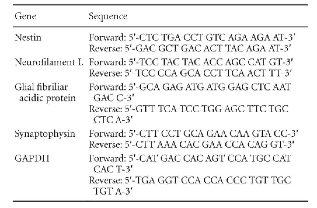
Table 1 Primer sequences speci fi c for neurons and glial cells
MTT assay
Differentiated mesenchymal stem cells were tested for their survival time in the presence or absence of citalopram as described previously (Shoae-Hassani et al., 2013a). MTT assays were performed at 0, 1, 3, 7, 14 and 21 days and at 1 and 2 weeks after citalopram treatment. Cells growing without citalopram treatment were used as controls. Brie fl y, 5 × 10³mesenchymal stem cells were seeded on 96-well plates and grown in the presence of citalopram (10 μmol/L). MTT (3-[4,5-dimethylthiazol-2-yl]-2,5-diphenyl tetrazolium bromide) (400 μg/mL) was added to each well for a 4 hour incubation period. At the end of the incubation period, the medium was removed and 100 μL dimethyl sulfoxide (DMSO) (Sigma) was added into each well. To dissolve the formazan crystals, the supernatant was pipetted several times. Absorbance was measured on an ELISA plate reader (Perkin Elmer, Waltham, MA, USA) at a wavelength of 540 nm.
Cumulative population doubling level
Citalopram-treated stem cells were continuously passaged in neurobasal media with and without retinoic acid (RA) for 30 days, and there was a 5-day interval between each passage. The cumulative population doubling level was calculated to determine their proliferation potential. Non-treated cells, cultured in the same condition but without citalopram, were used as controls. The cumulative population doubling level at each passage was calculated from the cell count using the equation ln(Nf/Ni)/ln2, where Ni and Nf are the initial and final cellnumbers, respectively, and ln is the natural log (Lin et al., 2005).

Table 2 BrdU-positive cells (percentage) derived from bone marrow mesenchymal stem cells after citalopram treatment versus control medium
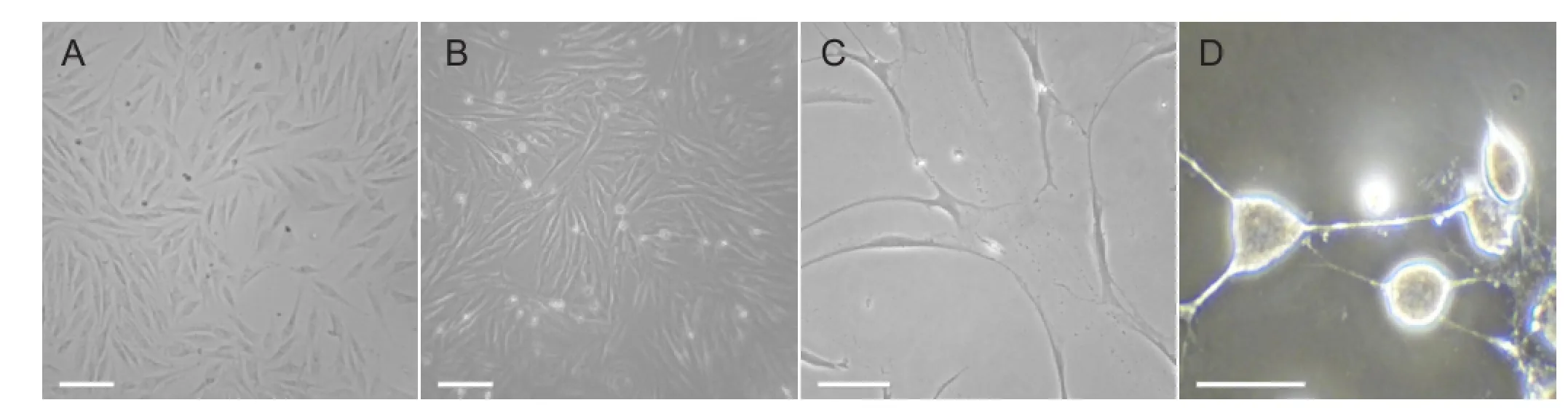
Figure 1 Morphological characteristics of bone marrow mesenchymal stem cells (BMSCs) and in vitro differentiation into neuronal-like cells.
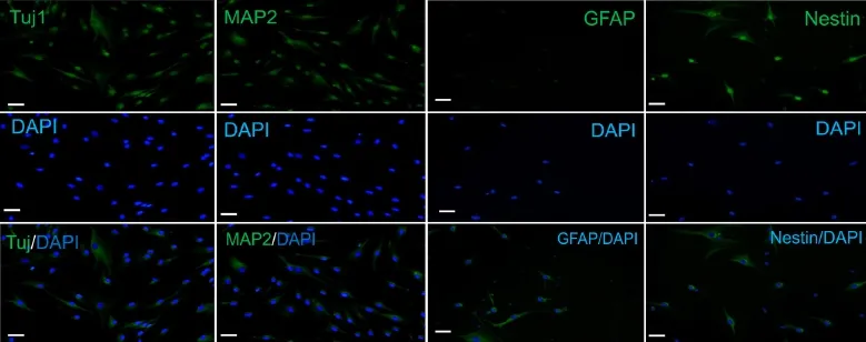
Figure 2 Expression of neuronal cell markers on the differentiated cells by BMSCs.
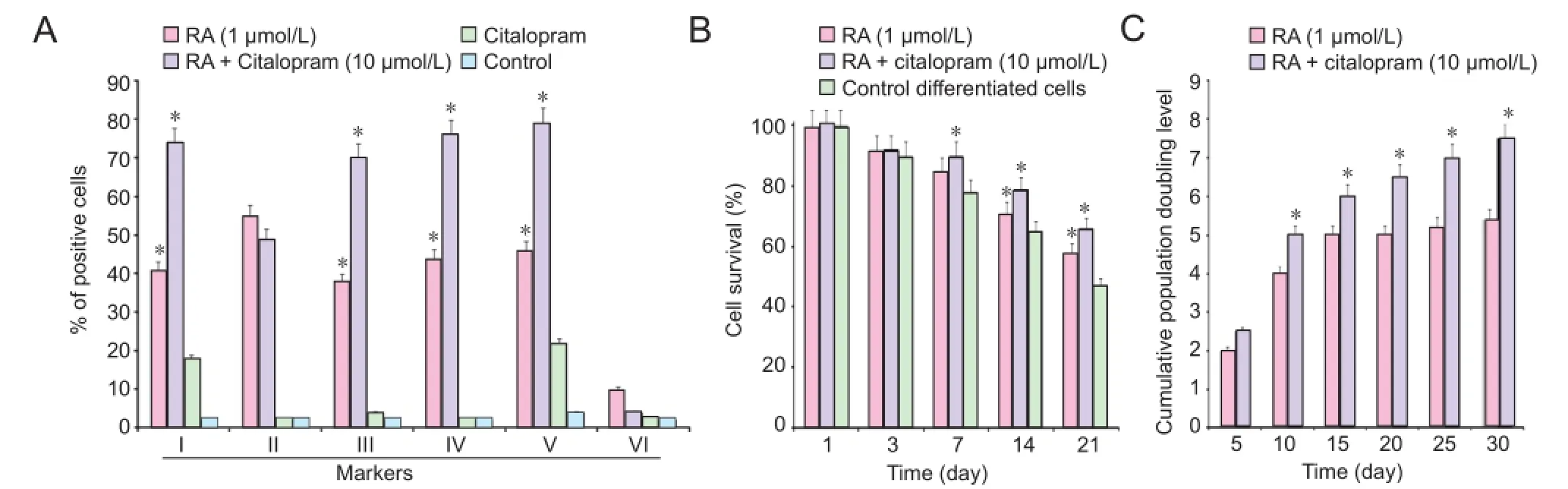
Figure 4 Characteristics of differentiated neuronal-like cells by BMSCs after 2 weeks of citalopram treatment.
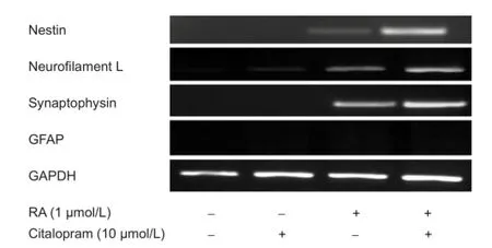
Figure 3 Expression of genes specific for neurons in differentiated cells by bone marrow mesenchymal stem cells.
5-Bromo-2-deoxyuridine (BrdU) incorporation assay
The BrdU assay was performed as described previously (Rahmani et al., 2013a). Citalopram and/or RA-treated cells and control cells were cultured overnight with 30 μg/mL of BrdU prior to immunostaining. Cells were fixed with 4% paraformaldehyde and then incubated with blocking buffer (PBS containing 0.05% Triton X-100, 0.5% bovine serum albumin) for 1 hour at room temperature. Following incubation in 2.0 mol/L HCl for 15 minutes at room temperature, cells were washed with 0.1 mol/L sodium borate buffer. Immunostaining was done using primary mouse anti-BrdU antibody (1:100; Roche, Welwyn Garden, Hertfordshire, UK), and the secondary antibody was FITC anti-mouse antibody (Molecular Probes, USA; 1:500). The nuclei were counterstained with ethidium bromide for 10 minutes at room temperature. The number and the percentage of BrdU-immunoreactive cells were calculated in relation to the total number of ethidium bromide cells.
Statistical analysis
One-way analysis of variance (ANOVA) and two-tailed Student’s t-test were used to analyze data using XLSTAT statistical software. Each experiment was conducted at least three times (n ≥ 3) and P-value of less than 0.05 was considered signi fi cant.
Results
Cell culture and differentiation
The in vitro study was conducted to determine whether the neurogenesis could be influenced by citalopram, a selective serotonin reuptake inhibitor antidepressant in cell culture system. After 7 days of culture, fi broblast-like cells with spindle-shaped morphology appeared on culture dishes (Figure 1A). After 7 days in vitro, stem cells growing in neuronal differentiation medium treated with citalopram displayed typical stem cell morphology (Figure 1B). Over the same time, stem cells treated with retinoic acid displayed neurite outgrowth (Figure 1C) and those treated with retinoic acid combined with citalopram increased neurite density (Figure 1D).
Immuno fl uorescence results
Citalopram and retinoic acid-treated human mesenchymal stem cells were visualized by fluorescence microscopy for detection of neuronal cell markers. Numerous cells immunoreactive for Tuj1, MAP-2 and nestin were seen in the immunostaining. There was no expression of glial fi brillary acidic protein (GFAP) in citalopram treated cells (Figure 2). The observed cellular characteristics and differentiation patterns demonstrate that citalopram enhanced the neuronal rather than the glial properties. The markers increased by citalopram were early neuronal cell markers, such as nestin.
RT-PCR analysis
RT-PCR detected the expression of neurofilament L, synaptophysin and nestin genes (Figure 3). There was no expression of glial cell marker GFAP as like as immunocytochemical staining. Expression of neuron-specific genes was up-regulated and remained elevated after 7 days in the citalopram-treated group.
MTT assay
The effect of citalopram on the survival period of the differentiated stem cells was observed directly and investigated by MTT assay. As shown in Figure 4B, cell survival rate was signi fi cantly higher (P < 0.05) in the citalopram + retinoic acid group than in the retinoic acid group. Retinoic acid increased the cell survival rate of differentiated neurons in this assay, and citalopram exhibited a preservative effect on differentiated stem cells. The continuance of citalopram treatment increased the time and percentage of cell viability (Figure 4B).
Cumulative population doubling level
Cumulative population doubling level was measured in the presence and absence of citalopram to determine the proliferative potential of citalopram-treated NPs. After 30 days of continuous passages, 7.41 and 5.63 cumulative population doubling level units were found in retinoic acid + citalopram-treated and only retinoic acid cells, respectively (Figure 4C). When the cells were passaged at regular intervals (every 4-5 days), compared with only retinoic acid-treated cells, the cumulative population doubling level of retinoic acid + citalopram-treated cells was significantly increased at day 15 (P < 0.05; Figure 4C). To examine the citalopram effects on DNA synthesis, the numbers of BrdU-positive cells were counted at days 0, 7, and 14 after citalopram and RA treatment. The percentage of BrdU-positive cells at days 7 (56%) and 14 (62%) was signi fi cantly increased in the retinoic acid + citalopram-treated cells compared with only retinoic acid-treated cells (P < 0.05; Table 2).
Discussion
Considering the obtained evidence, we showed that citalopram, a clinically effective antidepressant, increases stem cell survival and differentiation into a neuronal-like cell. Degenerative diseases are increasingly epidemic. Regenerative pharmacology is a form of therapy aimed at repairing the progressive cell destruction in degenerative diseases. Stem cellbased regenerative pharmacology supports the use of stem cells for derivation of novel therapeutics. Adult stem cells can provide a useful model to test the therapeutic compoundsand also can be used as models in regenerative pharmacology to test whether a specific drug promotes a lineage specific differentiation program (Shoae-Hassani et al., 2011a,c). Pharmacological agents could have inhibitory (Shoae-Hassani et al., 2011b) or stimulatory effect (Verdi et al., 2014) on the differentiation process of stem cells. There are many reports for selective serotonin reuptake inhibitor neurogenesis in vivo (Nandam et al., 2007; Newton and Duman 2007) but there was no study to survey the selective serotonin reuptake inhibitor effect on adult mesenchymal stem cell fate. The in vitro study was conducted to determine whether the neurogenesis could be influenced by citalopram, a selective serotonin reuptake inhibitor antidepressant in cell culture system.
Citalopram and retinoic acid-treated human mesenchymal stem cells were determined by fluorescence microscopy for detection of neuronal cell markers. Numerous cells immunoreactive for Tuj1, MAP-2 and nestin were seen in the immunostaining. There was no expression of GFAP in citalopram treated cells. The observed cellular characteristics and differentiation patterns demonstrate that citalopram enhanced the neuronal rather than the glial properties. The markers increased by citalopram were early neuronal cell markers, such as nestin. In 2011, Li et al. described rapid induction and long-term self-renewal of primitive neural precursors from human embryonic stem cells by small molecule inhibitors. It is obvious that these molecules have direct or indirect effects on some cellular pathways. The works by Menendez et al. (2011) confirmed that Wnt signaling and a Smad pathway blockade direct the differentiation of human pluripotent stem cells to multipotent neural crest cells. Previous studies by Rahmani et al. (2013a) showed that 10 μmol/L of fl uoxetine upregulated phosphorylation of Akt1 protein in induced stem cells signi fi cantly.
RT-PCR assay was used to detect the expression of neuro fi lament L, synaptophysin and nestin genes. There was no expression of glial cell marker GFAP, as confirmed by immunocytochemical staining. Expression of neuron-specific genes was up-regulated and remained elevated after 7 days in the retinoic acid + citalopram-treated group. Cells immunoreactive for neuronal cell markers showed neuronal-like cell appearance, i.e., large spherical cell body and extension. The percentage of differentiated neuronal cells reached to more than 70% in 14 days in the retinoic acid + citalopram-treated group compared to 50% of stem cells reaching a neuronal phenotype by 7 days in vitro induced just with retinoic acid (Figure 4A).
The effect of citalopram on the survival period of the differentiated stem cells was observed directly and investigated by MTT assay. We found that cell survival rate signi fi cantly higher following citalopram treatment than RA treated groups. While the retinoic acid increased differentiated neuronal cell survival in this assay, the effect of citalopram on differentiated stem cells was preservation. The continuance of citalopram treatment extended the time and percentage of cell viability. The obtained results are similar to those of Chen et al. (2007) who observed cell proliferation under in vitro conditions after 7 days, though they used different cell types and culture conditions. In 2001, Duman et al. reported that the new cell birth occurs in the brain and the rate of neurogenesis and survival of new neurons are regulated by a number of environmental and pharmacological treatments. The possibility that the cAMP signal transduction cascade and expression of brain derived neurotrophic factor (BDNF) contribute to the regulation of neurogenesis by antidepressants is supported by Thome et al. (2000). Up-regulation of brain derived neurotrophic factor could contribute to neurotrophic effects of antidepressants, including regulation of adult neurogenesis.
The cumulative population doubling level was measured with and without citalopram to determine the proliferative potential of citalopram-treated neural progenitors. After 30 days of continuous passages, 7.41 and 5.63 cumulative population doubling level units were found in retinoic acid + citalopram-treated and retinoic acid-treated cells, respectively. When the cells were passaged at regular intervals (every 4-5 days), the cumulative population doubling level of retinoic acid + citalopram-treated cells was significantly increased at day 15. For this analysis, the cells were passaged and they survived for up to 30 days. The proliferation rate of retinoic acid-treated cells exhibited a plateau from day 15 onward, while the plateau phase was on day 25 for retinoic acid + citalopram-treated cells. This fi nding suggests that the proliferation rate was higher in retinoic acid + citalopram-treated cells than in only retinoic acid-treated cells.
To examine the effects of citalopram on DNA synthesis, the numbers of BrdU-positive cells were counted at days 0, 7, and 14 after retinoic acid + citalopram treatment and only retinoic acid treatment. The percentage of BrdU-positive cells at days 7 (56%) and 14 (62%) was significantly increased in the retinoic acid + citalopram-treated cells compared to only retinoic acid-treated cells.
Studies of antidepressants have focused on cellular pathways, which are known to be activated by a number of extracellular signals, such as growth factors and neurotransmitters (Malberg 2004; Malberg and Blendy 2005). These signals regulate cellular processes associated with neuroplasticity and neurogenesis and seem necessary to mediate the therapeutic effects of citalopram.
To summary, our study elucidates the effect of citalopram on the neurogenesis of adult stem cells and con fi rms that it can increase the survival rate of the differentiated cells by adult stem cells as a therapeutic and regenerative drug.
Acknowledgments:We would like to thank Shoa-Hasani A for the helpful comments and are also grateful for the time and patience of the anonymous referees.
Author contributions:Verdi J, Mortazavi-Tabatabaei SA and Shoae-Hassani A conceived and designed this study and were responsible for data collection, analysis and interpretation, drafted the manuscript and provided critical revision of the manuscript for intellectual content. Verdi H and Sharif S provided suggestion on statistical expertise, experimental procedure and material application, obtained funds and supervised the study. All authors approved the final version of this paper.
Con fl icts of interest:None declared.
Atkinson S, Lako M, Armstrong L (2012) Potential for pharmacological manipulation of embryonic stem cells. Br J Pharmacol 169:269-289.
Bissonnette CJ, Lyass L, Bhattacharyya BJ, Belmadani A, Miller RJ, Kessler JA (2011) The controlled generation of functional basal forebrain cholinergic neurons from human embryonic stem cells. Stem Cells 29:802-811.
Boldrini M, Underwood MD, Hen R, Rosoklija GB, Dwork AJ, John Mann J (2009) Antidepressants increase neural progenitor cells in the human hippocampus. Neuropsychopharmacology 34:2376-2389.
Chen SJ, Kao CL, Chang YL, Yen CJ, Shui JW, Chien CS, Chen IL, Tsai TH, Ku HH, Chiou SH (2007) Antidepressant administration modulates neural stem cell survival and serotoninergic differentiation through bcl-2. Curr Neurovasc Res 4:19-29.
Chung S, Moon JI, Leung A, Aldrich D, Lukianov S, Kitayama Y (2011) ES cell-derived renewable and functional midbrain dopaminergic progenitors. Proc Natl Acad Sci USA 108:9703-9708.
Croce N, Mathé AA, Gelfo F, Caltagirone C, Bernardini S, Angelucci F (2014) Effects of lithium and valproic acid on BDNF protein and gene expression in an in vitro human neuron-like model of degeneration. J Psychopharmacol doi: 10.1177/0269881114529379.
Duman RS, Nakagawa S, Malberg J (2001) Regulation of adult neurogenesis by antidepressant treatment. Neuropsychopharmacology 25:836-844.
Hashimoto S, Inoue T, Muraki I, Koyama T (2009) Effects of acute citalopram on the expression of conditioned freezing in naïve versus chronic citalopram-treated rats. Prog Neuropsychopharmacol Biol Psych 33:113-117.
Huang B, Li K, Yu J, Zhang M, Li Y, Li W, Wang W, Guan L, Zhang W, Lin S, Huang X, Lin L, Lin Y, Zhang Y, Song X, Wang Z, Ge J (2013) Generation of human epidermis-derived mesenchymal stem cell-like pluripotent cells (hEMSCPCs). Sci Reports doi:10.1038/ srep01933.
Ilic D, Stephenson E, Wood V, Jacquet L, Stevenson D, Petrova A (2011) Derivation and feeder-free propagation of human embryonic stem cells under xeno-free conditions. Cytotherapy 14:122-128.
Kim J, Su SC, Wang H, Cheng AW, Cassady JP, Lodato MA (2011) Functional integration of dopaminergic neurons directly converted from mouse fi broblasts. Cell Stem Cell 9:413-419.
Kriks S, Shim JW, Piao J, Ganat YM, Wakeman DR, Xie Z (2011) Dopamine neurons derived from human ES cells ef fi ciently engraft in animal models of Parkinson’s disease. Nature 480:547-551.
Li W, Sun W, Zhang Y, Wei W, Ambasudhan R, Xia P (2011) Rapid induction and long-term self-renewal of primitive neural precursors from human embryonic stem cells by small molecule inhibitors. Proc Natl Acad Sci U S A 108:8299-8304.
Lin TM, Tsai JL, Lin SD, Lai CS, Chang CC (2005) Accelerated growth and prolonged lifespan of adipose tissue-derived human mesenchymal stem cells in a medium using reduced calcium and antioxidants. Stem Cells Dev 14:92-102.
Malberg JE (2004) Implications of adult hippocampal neurogenesis in antidepressant action. J Psychiatry Neurosci 29:196-205.
Malberg JE, Blendy JA (2005) Antidepressant action: to the nucleus and beyond. Trends Pharmacol Sci 26:631-638.
Malberg JE, Eisch AJ, Nestler EJ, Duman RS (2000) Chronic antidepressant treatment increases neurogenesis in adult rat hippocampus. J Neurosci 20:9104-9110.
Menendez L, Yatskievych TA, Antin PB, Dalton S (2011) Wnt signaling and a Smad pathway blockade direct the differentiation of human pluripotent stem cells to multipotent neural crest cells. Proc Natl Acad Sci U S A 108:19240-19245.
Nandam LS, Jhaveri D, Bartlett P (2007) 5-HT7, neurogenesis and antidepressants: a promising therapeutic axis for treating depression. Clin Exp Pharmacol Physiol 34:546-551.
Newton SS, Duman RS (2007) Neurogenic actions of atypical antipsychotic drugs and therapeutic implications. CNS Drugs 21:715-725.
Nori S, Okada Y, Yasuda A, Tsuji O, Takahashi Y, Kobayashi Y (2011) Grafted human-induced pluripotent stem-cell-derived neurospheres promote motor functional recovery after spinal cord injury in mice. Proc Natl Acad Sci U S A 108:16825-16830.
Noureddini M, Verdi J, Mortazavi-Tabatabaei SA, Sharif S, Azimi A, Keyhanvar P, Shoae-Hassani A (2012) Human endometrial stem cell neurogenesis in response to NGF and bFGF. Cell Biol Int 36:961-966.
Qin Y, Lin J, Zhou C, Yin Q, Xie Z, Zhang X, Liu XY, Gao W, Li J (2013) Mice cloned from white adipose tissue-derived cells. J Mol Cell Biol 5: 348-350.
Rahmani A, Kheradmand D, Keyhanvar P, Shoae-Hassani A, Darbandi-Azar A (2013a) Neurogenesis and increase in differentiated neural cell survival via phosphorylation of Akt1 after fluoxetine treatment of stem cells. BioMed Res Int ID 582526. dx.doi. org/10.1155/2013/582526.
Rahmani A, Shoae-Hassani A, Keyhanvar P, Kheradmand D, Darbandi-Azar A (2013b) Dehydroepiandrosterone stimulates nerve growth factor and brain derived neurotrophic factor in cortical neurons. Adv Pharmacol Sci ID 506191. dx.doi.org/10.1155/2013/506191.
Sasaki M, Honmou O, Akiayama Y, Uede T, Hashi K, Kocsis JD (2001) Transplantation of an acutely isolated bone marrow fraction repairs demyelinated adult rat spinal cord axons. Glia 35:26-34.
Schwaarz EJ, Alexander GM, Prockop DJ, Azizi SA (1999) Multipotential marrow stromal cells transduced to produce L-DOPA: engraftment in a rat model of Parkinson disease. Hum Gene Ther 10:2539-2549.
Shoae-Hassani A, Keyhanvar P, Seifalian AM, Mortazavi-Tabatabaei SA, Ghaderi N, Amirmozafari N (2013a) λ Phage nanobioparticle expressing apoptin ef fi ciently suppress human breast carcinoma tumor growth in vivo. PLoS One 8:e79907.
Shoae-Hassani A, Mortazavi-Tabatabaei SA, Sharif S, Rezaei-Khaligh H, Verdi J (2011a) DHEA provides a microenvironment for endometrial stem cells neurogenesis. Med Hypotheses 76:843-846.
Shoae-Hassani A, Mortazavi-Tabatabaei SA, Sharif S, Seifalian AM, Azimi A, Samadikuchaksaraei A, Verdi J (2013) Differentiation of human endometrial stem cells into urothelial cells on a three-dimensional nanofibrous silk-collagen scaffold: an autologous cell resource for reconstruction of the urinary bladder wall. J Tissue Eng Regen Med doi:10.1002/term.1632.
Shoae-Hassani A, Sharif S, Mortazavi-Tabatabaei SA, Verdi J (2011b) Could the endogenous opioid, morphine, prevent neural stem cell proliferation? Med Hypotheses 76:225-229.
Shoae-Hassani A, Sharif S, Seifalian AM, Mortazavi-Tabatabaei SA, Rezaie S, Verdi J (2013c) Endometrial stem cell differentiation into smooth muscle cell: a novel approach for bladder tissue engineering in women. BJU Int 112:854-863.
Shoae-Hassani A, Sharif A, Verdi J (2011c) The neurosteroid dehydroepiandrosterone could improve somatic cell reprogramming. Cell Biol Int 35:1037-1041.
Sollberger M, Rosen HJ, Shany-Ur T, Ullah J, Stanley CM, Laluz V, Weiner MW, Wilson SM, Miller BL, Rankin KP (2014) Neural substrates of socioemotional self-awareness in neurodegenerative disease. Brain Behav 4:201-214.
Thome J, Sakai N, Shin K, Steffen C, Zhang YJ, Impey S, Storm D, Duman RS (2000) cAMP response element mediated gene transcription is up-regulated by chronic antidepressant treatment. J Neurosci 20:4030-4036.
Verdi J, Shari S, Banafshe HR, Shoae-Hassani A (2014) Sertraline increases the survival of retinoic acid induced neuronal cells but not glial cells from human mesenchymal stem cells. Cell Biol Int doi:10.1002/cbin.10283.
Viswanathan S, Rao M, Keating A, Srivastava A (2013) Overcoming challenges to initiating cell therapy clinical trials in rapidly developing countries: India as a model. Stem Cells Transl Med 2:607-613.
Wang JW, David DJ, Monckton JE, Battaglia F, Hen R (2008) Chronic fluoxetine stimulates maturation and synaptic plasticity of adult born hippocampal granule cells. J Neurosci 28:1374-1384.
Copyedited by Widgerow A, Jin GH, Li YL, Li CH, Song LP, Zhao M
10.4103/1673-5374.131601
Alireza Shoae-Hassani , Italia St., Vesal Ave, Keshavarz Blv, Tohid Sq, Chamran Highway, Tehran 14185-615, Iran,
cell.therapy@yahoo.com.
http://www.nrronline.org/
Accepted: 2014-03-14
- 中国神经再生研究(英文版)的其它文章
- Virtual reality interface devices in the reorganization of neural networks in the brain of patients with neurological diseases
- Regulatory effects of anandamide on intracellular Ca2+concentration increase in trigeminal ganglion neurons
- Nasal mucosal inhalation of amyloid-beta peptide 3-10 defective adenovirus attenuates cytotoxicity induced by beta-amyloid (1-42)
- The synthetic thyroid hormone, levothyroxine, protects cholinergic neurons in the hippocampus of naturally aged mice
- Similar effects of substance P on learning and memory function between hippocampus and striatal marginal division
- Fusion protein of single-chain variable domain fragments for treatment of myasthenia gravis