Salvia fruticosa reduces intrinsic cellular and H2O2-induced DNA oxidation in HEK 293 cells; assessment using flow cytometry
Saleem Bani Hani, Mekki Bayachou
1Department of Medical Laboratory Sciences, Jordan University of Science and Technology, Irbid 22110, Jordan
2Department of Chemistry, Cleveland State University, 2399 Euclid Avenue, Cleveland, OH 44115, USA
Salvia fruticosa reduces intrinsic cellular and H2O2-induced DNA oxidation in HEK 293 cells; assessment using flow cytometry
Saleem Bani Hani1*, Mekki Bayachou2
1Department of Medical Laboratory Sciences, Jordan University of Science and Technology, Irbid 22110, Jordan
2Department of Chemistry, Cleveland State University, 2399 Euclid Avenue, Cleveland, OH 44115, USA
PEER REVIEW
Peer reviewer
Dr. Doaa Mohamed Abd El-Aziz, Department of Food Hygiene, Assiut University, Assiut, Egypt.
Tel: 002/0882334699
E-mail: doaassiut@yaho.com
Comments
This is a good research in which authors clear assess H2O2-induced DNA oxidation protection activity of the aqueous extract of S. fruticosa leaves based on measuring 8-oxoguanine moieties as a sensitive biomarkers oxidation for oxidative DNA lesions in HEK-293 cells, using flow cytometry. Details on Page 402
Objective:To investigate the role of water-soluble extract of Salvia fruticosa (Greek sage) (S. fruticosa) leaves in reducing both intrinsic cellular and H2O2-induced DNA oxidation in cultured human embryonic kidney 293 cells. S. fruticosa, native to the Eastern-Mediterranean basin, is widely used as a medicinal herb for treatment of various diseases.
Salvia fruticosa, DNA oxidation, Oxidative stress, Human embryonic kidney 293 cells, Flow cytometry
1. Introduction
Antioxidants are substances capable of counteracting the oxidative damage of the free radicals in body tissues, and reducing the cellular oxidative stress[1,2], thus, decreasing DNA, protein and lipid oxidation[3-5]. Enhancement of body defenses via oral antioxidant supplementation would seem to provide a reasonable and a practical approach to reduce the level of oxidative stress, and thus preventing the degenerative disorders such as cancer and diabetes[6,7].
Recently, the development of new technologies has revolutionized the screening of natural products for potent antioxidants and anticancer materials. Plant-derived extracts are emerging as major players in this field.
Salvia fruticosa(also called a Greek sage) (S. fruticosa), is a perennial herb native to the eastern Mediterranean region. Cohorts in the Mediterranean region have used water-soluble leaf extract ofS. fruticosato treat variousdiseases, especially digestive system diseases[8]. It has been suggested thatS. fruticosatreatment produces hypoglycemia mainly by reducing intestinal absorption of glucose[9,10]. Pitarokiliet al.(2003) showed that volatile metabolites ofS. fruticosaexhibited high antifungal activities[11].S. fruticosaoil extract as well as its alcoholic extract revealed a strong antioxidant activity[12,13]. Moreover, Orhanet al.(2008) showed thatS. fruticosahas a significant anticholinesterase activity[14]. Later reports showed thatS. fruticosastimulates DNA repair mechanism in cultured HeLa cells[15]. Recent study found that the crude ethanol extract ofS. fruticosahas antiproliferative activity against breast cancer cells[16]. A newly published study by Sevindik and Rencuzogullari (2013) concluded thatS. fruticosaleaf extract had no cytotoxic effect against human blood lymphocytes[17].
The vast majority ofS. fruticosaresearch has investigated the possible health benefits ofS. fruticosaoil and its constituents; little information is available about its watersoluble material. This study aimed to assess the H2O2-induced DNA oxidation protection activity of water-soluble extract ofS. fruticosaleaves in human embryonic kidney 293 cells (HEK-293 cells). To do this, we measured the 8-oxoguanine moieties, sensitive biomarkers for oxidative DNA lesions, using flow cytometry. This study is the first one that utilizes flow cytometry to measure directly the anti-DNA oxidation activity of a plant extract.
2. Materials and methods
2.1. Preparation of Salvia Fruticosa Extract
FreshS. fruticosaleaves were collected from the Marzoog garden in the northern Jordan. The leaves were dried in the shade for one week and stored in the dark for three months before use. The dried leaves were grounded by mortar and pestle to fine particles then dissolved in PBS buffer. A volume of 5 mL buffer per gram ofS. fruticosaleaves was added, and the final suspension was homogenized, transferred to a centrifuge tube, shaken overnight at room temperature and stored at 4 °C in the dark. The homogenized mixture was centrifuged at 10 000 r/min for 10 min and the supernatant was transferred to a new tube. The extract supernatant was further passed through an ultra-centrifugation membrane (<30 000 kDa; Amicon, Bedford) under high-pressure conditions (12 psi), in a filtration device (Amicon, Bedford). The extract passing the membrane was collected and stored at 4 °C in the dark for future use. Each mL of the preparation will contain (7.1± 1.0) mg of extract dry weight (0.7% w/v).
2.2. Cell culture
We used human kidney cells (the HEK-293 cell line, ATCC, Manassas, VA, USA) as a cell model in our investigation. HEK-293 cells have been extensively used in cell biology research for many years. The possible genotoxicity of the extracts were examined previously in these cells[18].
HEK-293 cells were incubated in the Dulbecco’s Modified Eagle Medium (DMEM) supplemented with non-essential amino acids, 2 mmol/L L-glutamine, 5% penicillin/ streptomycin and Earle’s BSS adjusted to contain 1.5 g/L sodium bicarbonate. Cells were kept at 37 °C in a humidified incubator containing 5% CO2in air.
2.3. H2O2treatment
HEK-293 cells (1×10⁶cells/mL) are plated and exposed to different treatments (a though c below) before flow cytometry analysis:
a. Addition of freshly prepared H2O2and incubation for 3 h at 37 °C. The final concentration of H2O2in the cultured cell plates was 0.1 mmol/L. Control assays were prepared in the absence of H2O2.
b. Addition of freshly prepared H2O2followed by 150 µLS. fruticosaextract and incubation for 3 h at 37 °C. The final concentration of H2O2in the cultured cell plates was 0.1 mmol/L. Control assays were prepared in the absence ofS. fruticosaextract.
c. Incubated for 3 h at 37 °C with only 100 µL ofS. fruticosaextract. Control assays were prepared in the absence of the extract.
2.4. Flow cytometry
The level of DNA oxidation was measured using a flow cytometric OxiDNA assay kit (Calbiochem, San Diego). The method used was adapted and standardized in our previous work to assess the oxidative DNA damage in human sperm[19]. This assay is based on utilizing a direct fluorescent protein binding method for detection the DNA oxidation moieties (8-oxoguanines). Briefly, HEK-293 cells were washed twice in PBS, resuspended in 1% paraformaldehyde at a concentration of (1-2)106cell/mL and placed on ice for 15 to 30 min. These cells were again washed and resuspended in 70% ice-cold ethanol by 5 min centrifugation at 1 600 r/min. The ethanol supernatant was removed and the cell pellets were washed twice in wash buffer and resuspended in 100 µL of the staining solution for 1 h at room temperature.
The staining solution contained fluorescein isothiocyanate (FITC) labeled protein conjugate, and deionized water. All cells were further washed using rinse buffer, resuspendedin 250 µL and incubated for 30 min in the dark on the ice for flow cytometry measurements.
Data acquisition was operated within 1 h on a flow cytometer equipped with a 515-nm argon laser as a light source (FACScan; Becton Dickinson, San Jose, CA). 10 000 cells were interrogated for each single assay at a flow rate of 100 cells/second. The FITC fluorescence (log green fluorescence) was measured on FL1 channel and data analysis was done using FlowJo v4.4.4 software (Tree Star Inc., Ashland, OR).
2.5. Statistical analysis
Differences in the mean values of FITC fluorescence were considered significant atP<0.05. Statistical analyses were performed using pairedt-test and two-tailed distribution by the SPSS/PC computer software (SPSS 10.0.7, SPSS Inc.).
3. Results
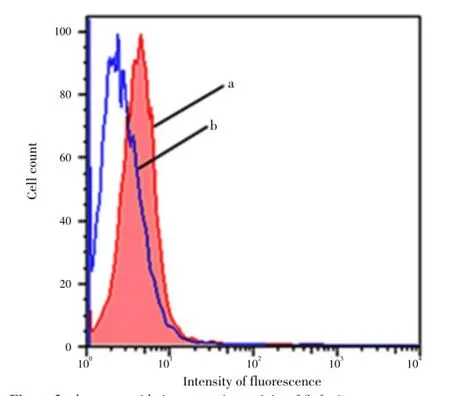
The direct effect of H2O2on DNA oxidation in HEK-293 cells is shown in Figure 1. Flow cytometry analysis of FITC-labeled HEK-293 showed that cells treated with 0.1 mmol/L H2O2and incubated 3 h at 37 °C exhibit increased intensity of FITC fluorescence (P<0.05), indicating an expected positive effect of H2O2on the intrinsic baseline of cellular DNA oxidation.

Figure 1. The effect of H2O2in inducing DNA oxidation in HEK-293 cells as evaluated by flow cytometry.a: Represents the flow cytometry histogram for cells without H2O2treatment; b: represents the flow cytomety histogram for cells incubated 3 h with 0.1 mmol/L H2O2. Data are representative of 6 independent experiments; the mean values of the histograms (a, and b) are statistically different (P<0.05).
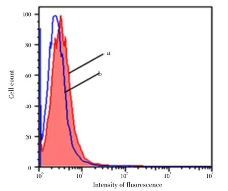
Figure 2 shows the effect of theS. fruticosaextract in decreasing DNA oxidation induced by 0.1 mm H2O2in HEK-293 cells. Cells incubated 3 h with 150 µL of the extract and exposed to 0.1 mmol/L H2O2showed lower intensity of fluorescence (P<0.05), and thus lower DNA damage.

Figure 2. The DNA-oxidation protection activity of S. fruticosa extract.a: Represents the flow cytometry histogram for cells incubated 3 h with 0.1 mmol/ L H2O2; b: represents the population incubated 3 h with 0.1mmol/L H2O2in the presence of 150 µL S. fruticosa extract. Data are representative of 6 independent experiments; the mean values of the histograms (a, and b) are statistically different (P<0.05).
Figure 3 shows the effect of theS. fruticosaextract in reducing the intrinsic cellular DNA oxidation in HEK-293 cells. As shown in the figure, cells incubated 3 h with 100 µL of the extract showed a lower intensity of fluorescence (P<0.05), and thus lower intrinsic cellular DNA damage compared to the control (in absence ofS. fruticosa).

Figure 3. The DNA-oxidation protection activity of S. fruticosa extract as evaluated by flow cytometry.a: Represents the flow cytometry histogram for cells without S. fruticosa extract treatment; b: Represents the flow cytomety histogram for cells incubated 3 h with 100 µL S. fruticosa extract. Data are representative of 6 independent experiments; the mean values of the histograms (a, and b) are statistically different (P<0.05).
4. Discussion
In cellular systems, in the presence of free ferrous and cuprous ions, and superoxide anion (·O2-), H2O2can produce hydroxyl radicals (·OH), a very short-lived reactive oxygen species, which recognized as Fenton’s reaction[20-22]. ·OH causes DNA lesion; when created adjacent to the DNA it strikes its main building blocks such as deoxyribose sugar and nitrogen bases (purines and pyrimidines), which may result in chemical alteration, and subsequently leads to injury, death, or enhancement of abnormal growth of cells (cancer development) [23].
The first flow cytometry experiment aimed to standardize the level of H2O2-induced DNA oxidation in HEK-293 cells as determined using 8-oxoguanine as oxidative damage marker. As expected for H2O2, our flow cytometry experiments showed an increase in the DNA oxidation after the addition of H2O2. However, in the presence ofS. fruticosaextract, we show a lower FITC-fluorescence intensity compared to H2O2alone, and hence, a lower level of DNA oxidation. Similar changes in DNA oxidation, have been reported in the study the protective effect of L-carnitine to reduce thein vitrooxidative stress on human spermatozoa using this same 8-oxoguanine marker in flow cytometry[19]. Although, the constituents of our extract in this study are unknown, the antioxidant effect may due to the presence of polyphenols such as rosmarinic acid and luteolin-7-glucoside[15].
In the last flow cytometry experiment, we intended to investigate the effect of water-soluble extract ofS. fruticosaon the basal-intrinsic cellular DNA oxidation in HEK-293 cells. Cells incubated 3 h with the extract showed lower levels of 8-oxoguanine moieties compared to controls in the absence of extract. This decrease in the cellular DNA oxidation may due to an increase in the activity of DNA repair machinery in the presence ofS. fruticosaextract. These results are in line with similar reports by Ramoset al.(2010)[15], who used the comet assay to investigate the antioxidant activity of the water-soluble extract ofS. fruticosa. The exact mechanism by whichS. fruticosaboosts the repair machinery of HEK-293 cells is not known at this point. However, like the recent reports about extracting polyphenols[24,25], a possible mechanism by which our water extract ofS. fruticosareduces the level of DNA oxidation is probably via alleviating the load of cellular oxidative stress by directly scavenging the reactive oxygen species.
In conclusion, our results suggest that water-soluble extract ofS. fruticosaleaves mediates protection against both intrinsic cellular and H2O2-induced DNA oxidation in HEK-293 cells. The reduction in the intrinsic cellular DNA oxidation may reflect an enhanced activity of the DNA repair machinery.
Conflict of interest statement
The authors declare that there are no conflicts of interest.
Acknowledgements
This work was supported by Cleveland State University and Jordan University of Science and Technology with grant number 20130097.
Comments
Background
The oxidation of lipid, DNA, protein, carbohydrate, and other biological molecules by toxic ROS may cause DNA mutation or/and serve to damage target cells or tissues, and this often results in cell senescence and death. Cancer chemoprevention by using antioxidant approaches has been suggested to offer a good potential in providing important fundamental benefits to public health. Antioxidants of plant extracts have formed the basis of many applications, including processed food preservation, pharmaceuticals, alternative medicine and natural therapies.
Research frontiers
This study is carried out to evaluate the water extract ofS. fruticosaleaves for its antioxidants activity in HEK293 cells by measuring 8-oxoguanine moieties as a sensitive biomarkers oxidation, using flow cytometry.
Related reports
L-carnitine has similar changes in DNA oxidation; to reduce thein vitrooxidative stress on human spermatozoa using this same 8-oxoguanine marker in flow cytometry.
Innovations and breakthroughs
In this study authors have demonstrated the antioxidant activity of the aqueous extract ofS. fruticosaleaves in HEK-293 cells by using flow cytometry.
Applications
Enhancement of body defenses via oral supplementation withS. fruticosaleaves protects against both H2O2-induced and intrinsic cellular DNA oxidation and so reduce the level of oxidative stress, and thus preventing the degenerative disorders such as cancer.
Peer review
This is a good research in which authors cleared assess H2O2-induced DNA oxidation protection activity of the aqueous extract ofS. fruticosaleaves based on measuring 8-oxoguanine moieties as a sensitive biomarkers oxidation for oxidative DNA lesions in HEK-293 cells, using flow cytometry.
[1] Sies H. Oxidative stress: oxidants and antioxidants. Exp Physiol 1997; 82: 291-295.
[2] Alok S, Jain SK, Verma A, Kumar M, Mahor A, Sabharwal M. Herbal antioxidant in clinical practice: a review. Asian Asian Pac J Trop Biomed 2014; 4: 78-84.
[3] Sivonová M, Tatarková Z, Duracková Z, Dobrota D, Lehotský J, Matáková T, et al. Relationship between antioxidant potential and oxidative damage to lipids, proteins and DNA in aged rats. Physiol Res 2007; 56: 757-764.
[4] Therond P. [Oxidative stress and damages to biomolecules (lipids, proteins, DNA)]. Ann Pharm Fr 2006; 64: 383-389. French.
[5] Banihani S, Agarwal A, Sharma R, Bayachou M. Cryoprotective effect of l-carnitine on motility, vitality and DNA oxidation of human spermatozoa. Andrologia 2013; doi: 10.1111/and.12130.
[6] Valko M, Leibfritz D, Moncol J, Cronin MT, Mazur M, Telser J. Free radicals and antioxidants in normal physiological functions and human disease. Int J Biochem Cell Biol 2007; 39: 44-84.
[7] Banihani S, Swedan S, Alguraan Z. Pomegranate and type 2 diabetes. Nutr Res 2013; 33: 341-348.
[8] Gurdal B, Kultur S. An ethnobotanical study of medicinal plants in Marmaris (Mugla, Turkey). J Ethnopharmacol 2013; 146: 113-126.
[9] Azevedo MF, Lima CF, Fernandes-Ferreira M, Almeida MJ, Wilson JM, Pereira-Wilson C. Rosmarinic acid, major phenolic constituent of Greek sage herbal tea, modulates rat intestinal SGLT1 levels with effects on blood glucose. Mol Nutr Food Res 2011; 55(Suppl 1): S15-25.
[10] Perfumi M, Arnold N, Tacconi R. Hypoglycemic activity of Salvia fruticosa Mill. from Cyprus. J Ethnopharmacol 1991; 34: 135-140.
[11] Pitarokili D, Tzakou O, Loukis A, Harvala C. Volatile metabolites from Salvia fruticosa as antifungal agents in soilborne pathogens. J Agric Food Chem 2003; 51: 3294-3301.
[12] Papageorgiou V, Gardeli C, Mallouchos A, Papaioannou M, Komaitis M. Variation of the chemical profile and antioxidant behavior of Rosmarinus officinalis L. and Salvia fruticosa Miller grown in Greece. J Agric Food Chem 2008; 56: 7254-7264.
[13] Senol FS, Orhan IE, Erdem SA, Kartal M, Sener B, Kan Y, et al. Evaluation of cholinesterase inhibitory and antioxidant activities of wild and cultivated samples of sage (Salvia fruticosa) by activityguided fractionation. J Med Food 2011; 14: 1476-1483.
[14] Orhan I, Kartal M, Kan Y, Sener B. Activity of essential oils and individual components against acetyl- and butyrylcholinesterase. Z Naturforsch C 2008; 63: 547-553.
[15] Ramos AA, Azqueta A, Pereira-Wilson C, Collins AR. Polyphenolic compounds from Salvia species protect cellular DNA from oxidation and stimulate DNA repair in cultured human cells. J Agric Food Chem 2010; 58: 7465-7471.
[16] Abu-Dahab R, Afifi F, Kasabri V, Majdalawi L, Naffa R. Comparison of the antiproliferative activity of crude ethanol extracts of nine Salvia species grown in Jordan against breast cancer cell line models. Pharmacog Mag 2012; 8: 319-324.
[17] Sevindik N, Rencuzogullari E. The genotoxic and antigenotoxic effects of Salvia fruticosa leaf extract in human blood lymphocytes. Drug Chem Toxicol 2013; doi:10.3109/01480545.2013.851689.
[18] Hudecová A1, Hašplová K, Miadoková E, Magdolenová Z, Rinna A, Collins AR, et al. Gentiana asclepiadea protects human cells against oxidation DNA lesions. Cell Biochem Funct 2012; 30: 101-107.
[19] Banihani S, Sharma R, Bayachou M, Sabanegh E, Agarwal A. Human sperm DNA oxidation, motility and viability in the presence of L-carnitine during in vitro incubation and centrifugation. Andrologia 2012; 44(Suppl 1): 505-512.
[20] Rodopulo AK. [Oxidation of tartaric acid in wine in the presence of heavy metal salts (activation of oxygen by iron)]. Izv Akad Nauk SSSR Biol 1951; 3: 115-128. [Article in Undetermined Language].
[21] Goldstein S, Meyerstein D, Czapski G. The Fenton reagents. Free Radic Biol Med 1993; 15: 435-435.
[22] Monroe EB, Heien ML. Electrochemical generation of hydroxyl radicals for examining protein structure. Anal Chem 2013; 85: 6185-6189.
[23] Miroslav F. Electrochemical sensors for DNA interactions and damage. Electroanalysis 2002; 14: 1449-1463.
[24] Tan X, Zhao C, Pan J, Shi Y, Liu G, Zhou B, et al. In vivo nonenzymatic repair of DNA oxidative damage by polyphenols. Cell Biol Int 2009; 33: 690-696.
[25] Keuser B1, Khobta A, Gallé K, Anderhub S, Schulz I, Pauly K, et al. Influences of histone deacetylase inhibitors and resveratrol on DNA repair and chromatin compaction. Mutagenesis 2013; 28: 569-576.
10.12980/APJTB.4.2014C1270
*Corresponding author: Saleem Bani Hani, MSc., Ph.D; Assistant Professor, Clinical Chemistry and Molecular Medicine, Department of Medical Laboratory Sciences, Jordan University of Science and Technology, P. O. Box 3030-Irbid-22110, Jordan.
E-mail: sabanihani@just.edu.jo
Fax: +962-2-7201087
Tel: +962-27201000 Ext. 23874
Foundation Project: Supported by Cleveland State University and Jordan University of Science and Technology; grant number 20130097.
Article history:
Received 4 Feb 2013
Received in revised form 12 Feb, 2nd revised form 19 Feb, 3rd revised form 6 Mar 2014
Accepted 12 Apr 2014
Available online 28 May 2014
Methods:Dried leaves of S. fruticosa were extracted in phosphate buffer saline and purified using both vacuum and high pressure filtrations. Each mL of the preparation contained (7.1±1.0) mg of extract. HEK-293 cells were incubated in one set with S. fruticosa extract in the presence of 0.1 mmol/L H2O2, and in the other set with the addition of the extract alone. The DNA oxidation was measured using fluorescence upon fluorescein isothiocyanate derivatization of 8-oxoguanine moieties. The fluorescence was measured using flow cytometry technique.
Results:Cells incubated 3 h with 150 µL extract and exposed to 0.1 mmol/L H2O2showed lower intensity of fluorescence, and thus lower DNA oxidation. Moreover, cells incubated 3 h with 100 µL of the extract showed lower intensity of fluorescence, and thus lower intrinsic cellular DNA oxidation compared to control (without S. fruticosa).
Conclusions:The results from this study suggest that the water-soluble extract of S. fruticosa leaves protects against both H2O2-induced and intrinsic cellular DNA oxidation in human embryonic kidney 293 cells.
 Asian Pacific Journal of Tropical Biomedicine2014年5期
- Asian Pacific Journal of Tropical Biomedicine的其它文章
- Ethnobotanical survey of folklore plants used in treatment of snakebite in Paschim Medinipur district, West Bengal
- Pharmacognostic studies of stem, roots and leaves of Malva parviflora L.
- Rapid detection of coliforms in drinking water of Arak city using multiplex PCR method in comparison with the standard method of culture (Most Probably Number)
- Antisickling activity of butyl stearate isolated from Ocimum basilicum (Lamiaceae)
- Antioxidant and antimicrobial properties of Litsea elliptica Blume and Litsea resinosa Blume (Lauraceae)
- Tamarind seed coat extract restores reactive oxygen species through attenuation of glutathione level and antioxidant enzyme expression in human skin fibroblasts in response to oxidative stress
