
Seroprevalence of bovine leptospiral antibodies by microscopic agglutination test in Southeast of Iran
2014-03-23 03:20ResearchCenterofTropicalandInfectiousDiseasesKermanUniversityofMedicalSciencesKermanIran
Research Center of Tropical and Infectious Diseases, Kerman University of Medical Sciences, Kerman, Iran
2Department of Pathobiology, Faculty of Veterinary Medicine, Shahid Bahonar University of Kerman, Kerman, Iran
3Department of Clinical Sciences, Faculty of Veterinary Medicine, Shahid Bahonar University of Kerman, Kerman, Iran
4Department of Clinical Sciences, Faculty of Veterinary Medicine, University of Tehran, Tehran, Iran
Seroprevalence of bovine leptospiral antibodies by microscopic agglutination test in Southeast of Iran
Mohammad Khalili1,2, Ehsanollah Sakhaee3, Mohammad Reza Aflatoonian1, Gholamreza Abdollahpour4, Saeed Sattari Tabrizi4, Elham Mohammadi Damaneh2, Sajad Hossini-nasab2
1Research Center of Tropical and Infectious Diseases, Kerman University of Medical Sciences, Kerman, Iran
2Department of Pathobiology, Faculty of Veterinary Medicine, Shahid Bahonar University of Kerman, Kerman, Iran
3Department of Clinical Sciences, Faculty of Veterinary Medicine, Shahid Bahonar University of Kerman, Kerman, Iran
4Department of Clinical Sciences, Faculty of Veterinary Medicine, University of Tehran, Tehran, Iran
PEER REVIEW
Peer reviewer
Yuki Eshita, Associate Professor, Department of Infectious Disease Control, Faculty of Medicine, Oita University, 1-1 Idaigaoka, Hasamamachi, Yufu-shi, Oita 879-5593, Japan. Tel: +81-97-586-5701
Fax: +81-97-586-5701
E-mail: yeshita@oita-u.ac.jp
Comments
This is a good epidemiological study in which the authors proved firstly the existence of leptospirosis in Southeast Iran by using MAT.
Details on Page 357
Objective:To evaluate serological findings of bovine leptospirosis which is a zoonotic disease with worldwide distribution caused by Leptospira interrogans.
Bovine leptospirosis, MAT, Serology, Iran
1. Introduction
Leptospirosis is the most common bacterial zoonosis worldwide, caused by spirochetes of the genusLeptospira. There are 20 species of leptospires, consisting of over 200 serovars, circulating in a wide range of animal reservoir hosts including rats, other rodents, livestock and domestic pets[1].Leptospira interrogans(L. interrogans) constitutes the major pathogenic leptospiral species that is responsible for human infection.L. interroganscan readily penetrate abraded skin and mucous membrane barriers to establish a systemic infection via haematogenous dissemination and subsequently colonizes multiple organs, particularly the kidneys and liver. While wild rodents serve as natural reservoirs, humans and a few other domesticated animals are accidental hosts in the transmission cycle of leptospirosis[2,3]. In rural areas, transmission is usually associated with farming and livestock, with increased risk during the warm and rainy months. In urban areas, infection is associated with overcrowding, poor hygiene standards, inadequate sanitation and poverty, all of which typically occur in urban slums in developing countries.
In developed countries, infection is now increasingly being associated with outdoor recreational exposure and international travel[4].
Suitability of the environment for the survival of leptospires appears to be a critical factor in maintaining the infection and transmission to humans. Leptospires have good affinity to areas where heavy rainfall results in water logging of the land. Human populations residing in such environment are at higher risk of acquiring leptospiral infection[5].
A basic knowledge of serovars and their maintenance hosts is required to understand the epidemiology of leptospirosis in a region. Though distinct variations in maintenance hosts and the serovars they carry can occur throughout the world. The general pattern is for serogroups Hardjo bovis, Pomona, and Grippotyphosa to be recoverable from cattle[6].
The diagnosis of leptospirosis is based on two principles which include the actual isolation of the leptospiral organisms and the detection of anti-leptospiral antibodies. Isolation by culture is very time-consuming and depends on the presence of live leptospira and their ability to grow on media provided, thus serological testing is a more widely used method. The detection of anti-leptospiral antibodies can be done using tests such as the microscopic agglutination test (MAT) and enzyme-linked immunosorbent assay[7]. The MAT is the gold standard test for diagnosing leptospirosis and is the most widely used method for detecting both leptospira IgG and IgM antibodies in animal sera. The sensitivity and specificity of the MAT reported in a recent study were 91.94% and 73.77%, respectively[8]. This test can be used qualitatively and quantitatively to detect the infecting serovar and titer (World Health Organization), but it requires the propagation of live leptospiral strains to be used as antigens for a hazardous and time-consuming process in which the interpretation of the results can be subjective[9].
To the authors’ knowledge, there is no report of leptospirosis in Southeast Iran; therefore, the aim of this study was to investigate the presence of anti-leptospira antibodies among dairy cattle farms by MAT, using five current reference strains ofL. interrogansin Jiroft suburb dairy farms, Kerman province, Southeast Iran.
2. Materials and methods
2.1. Sample taking
A total of 167 sera were collected from 9 commercial dairy herds in jiroft suburbs, from July to October 2011. Sera were separated after centrifugation at 3 000 g for 10 min at room temperature and kept at -20 °C until required. These samples were submitted to the Leptospira Research Laboratory of Teaching and Research Hospital of the Faculty of Veterinary Medicine at the University of Tehran, Iran.
2.2. Microscopic agglutination test (MAT)
MAT was carried out as described by Turner (1968) with some modification in Leptospira Research Laboratory as follows: Five reference strains ofL. interroganswhich were used as antigen includes:Leptospirahardjo(L. hardjo), Leptospira pomona(L. pomona), Leptospira icterohaemorrhagiae(L. icterohaemorrhagiae), Leptospira grippotyphosa(L. grippotyphosa) andLeptospiracanicola(L. canicola). All sera samples were serially diluted in phosphate buffer solution (PBS) in a microtiter plate (Greiner), starting from 1 in 50 dilution, using 2-fold dilution (1 in 100, 200 and 400). Then, 10 µL of serum dilution was added to 10 µL of appropriate antigen on a microscopic slide and incubated at 30 °C for 90 min. Finally the slide was examined under dark-field microscope (Olympus BX50). One antigen control and two (positive and negative) standard serum controls were used each time. Titers 1:100 or greater were considered positive. The end-point titer was determined as the highest serum dilution showing agglutination of at least 50% of the leptospires.
3. Results
Antibodies were found at least against one serovar ofL. interrogansin 29 samples (17.36%) among 167 sera at a dilution 1:100 or higher. Positive titers against more than one serovar were detected in 6 samples (20.68%) of the 29 positive sera (Table 1).

Table 1 Frequency (%) and number of positive serum samples by MAT at a dilution 1:100 or higher, among 167 samples.
According to Table 2, the highest prevalence of positive sera by MAT was found in farm 3 (4.79%), followed by farm 5 and 9 (3.59%), farm 4 and 7 (1.80%), farm 6 (1.20%) and farm 1 (0.6%), while no positive serum was find in farm 2 and 8.

Table 2 Number and frequency (%) of total and positive sera in each farm by MAT.
Positive titers were detected against serovarL. pomona(16sero),L. grippotyphosa(11 sera),L. canicola(6 samples), andL. hardjo(2 samples). There is no positive sample againstL. ichterohaemorrhagiae(Table 3). Table 4 presents the number and frequency of each serovar, in different farms.
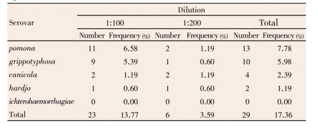
Table 3 Number and frequency (%) of serum samples with positive titer against each serovar, at each dilution.

Table 4 Number and frequency (%) of each serovar, in different farms.
4. Discussion
Different researchers have demonstrated the presence of antibodies anti leptospira species and variations in clinical manifestations for the zoonoses in different regions of Iran[10-12].
Jiroft is located in a vast plain, Halil River, on the southern outskirts of the Jebal Barez mountain chain, surrounded by two rivers. The mean elevation of the city is about 650 m above sea level. The weather of the city is very warm and humid in summer and temperatures are moderate in winter. Heavy rainfall occurs in the Jiroft.
Heavy rainfall and flooding increase the risk of leptospirosis by bringing bacteria and their animal hosts into closer contact with humans. Numerous outbreaks of leptospirosis have been reported following extreme weather events around the world, in geographically diverse areas including India, Laos, Indonesia, Italy, Brazil, Guyana Nicaragua, Puerto Rico, the USA, New Caledonia and Australia[13].
The prevalence of serum positive titer against five reference strains ofL. interrogansincludes:L. hardjo, L. pomona, L. icterohaemorrhagiae, L. grippotyphosaandL. canicolain in Jiroft suburb dairy farms by MAT in present study (17.36%) is lower than the previous studies. It may probably due to our samples, because serum samples were taken from full industrial and advanced dairy farms which managed under veterinary specialist surveillance, and it seems that there is high correlation between consideration to hygiene and prevalence of leptospirosis in dairy farms, in addition to this the rainfall rate has decreased in recent years.
Results of previous studies about prevalence of each serovar in Iran show thatL. hardjowas the most (67.7%) andL. icterohaemorrhagiaethe least (0.8%) prevalent serovars in Tehran suburb,L. grippotyphosawas the most prevalent serovar in UrmiaL. canicolawas the most (39.9%) andL. hardjothe least (4.7%) prevalent serovars in Karadj suburb,L. grippotyphosawas the most prevalent serovar in Gilan province[10,11],L. canicolawas the most andL. grippotyphosathe least prevalent serovars in Shiraz suburb[14],L. canicolawas the most prevalent serovar in tribal area of west central of Iran[15], and finallyL. grippotyphosawas the most andL. ballumthe least prevalent serovars in Ahvaz[16].
Durham and Paine believed that there is a significant difference between prevalence ofL. hardjoandL.pomonain industrial and traditional herds[16]. They reported that prevalence of serum positive titer againstL. hardjois 7.27% andL. pomonais 16.13% in industrial dairy farms, while prevalence of serum positive titer againstL. hardjois 16.13% andL. pomonais 8.1% in traditional dairy farms[17]. Hajikolaeiet al.(2005) believe that there is a significant difference in leptospiral prevalence between industrial and traditional dairy farms, too[16].
The MAT has many disadvantages which indicate the need for an alternative test for routine diagnosis of leptospirosis. One major problem with the MAT is its use of live organisms as antigens. This requires the continuous culture and handling of these hazardous bacteria in laboratories and the subjective assessment of results can also make quality assurance of the MAT difficult. Another problem associated with the MAT is that it only detects agglutinating antibodies and, non-agglutinating antibodies may go undetected.
In this study the most prevalent (L. pomona) and the least prevalent (L. icterohaemorrhagiae) serovar are different to previous studies. It may be depend on important items: there is a significant difference between prevalence of mentioned serovars in commecial and traditional herds, on the other hand, kind and prevalence of serovars change during the time in one area and between regions.
Conflict of interest statement
We declare that we have no conflict of interest.
Acknowledgements
This research was financially supported by Research Center for Tropical and Infectious Diseases, Kerman University of Medical Sciences (Grant No.: 91/43).
Comments
Background
Leptospirosis is the most common bacterial zoonosis worldwide, caused by spirochetes of the genus Leptospira. There are 20 species of leptospires, consisting of over 200 serovars. In rural areas, transmission is usually associated with farming and livestock, and also in urban areas, infection is associated with overcrowding, poor hygiene standards,etc., especially in urban slums. Even in developed countries, infection is now increasingly being associated with outdoor recreational exposure and international travel. In this paper, serological findings of bovine leptospirosis were evaluated by MAT in Southeast Iran.
Research frontiers
The MAT was applied to detect the antibodies. This study was the first survey report in Southeast Iran, and the antibodies were detected by MAT at least against one serovar ofL. interrogansin 29 sera (17.36%) among 167 samples, andL. pomonawas the most prevalent serovar. And also, the authors determined the prevalence of human leptospiral infections by MAT.
Related reports
Previous study on leptospirosis prevalence in Iran showed 31% serum positive titer againstL. interrogansin cattle and 17% in sheep (Maghami 1967), 24.6% in Tehran suburb dairy farms (Maghami, 1980), 3.0% to 30.7% in Tehran suburb (Moharramiet al.1992), and 53.73% in Ahvaz suburb (Hajikolaeiet al.2005). The prevalence of serum positive samples in Jiroft suburb dairy farms by MAT in present study (17.36%) was lower than the previous studies. It seems that there is high correlation between consideration to hygiene and prevalence of leptospirosis in dairy farms, in addition to this the rainfall rate has decreased in recent years. In this study the most prevalent (L. pomona) and the least prevalent (L. icterohaemorrhagiae) serovar are different to previous studies. It may be depend on two factors: there is a significant difference between prevalence of serovars in industrial and traditional dairy farms, on the other hand, kind and prevalence of serovars change during the time in one area and between regions.
Innovations and breakthroughs
This sutdy was the first report of leptospirosis in Southeast Iran and showed thatL. pomonawas the most andL. icterohaemorrhagiaethe least prevalent serovars in Southeast Iran.
Applications
The MAT as one of serodiagnostic methods, is particularly useful in differentiation between infective serovars.
Peer review
This is a good epidemiological study in which the authors proved firstly the existence of leptospirosis in Southeast Iran by using MAT.
[1] Ko AI, Goarant C, Picardeau M. Leptospira: the dawn of the molecular genetics era for an emerging zoonotic pathogen. Nat Rev Microbiol 2009; 7: 736-747.
[2] Adler B, de la Peña Moctezuma A. Leptospira and leptospirosis. Vet Microbiol 2010; 140: 287-296.
[3] Cinco M. New insights into the pathogenicity of leptospires: evasion of host defences. New Microbiol 2010; 33: 283-292.
[4] Lau C, Smythe L, Weinstein P. Leptospirosis—an emerging disease in travellers. Travel Med Infect Dis 2010; 8: 33-39.
[5] Sharma S, Vijayachari P, Sugunan AP, Natarajaseenivasan K, Sehgal SC. Seroprevalence of leptospirosis among high-risk population of andaman islands, India. Am J Trop Med Hyg 2006; 74: 278-283.
[6] Levett PN. Leptospirosis: a forgotten zoonosis? Clin Appl Immunol Rev 2004; 4: 435-448.
[7] Brandão AP, Camargo ED, da Silva ED, Silva MV, Abrão RV. Macroscopic agglutination test for rapid diagnosis of human leptospirosis. J Clin Microbiol 1998; 36: 3138-3142.
[8] Dassanayake DLB, Wimalaratna H, Agampodi SB, Liyanapathrirana VC, Piyarathna TACL, Goonapienuwala BL. Evaluation of surveillance case definition in the diagnosis of leptospirosis, using the microscopic agglutination test: a validation study. BMC Infect Dis 2009; 9: 48.
[9] Suepaul SM, Carrington CV, Campbell M, Borde G, Adesiyun AA. Seroepidemiology of leptospirosis in livestock in Trinidad. Trop Anim Health Prod 2011; 43: 367-375.
[10] Sakhaee E, Abdollahpour GHR. Detection of leptospiral antibodies by microscopic agglutination test in north-east of Iran. Asian Pac J Trop Biomed 2011; 1: 227-229.
[11] Sakhaee E, Abdollahpour GHR, Bolourchi M, Hasani Tabatabayi AM, Sattari Tabrizi S. Serologic and bacteriologic diagnosis of bovine leptospirosis in tehran suburb dairy farms. J Vet Res 2007; 8: 325-332. [12] Bahari A, Abdollahpour G, Sadeghi-Nasab A, Sattari Tabrizi S, Yavari M, Dadmehr B. A serological survey on leptospirosis in aborted dairy cattle in industrial farms of Hamedan suburb, Iran. Iranian J Vet Res 2011; 12: 337-339.
[13] Lau CL, Smythe LD, Craig SB, Weinstein P. Climate change, flooding, urbanisation and leptospirosis: fuelling the fire. Trans R Soc Trop Med Hyg 2010; 104: 631-638.
[14] Firouzi R, Vandeyousefi J. Serological survey on bovine leptospirosis in Shiraz suburb dairy herds. Iranian J Vet Res 2000; 1(2): 118-123.
[15] Ebrahimi A, Alijani L, Abdollahpour GR. Serological survey of human leptospirosis in tribal area of west central Iran. IJMS 2003; 28: 93-95.
[16] Haji Hajikolaei MR, Ghorbanpour M, Abdollahpour GR. Serological study of leptospirosis in cattle in Ahvaz. J Fac Vet Med Univ Tehran 2005; 60: 7-14.
[17] Durham PJK, Paine GD. Serological survey for antibodies to infective agents in beef cattle in northern South Australia. Aust Vet J 1997; 75: 139-140.
10.12980/APJTB.4.2014C1206
*Corresponding author: Mohammad Khalili, D.V.M, Ph.D., Associate Prof. of Microbiology, Department of Pathobiology, Faculty of Veterinary Medicine, Shahid Bahonar University of Kerman, Kerman, P.O. Box: 76169 133, Iran.
Tel: +98 341 3202909 Fax: +98 341 3222047
E-mail: mdkhalili1@yahoo.com; mdkhalily@uk.ac.ir
Foundation Project: Supported by Research Center for Tropical and Infectious Diseases, Kerman University of Medical Sciences (Grant No.: 91/43).
Article history:
Received 22 Feb 2013
Received in revised form 27 Feb, 2nd revised form 3 Mar, 3rd revised form 10 Mar 2014
Accepted 24 Mar 2014
Available online 28 May 2014
Methods:One hundred and sixty seven sera were collected from 9 commercial dairy herds in jiroft suburbs, from July to October 2011. Microscopic agglutination test (MAT) was used to evaluates serological findings of bovine leptospirosis in Jiroft suburb dairy farms, Kerman province, Iran.
Results:Antibodies were found by MAT at least against one serovar of Leptospira interrogans in 29 samples (17.36%) among 167 sera at a dilution 1:100 or higher, and Leptospira pomona was the most prevalent serovar. Positive titers against more than one serovar were detected in 6 sera of the positive samples.
Conclusion:This study is the first report of leptospirosis in Southeast Iran and showed that Leptospira pomona was the most and Leptospira icterohaemorrhagiae the least prevalent serovars in Southeast Iran.
 Asian Pacific Journal of Tropical Biomedicine2014年5期
- Asian Pacific Journal of Tropical Biomedicine的其它文章
- High prevalence of soil-transmitted helminths in Southern Belizehighlighting opportunity for control interventions
- Antibacterial properties of lucifensin in Lucilia sericata maggots after septic injury
- Geophagy (rock eating), experimental stress and cognitive idiosyncrasy
- Hypericum caprifoliatum and Hypericum connatum affect human trophoblast-like cells differentiation and Ca2+influx
- Isolation of antileishmanial, antimalarial and antimicrobial metabolites from Jatropha multifida
- Tamarind seed coat extract restores reactive oxygen species through attenuation of glutathione level and antioxidant enzyme expression in human skin fibroblasts in response to oxidative stress