Calcinosis circumscripta in a captive African cheetah (Acinonyx jubatus)
Chisoni Mumba, David Squarre, Maxwel Mwase, John Yabe, Tomoyuki Shibahara
1University of Zambia, School of Veterinary Medicine, Box 32379, Lusaka 10101, Zambia
2Zambia Wildlife Authority (ZAWA), Private Bag 1, Chilanga, Zambia
3National Institute of Animal Health, 3-1-5 Kannondai, Tsukuba, Ibaraki 305-0856, Japan
Calcinosis circumscripta in a captive African cheetah (Acinonyx jubatus)
Chisoni Mumba1*, David Squarre2, Maxwel Mwase1, John Yabe1, Tomoyuki Shibahara3
1University of Zambia, School of Veterinary Medicine, Box 32379, Lusaka 10101, Zambia
2Zambia Wildlife Authority (ZAWA), Private Bag 1, Chilanga, Zambia
3National Institute of Animal Health, 3-1-5 Kannondai, Tsukuba, Ibaraki 305-0856, Japan
PEER REVIEW
Peer reviewer
Dr. Palanivelu M, Scientist, Division of Pathology, Indian Veterinary Research Institute, Izatnagar.
Tel: +915812310074
E-mail: drpalvet@gmail.com
Comments
This is a rarely documented condition in wild cats and appears to be the first recorded case in cheetah. The authors have neatly described the gross and histopathological lesions, supported with adequately good quality images or photomicrographs.
Details on Page 834
This article reports a first case of calcinosis circumscripta in a captive African cheetah (Acinonyx jubatus). Histopathology demonstrated well defined multiple cystic structures containing granular, dark basophilic materials with peripheral granulomatous reaction, characterized by presence of multinucleated giant cells surrounded by a varying amounts of fibrous connective tissues. Special staining with von Kossa revealed black stained deposits confirming the presence of calcium salts.
Calcinosis circumscripta, von Kossa, Cheetah, Zambia
1. Introduction
Calcinosis circumscripta is an uncommon syndrome of ectopic mineralization characterized by deposition of calcium salts in soft tissues. It has been thought to be associated with cystic apocrine glands of the skin, and some researchers called this lesion ‘‘cystic apocrine calcinosis’’[1]. However, calcinosis circumscripta has been found in the tongue, which lacks apocrine glands[1]. The focal mineralized lesions are most frequently located in the region of extremity joints as well as cervical and thoracic spine segments[2]. The other sites predisposed to develop such pathological changes, with very few references in the literature, may include the mouth, gingiva, frenulum of the tongue, salivary glands, pinna, mandible, chest region and jejunum[2].
Dystrophic calcinosis circumscripta occurs when the serum calcium and phosphate levels are normal and the calcification is localized to a specific area of tissue damage. The primary lesion can be due to injury, necrosis, inflammation or neoplasia. Tissue damage may be due to mechanical, chemical, infectious or other factors[3]. Calcinosis circumscripta has not previously been reported in a cheetah, hence necessitating the publication of thisarticle.
2. Case report
A growth on the skin, covering the upper left hind leg of a male cheetah was reported during routine deworming of captive cheetahs. The attending veterinarians were informed that the cheetah was one year old and born as 4 cubs (2 males and 2 females). At the age of one year, the 2 males were separated and taken to Chaminuka Lodge and game reserve where they were used for cheetah walks (an event called ‘The Cheetah Experience’). The cheetahs are fed on 3 kg of chicken each day. They were vaccinated against rabies and cat flu at the age of 3 months and then booster vaccine administered at 6 months. The growth was observed on the bone prominence of the femur (gluteal area) of the left hind leg. The growth was freely movable under the skin but attached to the facia on upper thigh muscles. Surgery was done after anaesthetising the cheetah with xylazine and ketamine. The site was aseptically prepared and incision made around the mass. Blunt dissection using haemostatic forceps was used to detach the mass from underlining muscles and fascia until the mass was freely detached. Three layers of suture were used to close the surgical incision and cheetah was premedicated with procaine penicillin intramuscularly and topical terramycin wound spay was applied on the surgical wound. Wound healing progressed well and the cheetah is now in a good condition. Blood samples were collected for serum biochemistry at the University of Zambia and the biopsy sample was sent to the laboratory for histopathological examination at National Institute of Animal Health, Tsukuba, Japan.
The biopsy sample was fixed in 10% neutral buffered formalin, embedded in paraffin wax, trimmed, and sections were stained with hematoxylin and eosin. Special staining with von Kossa was used to demonstrate calcium deposition. The von Kossa (Calcium Stain) was intended for use in the histological visualization of calcium deposits in paraffin sections. This method is not specific for calcium itself, instead it stains calcium deposits or salts.
On physical examination, a mass was observed on the bone prominence of the head of femur of the left hindleg (Figure 1). After shaving the site, a long scar on the skin across the mass was observed, which was suggestive of mechanical trauma by a sharp object (foreign body).
After surgical removal of the mass, gross pathology demonstrated a mass measuring 5 cm×3 cm in diameter, which was very firm, white in colour and hard to cut. The cut surface had several areas of white chalky materials which were git-like in appearance.
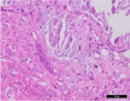
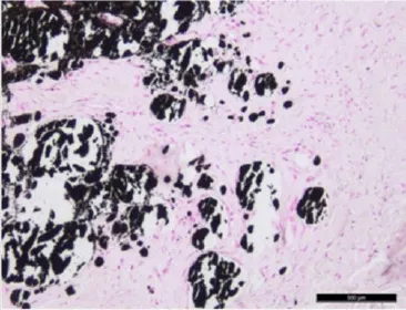
Histopathology demonstrated well defined multiple cystic structures containing granular, darkly basophilic materials with peripheral granulomatous reaction, characterized by presence of multinucleated giant cells surrounded by a varying amounts of fibrous connective tissues (Figure 2). The lesion reacted positive to van Kossa special stain as black coloured calcium deposits were seen (Figure 3). Serum calcium levels did not show any significant pathological finding.

Figure 1. Mass on the bone prominence of the head of femur of the left hind leg.

Figure 2.Multiple cystic structures containing granular, dark basophilic materials (hematoxylin and eosin ×400).

Figure 3.Special staining (von Kossa) showing black coloured calcium deposits (positive reaction) (von Kossa ×400). Furthermore, serum calcium levels were normal.
3. Discussion
Calcinosis circumscripta has been reported in nonhuman primates and humans[4]. Calcinosis circumscripta in humans is more common in females than males and may be associated with various connective tissue disorders such as calcinosis, Raynaud’s phenomenon, esophageal dysmotility, sclerodactyly, telangiectasia (CREST syndrome), and dermatomyositis. In addition, trauma, insect bites, metabolic calcification (e.g., renal failure), and inherited disorders (e.g., pseudoxanthoma elasticum, Werner’s syndrome, and Ehlers-Danlos syndrome) may cause calcinosis circumscripta[5]. In animals, the syndrome has been reported in several species. According to Jeonget al.[6], calcinosis in general is the deposition of calcium salts in tissues other than bone and teeth and may be divided into localised (dystrophic), systemic (metastatic) and calcinosis circumscripta (skin/subcutis). In localised (dystrophic) calcinosis, the pre-requisite is injury to tissues or cells which result into deposition of calcium and phosphorus in a localised area[3]. Idiopathic calcinosis may also occur in absence of localised tissue or systemic tissue injury. In the dermis and subcutis, partial calcium deposition that has a cystic-like structure and contains calcium phosphate or calcium carbonate is called calcinosis circumscripta[5], which is typical of this case. Body serum calcium levels were within normal limits which ruled out the possibility of systemic (metastatic) calcinosis, the condition which occurs in hypercalcemic state[7]. According to Taftiet al.[3], dystrophic calcinosis circumscripta occurs in the setting of normal serum calcium and phosphate levels and the calcification is localized to a specific area of tissue damage.
To conclude, the diagnosed case of dystrophic calcinosis circumscripta in a male cheetah under report might be due to repetitive trauma by a sharp object, probably needle as evidenced by presence of long scar mark and history of repeated injection of vaccines against rabies and cat flu. We also used the same site for subcutaneous administration of ivermectin and sedating the cheetah during surgery.
Conflict of interest statement
We declare that we have no conflict of interest.
Comments
Background
Although these localized tissue calcification are not life-threatening, they may significantly interfere with the functional ability particularly in wild cats which solely depends on hunting for their survival. So these types of cases are needed to be reported to take necessary therapeutic measures before it get complicated.
Research frontiers
Since there are only a few reports are available in wild animals per se, a rare case of calcinosis circumscripta in a cheetah warrants to put in the record.
Related reports
There are very meager reports available on this condition in canines, non-human primates and humans, but no reports are available in wild cats or cheetah as such.
Innovations and breakthroughs
It is a rare and probably a first documented report of calcinosis circumscripta in cheetah correlated to chronic/ repeated trauma.
Applications
This type of case report would potentially help wild life authorities and veterinarians associated with wild animals to identify this type of conditions and take therapeutic measures well in advance before it gets complicated.
Peer review
This is a rarely documented condition in wild cats and appears to be the first recorded case in cheetah. The authors have neatly described the gross and histopathological lesions, supported with adequately good quality images or photomicrographs.
[1] Yanai T, Noda A, Kawakami S, Sakai H, Lackner AA, Masegi T. Lingual calcinosis circumscripta in a captive sitatunga. J Wildl Dis 2001; 37: 813-815.
[2] Lojszczyk-Szczepaniak A, Orzelski M, Smiech A. Canine calcinosis circumscripta- retrospective studies. Medycyna Wet 2008; 64: 1397-1400.
[3] Tafti AK, Hanna P, Bourque AC. Calcinosis circumscripta in the dog: a retrospective pathological study. J Vet Med A Physiol Pathol Clin Med 2005; 52: 13-17.
[4] Radi ZA, Sato K. Bilateral dystrophic calcinosis circumscripta in a cynomolgus macaque (Macaca fascicularis). Toxicol Pathol 2010; 38: 637-641.
[5] Olsen KM, Chew FS. Tumoral calcinosis: pearls, polemics, and alternative possibilities. Radiographics 2006; 26(3): 871-885.
[6] Jeong W, Noh D, Kwon OD, Williams BH, Park SC, Lee M, et al. Calcinosis circumscripta on lingual muscles and dermis in a dog. J Vet Med Sci 2004; 66(4): 433-435.
[7] Thompson RG. General veterinary pathology. 2nd edition. Philadelphia: WB Saunders Company; 1984.
10.12980/APJTB.4.2014C1332
*Corresponding author: Chisoni Mumba, University of Zambia, School of Veterinary Medicine, Box 32379, Lusaka 10101, Zambia.
E-mail: sulemumba@yahoo.com
Article history:
Received 1 Jan 2014
Received in revised form 7 Feb, 2nd revised form 27 Mar, 3rd revised form 10 May 2014
Accepted 23 Jun 2014
Available online 11 Sep 2014
 Asian Pacific Journal of Tropical Biomedicine2014年10期
- Asian Pacific Journal of Tropical Biomedicine的其它文章
- Disseminated toxocariasis in an immunocompetent host
- Human ophthalmomyiasis externa caused by the sheep botfly Oestrus ovis: a case report from Karachi, Pakistan
- Production and purification of a bioactive substance against multi-drug resistant human pathogens from the marine-sponge-derived Salinispora sp.
- In vitro antibacterial activity of leaf extracts of Zehneria scabra and Ricinus communis against Escherichia coli and methicillin resistance Staphylococcus aureus
- In vitro antioxidant and anti-inflammatory activities of Korean blueberry (Vaccinium corymbosum L.) extracts
- Efficacy of seed extracts of Annona squamosa and Annona muricata (Annonaceae) for the control of Aedes albopictus and Culex quinquefasciatus (Culicidae)
