Investigation of the interaction between two phenylethanoid glycosides and bovine serum albumin by spectroscopic methods
Ai-Zhi Wu, Chao-Zhan Lin, Ya-Jing Zhai, Jia-Lin Zhuo, Chen-Chen Zhu
School of Chinese Materia Medica, Guangzhou University of Chinese Medicine, Guangzhou 510006, PR China
1. Introduction
Phenylethanoid glycosides are an interesting group of phenolic natural products that are widely distributed in Callicarpa plants.Structurally, they are characterized by a hydroxyphenylethyl moiety attached to β-glucopyranose through glycosidic linkage.
Such compounds possess broad pharmacological activities, very low toxicity and good safety features [1].
BSA consists of 583 amino acids with 20 tyrosyl residues,which is largely α-helical and contains three structurally homologous domains (I-III). Each domain in turn is the product of two sub-domains(A and B).The principal binding sites with different specificities are referred to as site I and site II [2], and located in the hydrophobic cavities of sub-domains IIA and IIIA, respectively. Many drugs specifically bind to serum albumin,such as phenylbutazone in site I and ibuprofen in site II [3].
Spectral methods are powerful tools for the study of drug binding with proteins since they allow nonintrusive measurements of substances in low concentration [4]. Studies on the binding of drugs to albumins may provide information of structural features, which determine the therapeutic effectivity of drugs, and become an important research field in life sciences. In this work, two glycosides, acteoside and forsythoside B, were first isolated from the traditional Chinese herb Callicarpa peii H.T.Chang, and to the best of our knowledge,it was also the first investigation about the interaction between phenylethanoid glycosides and serum albumin. This study is expected to provide a qualitative understanding of the binding effect of acteoside or forsythoside B with BSA.
2. Experimental
2.1. Materials and apparatus
BSA (Fraction V) was obtained from Sigma Chemical Company. Acteoside and forsythoside B were isolated by the authors from Callicarpa peii H.T. Chang. Their purities were over 98% by normalization of the peak areas detected by HPLC-UV. Steady-state fluorescence measurements were carried out through a F2500 spectrophotometer.UV-vis and CD measurements were performed with a UV1000 UV-vis spectrophotometer and Chirascan spectropolarimeter,respectively.
2.2. Spectroscopic measurement
Fluorescence measurements were carried out keeping the concentration of BSA fixed at 4.0×10⁻⁷M and the drugs varied from 0 to 3.5×10⁻⁵M. The excitation wavelength was 280 nm and the intrinsic fluorescence emission spectra of BSA were recorded at three different temperatures (298, 304 and 310 K). Absorption spectra were recorded at 0.5 nm intervals keeping the concentration of BSA fixed at 1.0×10⁻⁵M and the drugs varied from 0 to 4.0×10⁻⁵M. CD spectra were recorded at 0.5 nm intervals under constant nitrogen flush keeping the concentration of BSA fixed at 2.0×10⁻⁶M and the molar ratio of the drugs to BSA varied as 0:1 and 10:1.
Displacement experiments were performed keeping phenylbutazone and ibuprofen as site probes of sites I and II,respectively.The concentrations of BSA and the probe were fixed at 1.13×10⁻⁶M. Then the fluorescence titration experiments in the presence of phenylbutazone or ibuprofen were performed.
3. Results and discussion
3.1. Fluorescence quenching of BSA by drugs
As shown in Fig.1A, upon the addition of the drug into BSA solution, the fluorescence intensity of BSA at around 340 nm regularly decreased and the fluorescence intensity decreased tardily in each titration curve which indicated that the drug could interact with BSA and the gradual saturation of the BSA binding site. Furthermore, the maximum wavelength of BSA shifted from about 340 to 347 nm after the addition of the drugs. A redshift of the emission peak was observed when the tryptophan residue of BSA was placed in a more hydrophilic environment [5]. The reason may be that the glycosyl groups of two phenylethanoid glycosides resulted in the increasing polarity of microenvironment around the tryptophan residue of BSA after the addition of the drug. A concomitant increase of emission at about 460 nm is the result of the radiationless energy transfer between the tryptophan residue and the drug bound to BSA. The occurrence of an isosbestic point at about 423 nm indicated that the quenching of protein fluorescence depended on the formation of acteoside-BSA and forsythoside B-BSA compounds.
In order to determine the possible quenching mechanism of the drug binding to BSA,the fluorescence quenching constants are usually analyzed by the Stern-Volmer equation[6]and the results are listed in Table 1. The values of Kqdecrease with rising temperature, and are larger than the limiting diffusion constant Kdiff(2.0×10¹⁰M-1s-1) [7], which suggests that the possible quenching mechanism is a static quenching process accompanied with the formation of complexes, while dynamic collision could be negligible.

Fig.1 (A) Effects of acteoside on the fluorescence spectra of BSA in Tris-HCl buffer of pH 7.40. (B) Spectral overlap of BSA fluorescence (a) with the absorption spectra (b) of acteoside. c(BSA)=c(acteoside)=1.0×10⁻⁵ M.

Table 1 Thermodynamic parameters between the drugs and BSA at different temperatures.

Table 2 Binding constants of drugs to BSA before and after the addition of site probe at 298 K.

Fig.2 UV-vis absorbance spectra of BSA in the presence of (A) acteoside and (B) forsythoside B. Curve a: c(BSA)=0.0;c(drug)=1.0×10⁻⁵ M, curves b-j: c(BSA)=1.0×10⁻⁵ M, c(drug)=0, 0.5×10⁻⁵, 1.0×10⁻⁵, 1.5×10⁻⁵, 2.0×10⁻⁵, 2.5×10-5,3.0×10-5, 3.5×10⁻⁵, and 4.0×10⁻⁵ M. The inset shows the relationship of absorbance spectra maximum wavelength about 278 nm and the molar concentration ratio of the drug to BSA.
Because the concentrations of drugs were far more than that of BSA, the double logarithm equation used to calculate the binding constant Kaand the number of binding sites n are reasonable for a static quenching process(Table 1)[8].The Kaof acteoside with BSA is larger than that of forsythoside B with BSA, which may be because forsythoside B has one additional apiose group that is unfavorable for the binding of this drug with BSA. Our previous experimental results [9]reported that the Kaof cistanoside F binding to BSA is 4.36×10⁴M-1at 298 K, less than that of phenylethanoid glycosides,which may be because the latter has one additional hydrophobic phenylethyl moiety that is favorable for the drug binding to BSA. n is approximately equal to 1, indicating that the drugs can be stored and carried by BSA under physiological conditions via formation of the molar concentration ratio 1:1 complexes. The facts ΔG<0, ΔH>0 and ΔS>0 indicate that the drug binding to BSA is a spontaneous intermolecular reaction. The positive ΔH and ΔS values are frequently taken as mainly entropy driven while enthalpy is unfavorable for it. Therefore, hydrophobic forces between aromatic rings of the drugs and hydrophobic pocket of BSA play a major role in the binding process [10].
To determine the specificity of the drug binding, competition experiments were performed with phenylbutazone and ibuprofen. The results (Table 2) showed that phenylbutazone(site I) gave a significantly higher displacement of drugs than ibuprofen (site II) after the addition of the site probes, suggesting that the drug binding site on BSA is the hydrophobic pocket of site I,which is the main binding site for the drugs binding to BSA through hydrophobic forces [11].
3.2. Energy transfer from BSA to drugs

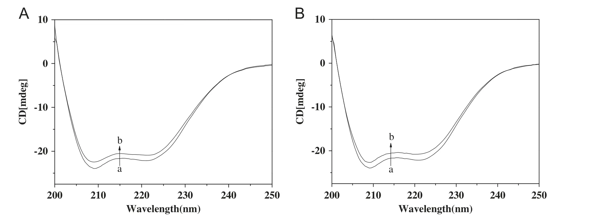
Fig.3 Circular dichroism spectra of (A) acteoside-BSA and (B) forsythoside B-BSA systems at pH=7.40. Concentration ratios of acteoside or forsythoside B to BSA are 0:1 and 10:1 (from curves a and b), respectively.
3.3. Conformation investigations
To further investigate whether any conformational changes of BSA molecules occurred in the binding reaction, UV-vis and CD spectra of BSA were measured. UV-vis spectra (Fig.2)showed that the absorption peaks at 278 nm had a slight redshift toward long wavelengths with increasing addition of the drug, which may be the result of the increase of conjugation around the chromophore of BSA due to the specific interaction between the drugs and BSA [13]. The absorption peak of acteoside shifted from 278 to 280 nm and that of forsythoside B shifted from 278 to 280.5 nm, which showed that the microenvironments around the chromophore of BSA were more affected by forsythoside B than acteoside at the same drug concentrations. CD spectra (Fig.3) exhibited that the band intensity of BSA decreased in the presence of the drugs without significant shift of the peaks, indicating that the structure of BSA after the addition of the drugs is also predominantly α-helix. The α-helix contents of BSA were calculated to be 55.67%in free BSA,51.88%for acteoside and 52.39% for forsythoside B [14] in bound form at molar ratio of drug to BSA 10:1, respectively, which may be the result of binding constant Kaof acteoside with BSA being larger than that of forsythoside B with BSA. A small decrease of α-helix percentage indicated the drugs bound with BSA, which induced slight unfolding of the constitutive polypeptides of BSA and increased the exposure of some hydrophobic regions previously buried.
4. Conclusions
Based on the above analyses, two relationships may exist in the interaction of phenylethanoid glycoside with BSA: (1) the increase of sugar numbers among phenylethanoid glycosides is unfavorable for the drug binding to site I of BSA through hydrophobic interaction due to the increase of drug polarity and volume, and (2) hydrophobic phenylethyl moiety in the drugs is favorable for the drug binding to BSA through hydrophobic interaction. In conclusion, the binding interaction of drug with BSA was enhanced by less sugar groups and more aromatic rings;however,the effect of hydroxyl and drug volume was not negligible.
This work was financially supported by the National Natural Science Foundation of China(Grant nos.81173535 and 81001613).
[1] G.M. Fu, H.H. Pang, Y.H. Wong, Naturally occurring phenylethanoid glycosides: potential leads for new therapeutics, Curr.Med. Chem. 15 (2008) 2592-2613.
[2] G.Sudlow,D.J.Birkett,D.N.Wade,Further characterization of specific drug binding sites on human serum albumin, Mol.Pharmacol. 12 (1976) 1052-1061.
[3] Y.T. Sun, Y.P. Zhang, S.Y. Bi, et al., Studies on the interaction for bovine serum albumin with phenylbutazone and ibuprofen by fluorescence spectrometry, Chem. J. Chin. Univ. 30 (2009)1095-1100.
[4] M. Guo, L. He, X.W. Lu, Binding mechanism of traditional Chinese medicine active component 5-hydroxymethyl-furfural and HSA or BSA, Acta. Pharm. Sin. 47 (2012) 385-392.
[5] S.Y. Bi, L.L. Yan, B.B. Wang, et al., Spectroscopic and voltammetric characterization of the interaction of two local anesthetics with bovine serum albumin, J. Lumin. 131 (2011) 866-873.
[6] J.R. Lakowicz,Fluorescence quenching:theory and applications,Principles of Fluorescence Spectroscopy, Kluwer Academic/Plenum Publishers, New York, 1999.
[7] G.R. Eckenhoff, J.S. Johansson, Molecular interactions between inhaled anesthetics and proteins, Pharmacol. Rev. 49 (1997)343-368.
[8] G.Z. Li, Y.M. Liu, X.Y. Guo, et al., Studies on the interaction between bovine albumins and topotecan hydrochloride or lrinotecan hydrochloride, Acta. Chim. Sin. 64 (2006) 679-685.
[9] A.Z.Wu,C.Z.Lin,X.N.Zhao,et al.,Interaction between bovine serum albumin and cistanoside F isolated from Callicarpa peii H.T. Chang, China J. Chin. Mater. Med. 37 (2012) 1392-1398.
[10] D.P. Ross, S. Subramanian, Thermodynamic of protein association reactions: forces contributing to stability, Biochemistry 20(1981) 3096-3102.
[11] J.N. Tian, J.Q. Liu, Z.D. Hu, et al., Interaction of wogonin with bovine serum albumin, Biol. Med. Chem. 13 (2005) 4124-4129.
[12] L. Matei, M. Hillebrand, Interaction of kaempferol with human serum albumin: a fluorescence and circular dichroism study,J. Pharm. Biomed. Anal. 73 (2010) 768-773.
[13] Z.D. Qi, Y. Zhang, F.L. Liao, et al., Probing the binding of morin to human serum albumin by optical spectroscopy, J.Pharm. Biomed. Anal. 46 (2008) 699-706.
[14] Z.X. Lu, T. Cui, Q.L. Shi, Applications of circular dichroism(CD) and optical rotatory dispersion(ORD),Molecular Biology, Science Press, Beijing, China, 1987.
 Journal of Pharmaceutical Analysis2013年1期
- Journal of Pharmaceutical Analysis的其它文章
- On-line coupling of derivatization with pre-concentration to determine trace levels of methotrexate
- Simultaneous determination of atorvastatin, metformin and glimepiride in human plasma by LC-MS/MS and its application to a human pharmacokinetic study
- JPA Prize in 2012
- Study on changes of polyamine levels in mice with the development of U14 cervical cancer
- Quantitation of bivalirudin, a novel anticoagulant peptide,in human plasma by LC-MS/MS: Method development,validation and application to pharmacokinetics
- A rapid and sensitive liquid chromatography-tandem mass spectrometric assay for duloxetine in human plasma: Its pharmacokinetic application
