12-羟基二十碳四烯酸与细胞增殖及凋亡关系研究进展
孙吉君 宋宗明
●综 述
12-羟基二十碳四烯酸与细胞增殖及凋亡关系研究进展
孙吉君 宋宗明
细胞维持正常增殖需要体内复杂而精密的调控,其中细胞内信号转导是极其重要的一方面,若细胞内的信号转导调控失调导致细胞恶性增殖就会形成肿瘤。众多研究表明,花生四烯酸的代谢产物12-羟基二十碳四烯酸(12-HETE)与正常细胞及癌变细胞的增殖、迁移密切相关。大多数肿瘤细胞都可产生12-HETE,在肿瘤发生时,较高的12-HETE浓度是重要的信号分子,并在多条信号途径中起重要作用。本文现就12-HETE与细胞增殖及凋亡关系的研究进展作一综述。
1 12-HETE的生成
花生四烯酸是细胞膜的结构组成成分,随着磷脂酶A2的激活而释放,经过12-脂氧化酶(12-LOX)介导形成不稳定的12-过氧化氢二十碳四烯酸,继之被谷胱甘肽过氧化物酶催化为稳定的12-HETE,在体内发挥各种生物学作用[1]。人体15-脂氧化酶-1(15-LOX-1)和白细胞型12-LOX因具有高度同源性被称为12/15-脂氧化酶(12/15-LOXs),12-HETE同样可以通过此酶氧化花生四烯酸等游离多烯脂肪酸而生成[2]。此外,花生四烯酸等不饱和脂肪酸通过细胞色素p450也可生成12-HETE。12-HETE对映体12(R)-HETE和12(S)-HETE的出现归于自氧化作用,虽然12-(S)-HETE对映体可以被12-LOX代谢形成,但12(R)-HETE是经哪种酶形成至今尚不清楚[3]。目前,研究发现12(S)-HETE在血小板聚集、肿瘤细胞转移、细胞凋亡中起重要作用,12(S)-HETE水平的升高与免疫功能紊乱有关[4]。
2 12-HETE在细胞中的表达
12-HETE在正常组织如巨噬细胞、成纤维细胞、血管平滑肌细胞中存在基础水平的表达,对细胞生长有促进作用,但过多的12-HETE则会导致细胞增殖恶性循环形成肿瘤。12-HETE在前列腺癌[5]、黑色素瘤[6]、肾癌[7]、膀胱癌[8]、睾丸癌[9]等及其他肿瘤细胞株中的水平都有所增高。在乳腺癌组织较相对未累及的正常组织12-HETE水平增高,同样在体外培养的乳腺癌细胞较正常乳腺上皮细胞仍有此现象,而乳腺癌细胞的增殖效应可以被12-HETE所增加,12-LOX通路抑制剂可以阻断一些乳腺癌细胞的增殖[10]。在胰腺癌[11]、卵巢癌[12]细胞组织中,12-HETE也被证实有类似作用。此外,12-HETE在肿瘤发生的不同阶段其产生水平有所差别。Matsuyama等[13]对前列腺癌的研究发现,12-HETE在正常前列腺和良性前列腺增生组织中呈较低水平,而在前列腺的上皮瘤和前列腺癌中水平显著升高。另外,还有研究表明,12-HETE在正常皮肤及混合痣的黑色素细胞中产生无明显区别,在发育不良痣及黑色素瘤的黑色素细胞浓度显著增加[6]。
3 12-HETE细胞增殖信号转导作用
12-HETE可参与细胞生成的调节,发挥其致有丝分裂作用。文献报道,12-HETE可直接调节人晶状体上皮细胞[14]、内皮细胞[15]和平滑肌细胞[16]DNA的合成,促进细胞增殖。12-HETE对成纤维细胞[17]、乳腺上皮细胞[18]、心肌细胞[19]及胃癌细胞[20]等[11,21-22]肿瘤细胞起致有丝分裂作用。
国内外研究表明,12-HETE可能参与部分细胞因子的细胞增殖作用[23-24]。IL-1、IL-4和IL-8对血管平滑肌细胞有致有丝分裂作用,这些细胞因子作用后的血管平滑肌细胞12-HETE水平显著升高,而12-LOX抑制剂baicalein可以使生成的12-HETE减少,从而减弱这种反应[23]。由此可见,花生四烯酸经12-LOX通路产生的12-HETE,至少可以部分调节IL-1、IL-4和IL-8的致平滑肌细胞有丝分裂作用。此外,内皮素-1作用的血管平滑肌细胞,12-HETE的表达水平增高,baicalein同样可使内皮素-1诱导血管平滑肌细胞增殖效应被抑制,12-HETE也可经12-LOX途径调节内皮素-1诱导的血管平滑肌细胞增殖[24],可见12-LOX的活化和12-HETE的表达可能是血管平滑肌细胞迁移和增殖的关键。
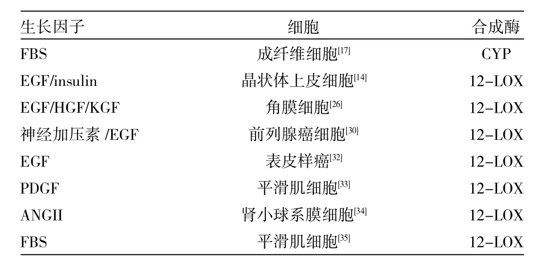
另外,12-HETE也参与部分生长因子诱导的细胞信号传导,如神经加压素、表皮生长因子、血小板源性生长因子、血清、血管紧张素Ⅱ及胰岛素等。如表1所示的生长因子通过增强12-LOX或细胞色素(CYP)的表达及活性诱导12-HETE的合成直到达到比前列腺素类高的生理浓度[25,17]。因此,12-LOX和CYP抑制剂能调节众多生长因子诱导的细胞生长。已有研究表明,12-HETE在对晶状体上皮细胞[14]、角膜上皮细胞[26]、肾小球系膜细胞[27]具有重要的致有丝分裂作用,组织损伤后生长因子的释放可以通过诱导12-HETE的合成增加细胞增殖和损伤修复[28]。由此可见,12-HETE在角膜损伤和角膜移植排斥反应时含量明显增多。这解释了大鼠角膜LOX的抑制剂可以延缓角膜上皮迁移和伤口愈合的原因。此外,12-HETE也能调节肿瘤细胞生长因子[如PC3/DU145前列腺癌细胞[29-30]、U251神经胶质瘤细胞[31]及其它细胞(见表1)]的作用。

表1 不同生长因子在不同细胞诱导12-HETE合成
目前,12-HETE细胞受体尚未见报道,但其亚细胞结合位点已有报道,如12(S)-HETE在肺癌细胞的细胞液,细胞核,线粒体的结合位点[36]。肌动蛋白及线粒体ATP合成酶的α亚基也被确定为HETEs潜在的结合位点。这些位点可能涉及12-HETE细胞增殖、迁移的作用。
此外,花生四烯酸代谢产物刺激细胞生长的可能的传导通路曾被探讨过。首先是细胞外信号调节激酶(MAPK/ERK)传导通路,MAPK/ERK信号通路可以引起非常广泛的细胞反应,如参与细胞的生长、发育、分裂及细胞间的功能同步等多种生理过程,并在细胞恶性转化等病理过程中起重要作用。12-HETE可以通过激活MAPK/ERK通路而刺激肿瘤的生长。有研究表明,外源性12(S)-HETE显著刺激胰腺癌细胞增殖呈时间及浓度依赖性,并且12(S)-HETE能诱导胰腺癌细胞蛋白酪氨酸磷酸化,而当蛋白酪氨酸激酶活性被抑制后,12(S)-HETE诱导的增殖效应消失。ERK通路抑制剂U0126同样也能消除12(S)-HETE刺激胰腺癌细胞增殖的作用且12(S)-HETE刺激ERK磷酸化作用能被蛋白酪氨酸激酶抑制剂阻断。由此可见,ERK及蛋白酪氨酸激酶活化参与12(S)-HETE诱导的胰腺癌细胞增殖[11]。在3T6成纤维细胞培养中,12(S)-HETE通过激活ERK1/2和p38MARK通路,发挥其促细胞增殖作用[17]。12-HETE介导的第二条促进细胞增殖的通路是PI3K-Akt/PKB信号通路,磷脂酰肌醇3-激酶(PI3K)是一双重的特异性激酶,同时具有使脂类及蛋白的丝氨酸/苏氨酸磷酸化的能力,Akt/PKB是与PKA和PKC家族同源丝氨酸/苏氨酸蛋白激酶,该途径也被认为具有抑制细胞凋亡,维持细胞生存的作用。近年来,关于胰腺癌的研究还显示,PI3K-Akt/PKB信号通路介导了12-HETE刺激肿瘤的生长[11]。
据报道12-HETE通过Jak-2和PI3K依赖诱导成纤维细胞生长因子-2(FGF-2)表达刺激内皮生长[37]。另一方面,12-HETE还能通过增殖的上皮细胞表达c-fos/c-myc[14],通过内皮细胞增殖诱导转录因子cAMP反应元件(CRE)结合蛋白的超激活物p38MAPK[38]。研究表明,12-HETE调节细胞信号通路而控制细胞增殖的过程包括调控细胞周期。Pidgeon等[39]最初发现,在G1早期细胞周期蛋白D1和cdk4/6可以协同限制一些刺激因素诱导的细胞增殖速率的增加,而肿瘤细胞中细胞周期蛋白D1存在过表达,且Ras/ERK信号级联反应能调节细胞周期蛋白D1的水平。他们用12-LOX抑制剂baicalein或BHPP处理前列腺癌细胞系DU-145和PC3细胞,发现细胞生长停滞在细胞周期G0/G1期,且这种阻滞作用呈剂量依赖性。而后的研究发现,细胞周期蛋白D1和D3的抑制水平与肿瘤抑制性蛋白-Rb蛋白的磷酸化水平有关[39]。此外,在12-LOX被抑制时也有报导细胞周期停滞在S期[40]。
4 12-HETE抑制细胞凋亡
目前,已有的实验结果表明,12-LOX抑制剂可以显著诱导肿瘤细胞发生凋亡,在实验中还可以观察到典型的凋亡形态[41]。据Wong等[42]在胃癌的研究中发现,用12-LOX反义寡聚核苷酸转染AGS和MKN-28这2个胃癌细胞系后,能诱导胃癌细胞的凋亡。而以12-LOX抑制剂Baicalein处理胰腺癌细胞株可以显著影响抗凋亡蛋白(如Bcl-2、Mcl-1)和促凋亡蛋白(Bax)之间的平衡,使促凋亡蛋白/抗凋亡蛋白比值增高触发线粒体释放细胞色素C,继而激活Caspase级联而导致凋亡[41],这种12-LOX的抑制剂抑制细胞凋亡的现象可以被外源性添加12-HETE反转;在人类乳腺癌细胞,也发现有类似现象[43]。此外,12-LOX抑制剂可以通过阻断细胞周期而抑制细胞生长并诱导细胞发生凋亡。
诱导细胞凋亡往往发生在细胞周期停滞之前,调控的关键步骤通常发生在G1期[41]。由于12-HETE可抑制程序性细胞死亡,12-LOX抑制剂造成12-HETE合成下降导致细胞停滞和凋亡。其机制比较复杂,可能涉及Akt磷酸化水平降低、survivin表达水平下调和随后caspase-3与caspase-7的活化,以及Bcl-2和Bcl-XL表达水平的降低等[44]。在这方面,Wong等[42]发现,抑制12-HETE合成、减少Akt磷酸化水平及增加caspase 3活性是参与细胞凋亡的发展的重要因素。据报道,12-HETE可以下调bcl-2和上调Bax从而影响α(V)β(5)整合素、玻璃黏附蛋白受体及肌动蛋白微丝的表达和定位从而防止细胞凋亡[29]。
对于肿瘤细胞,抑制凋亡是促进肿瘤细胞生长的另一种机制。细胞凋亡严重失调与肿瘤的发生、发展与转归有着密切关系,引发细胞凋亡是由于促凋亡和抗凋亡信号蛋白之间的平衡紊乱导致促凋亡信号占优势。12-HETE的抗凋亡活性可能通过妨碍清除基因改变的癌前病变细胞实现,从而在肿瘤的出现中起重要作用。HETEs可阻止发生染色体重排或其他类型的带有DNA损伤的细胞死亡[45],这种细胞在正常自检情况下(如p53途径)会被清除。但目前尚无明确证据表明HETEs和p53途径之间存在转录拮抗作用。
5 展望
目前的研究表明,12-HETE与细胞的增殖凋亡特别是肿瘤细胞的增殖凋亡密切相关,12-HETE通过刺激EGF和其他生长因子的转录激活参与细胞生长的众多信号转导通路,发挥增强细胞增殖的作用。尽管12-LOX抑制剂已成为目前治疗肿瘤的新方法,但仍有较多问题尚待解决,确定正常细胞和癌变细胞12-LOX/CYP的靶基因将是未来研究的重要领域。当了解哪些基因参与12-HETE的生成,哪些受体、转录因子和信号通路参与12-HETE合成,并能够确定12-HETE的生物效应后,设计能够调节细胞增殖、创面修复及防治癌症发生、发展的新治疗策略将成为现实。
[1]Kuhn H,Thiele B J.The diversity of the lipoxygenase family Many sequence data but little information on biological signi?cance[J].FEBS Lett,1999,449:7-11.
[2]Yamamoto S.Mammalian lipoxygenases:molecular structures and functions[J].Biochim Biophys Acta,1992,1128:117-131.
[3]Bylund J,Kunz T,Valmsen K,et al.Cytochromes P450 with bisallylic hydroxylation activity on arachidonic and linoleic acids studied with human recombinant enzymes and with human[J].J Pharmacol Exp Ther,1998,284:51-60.
[4]Dailey L A,Imming P.12-Lipoxygenase:classification,possible therapeutic benefits from inhibition,and inhibitors[J].Curr Med Chem,1999,6:389-398.
[6]Winer I,Normolle D P,Shureiqi I,et al.Expression of 12-lipoxygenase as a biomarker for melanoma carcinogenesis[J].Melanoma Res,2002,12:429-434.
[7]Yoshimura R,Inoue K,Kawahito Y,et al.Expression of 12-lipoxygenase in human renal cell carcinoma and growth prevention by its inhibitor[J].Int JMol Med,2004,13:41-46.
[8]Yoshimura R,Matsuyama M,Tsuchida K,et al.Expression of lipoxygenase in human bladder carcinoma and growth inhibition by itsinhibitors[J].J Urol,2003,170:1994-1999.
[9]Yoshimura R,Matsuyama M,Mitsuhashi M,et al.Relationship between lipoxygenase and human testicular cancer[J].Int J Mol Med, 2004,13:389-393.
[10]Natarajan R,Nadler J.Role of lipoxygenases in breast cancer[J].Front Biosci,1998,3:E81-88.
[11]Ding X Z,Tong W G,Adrian T E.12-lipoxygenase metabolite 12 (S)-HETE stimulates human pancreatic cancer cell proliferation via protein tyrosine phosphorylation and ERK activation[J].Int J Cancer,2001,94:630-636.
[12]Guo A M,Liu X,Al-Wahab Z,et al.Role of 12-lipoxygenase in regulation of ovarian cancer cell proliferation and survival[J].Cancer Chemother Pharmacol,2011,68:1273-1283.
[13]Matsuyama M,Yoshimura R,Mitsuhashi M,et al.Expression of lipoxygenase in human prostate cancer and growth reduction by itsinhibitors[J].Int JOncol,2004,24:821-827.
[14]Arora J K,Lysz T W,Zelenka P S.A role for 12(S)HETE in the response of human lens epithelial cells to epidermal growth factor and insulin[J].Invest Ophthalmol VisSci,1996,37:1411-1418.
[15]Tang D G,Renaud C,Stojakovic S,et al.12-S-HETE is a mitogenic factor for microvascular endothelial cells:its potential role in angiogenesis[J].Biochem Biophys Res Commun,1995,211: 462-468.
[16]Preston I R,Hill N S,Warburton R R,et al.Role of 12-lipoxygeasein hypoxia induced rat pulmonary arterysmooth muscle cell proliferation[J].AmJ Physiol CellMol Physiol,2006,290:367-374.
[17]Nieves D,Moreno J J.Hydroxyeicosatetraenoic acids released through the cytochrome P450 pathway regulate 3T6 fibroblast growth[J].J Lipid Res,2006,47:2681-2689.
[18]Fujita H,Saito F,Sawada T,et al.Lipoxygenase inhibition decreases neointimal formation following vascular injury[J].Atherosclerosis,1999,147:69-75.
[19]Wen Y,Gu J,Peng X,et al.Overxpression of 12-lipoxygenase and cardiac fibroblast hypertrophy[J].Trends Cardiovasc Med, 2003;13:129-136.
[20]Chen F L,Wang X Z,Li J Y,et al.12-lipoxygenase induces apoptosis of human gastric cancer AGS cells via the ERK 1/2 signalpathway[J].Dig DisSci,2008,53:181-187.
[21]Hussey H J,Tisdale M J.Inhibition of tumour growth by lipoxygenase inhibitors[J].Br J Cancer,1996,74:683-687.
[22]Nie D,Krishnamoorthy S,Jin R,et al.Mechanisms regulating tumor angiogenesis by 12-lipoxygenase in prostate cancer cells [J].J Biol Chem,2006,281:18601-18609.
[23]Natarajan R,Rosdahl J,Gonzales N,et al.Regulation of 12-lipoxygenase by cytokines in vascular smooth muscle cells [J].Hypertension,1997,30:873-879.
[24]Ljuca F,Drevensek G.Endothelin-1 induced vascular smooth muscle cell proliferation is mediated by cytochrome p-450 arachidonic acid metabolites[J].Bosn J Basic Med Sci,2010,10: 223-226.
[25]Bandyopadhyay G K,Imagawa W,Wallace D R,et al.Proliferative effects of insulin and epidermal growth factor on mouse mammary epithelial cells in primary culture.Enhancement by hydroxyeicosatetraenoic acids and synergism with prostaglandin E2[J].JBiol Chem,1988,263:7567-7573.
[26]Ottino P,Taberi F,Bazan H E.Growth factor-induced proliferation in corneal epithelial cells is mediated by 12(S)-HETE[J].Exp Eye Res,2003,76:613-622.
[27]Reddy M A,Adler S G,Kim Y S,et al.Interaction of MAPK and 12-lipoxygenase pathways in growth and matrix protein expression in mesangial cells[J].Am J Physiol Renal Physiol,2002, 283:F985-994.
[28]Wilson S E,Chen L,Mohan R R,et al.Effect of HGF,KGF,EGF and receptor messenger RNAs following corneal epithelial wounding [J].Exp Eye Res,1999,68:377-397.
[29]Pidgeon G P,Tang K,Cai Y L,et al.Overexpression of platelet-type 12-lipoxygenase promotes tumor cell survival by enhancing alpha(v)beta(3)and alpha(v)beta(5)integrin expression[J].Cancer Res,2003,63:4258-4267.
[30]Hassan S,Carraway R E.Involvement of arachidonic acid metabolism and EGF receptor in neurotensin-involved prostate cancerPC3 cell growth[J].RegulPept,2006,133:105-114.
[31]Guo M,Roman R J,Falck J R,et al.Human U251 glioma cell proliferation is suppressed by HET0016[N-hydroxy-N'-(4-butyl-2-methylphenyl)formamidine],a selective inhibitor of cytochrome P4504A[J].J Pharmacol Exp Ther,2005,315:526-533.
[32]Chang W C,Ning C C,Lin M T,et al.Epidermal growth factor enhances a microsomal 12-lipoxygenase activity in A431 cells[J].J Biol Chem,1992,267:3657-3666.
[33]Natarajan R,Bai W,Rangarajan V,et al.Platelet-derived growth factor BB mediated regulation of 12-lipoxygenase in porcine aortic smooth muscle cells[J].J Cell Physiol,1996,169:391-400.
[34]Reddy M A,Adler S G,Kim Y S,et al.Interaction of MAPK and 12-lipoxygenase pathways in growth and matrix protein expression in mesangial cells[J].Am J Physiol Renal Physiol, 2002,283:985-994.
[35]LimorR,Sharon O,Knoll E,et al.Lipoxygenase-derived metabolites are regulators of peroxisome proliferator-activated receptor gamma-2 expression in human vascular smooth muscle cells[J].Am JHypertens,2008,21:219-223.
[36]Kang L T,Phillips T M,Vanderhoek J Y.Novel membrane target proteins for lipoxygenase-derived mono(S)hydroxyl fatty acids [J].Biochim Biophys Acta,1999,1438:388-398.
[37]McCabe N P,Selma S H,Jankun J.Vascular endothelial growth factor production in human prostate cancer cells in stimulated by overexpression of platelet 12-lipoxygenase[J].Prostate,2006,66: 779-787.
[38]Reddy M A,Thimmalapura P R,Lanting L,et al.The oxidize lipid and lipoxygene product 12(S)hydroxyeicosatetraenoic acid induces hypertrophy and?bronectin transcription in vascular smooth muscle cells via p38 MAPK and cAMP response element-binding protein activation Mediation of angiotensin II effects[J].JBiol Chem,2002,277:9922-9928.
[39]Pidgeon G P,Kandouz M,Meram A,et al.Mechanisms controlling cell cycle arrest in induction of apoptosis after 12-lipoxygenase inhibition in prostate cancer cells[J].Cancer Res,2002,62:2721-2727.
[40]黄彩云,王小众.12-脂氧合酶对胃癌细胞AGS增殖与凋亡的影响[D].福建医科大学,2006.
[41]Tong W G,Ding X Z,Witt R C,et al.Lipoxygenase inhibitors attenuate growth of human pancreatic cancer xenografts and induce apoptosis through the mitochondrial pathway[J].Mol Cancer Ther,2002,1:929-935.
[42]Wong B C,Wang W P,Cho C H,et al.12-Lipoxygenase inhibition induced apoptosis in human gastric cancer cells[J].Carcinogenesis,2001,22:1349-1354.
[43]Lovat P E,Oliverio S,Ranalli M,et al.GADD153 and 12-lipoxygenase mediate fenretinide-induced apoptosis of neuroblastoma[J].CancerRes,2002,62:5158-5167.
[44]王志成,郭德玉,孙慧勤.血小板型12-脂加氧酶与肿瘤[J].肿瘤防治杂志,2005,20(5):387-390.
[45]Moreno J J.New aspects of the role of hydroxyeicosatetraenoic acids in cell growth and cancer development[J].Biochem Pharmacol,2009,77:1-10.
2012-03-02)
(本文编辑:欧阳卿)
325027 温州医科大学附属眼视光医院
宋宗明,E-mail:szmeyes@126.com

