
Late-onset Lens Particle Glaucoma as a Consequence of Posterior Capsule Rupture after Pars Plana Vitrectomy
2012-08-24 10:15:02YihuaSuZhenMaoYiLiuYuxiaYangXingLiu
眼科学报 2012年1期
Yihua Su,Zhen Mao, Yi Liu,Yuxia Yang, Xing Liu*
1.The First Affiliated Hospital of Sun Yat-sen University, Guangzhou 510080, China
2.State Key Laboratory of Ophthalmology,Zhongshan Ophthalmic Center, Sun Yat-sen University,Guangzhou 510060,China
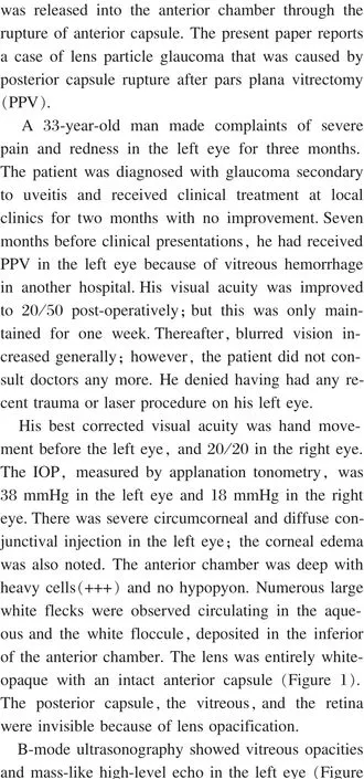
Figure 1 The slit lamp photograph of the left eye showed white floccule in the anterior chamber.
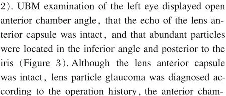
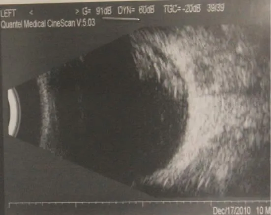
Figure 2 Type-B ultrasound showed vitreous opacities and mass-like high-level echo in the left eye.
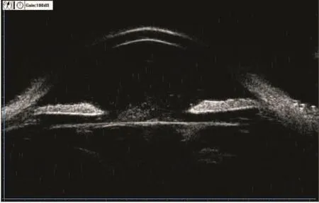
Figure 3 UBM of left eye showed presence of wide-open angles,and echo of the lens was not intact.There was abundant exudation around the back of iris and inferior angle.
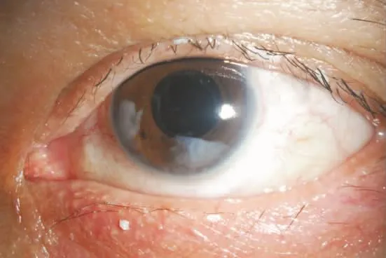
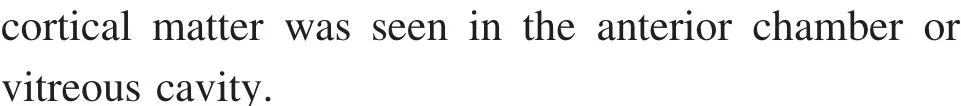
Figure 4 One week after operation,some cortical matter was seen in the anterior chamber.
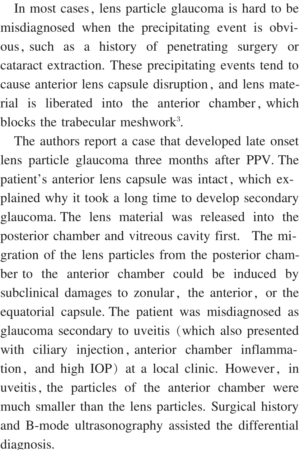
Discussion

- 眼科学报的其它文章
- New Applications of Femtosecond Laser in Cataract Surgery
- Eyelid Basal Cell Carcinoma Arising on the Site of a Congenital Port Wine Hemangioma
- Comparison of Corneal Thickness of Young People with Middle and High Myopia between Shantou and Zhengzhou
- Efficacy of Cytidine-5′-diphosp-bocholine Combined with Compound Anisodine in the Treatment of Early Optic Nerve Contusion
- Efficacy of Removing Dislocated Lens using Intravitreal Phacoemulsification
- Relationship between the Alignment of a Non-Mydriatic Fundus Camera,Anterior Chamber Depth and Axial Length