
Hydrophobic Acrylic Intraocular Lens in Both Eyes
2012-08-24 10:40:02ShengliHaoHongZhang
眼科学报 2012年3期
Shengli Hao, Hong Zhang
1 Department of Ophthalmology, Tianjin Binhai New Area Dagang Hospital, Affiliated Hospital of Tianjin Medical University, School of Clinical Medicine, Tianjin 300270, China
2 Tianjin Medical University Eye Centre, Tianjin 300070, China

Case report
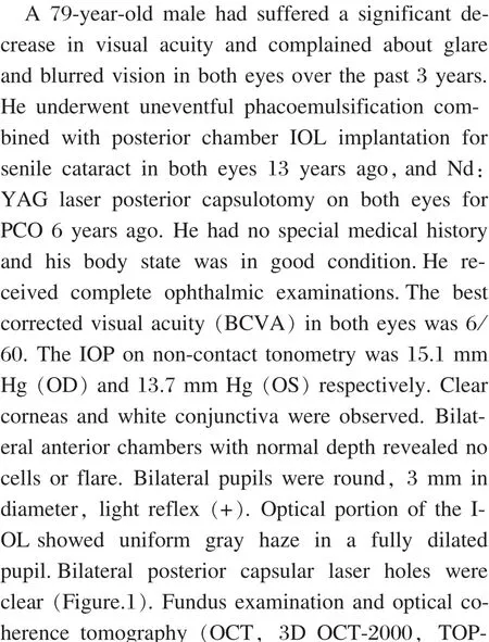
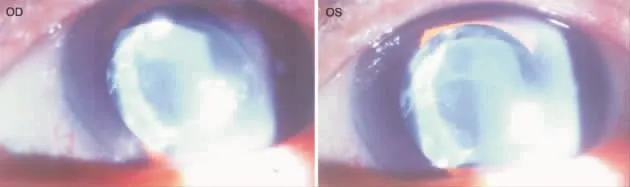
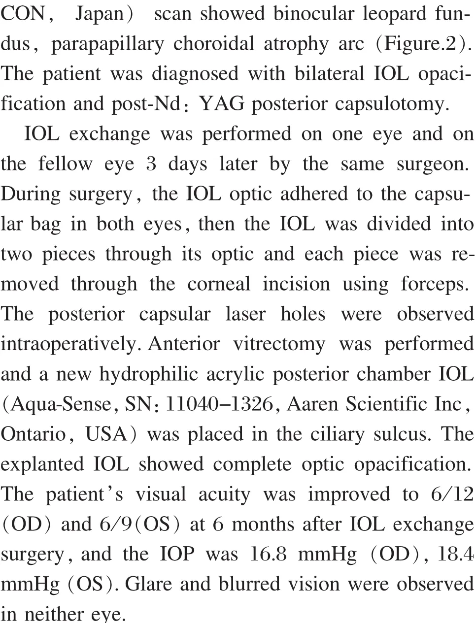
Figure 1 Slit-lamp photograph showing the optic portion of the IOL displayed uniform gray haze in a fully dilated pupil in both eyes.Binocular posterior capsular laser holes were clear.
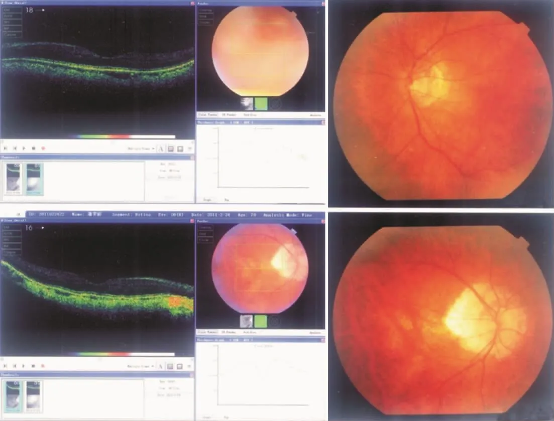
Figure 2 The fundus photograph and optical coherence tomography (OCT,3D OCT-2000,TOPCON,Japan)scan showed binocular leopard fundus, parapapillary choroidal atrophy arc.
Discussion


- 眼科学报的其它文章
- Different Dosages of Intravitreal Triamcinolone Acetonide Injections for Macular Edema Secondary to Central Retinal Vein Occlusion
- Clinical Analysis of Cataract Surgery Complicated by Endophthalmitis
- Efficacy of Progressive Addition Lenses in the Treatment of Ametropia after the Single Eye's IOL Implantation
- Lipofuscin-and Melanin-related Fundus Autofluorescence in Patients with Submacular Idiopathic Choroidal Neovascularization
- Application Value of Topical Aneasthesia in Children Strabismus Surgery
- Histological and Ultrastructural Features for Proliferation Inhibition by Delivery of Exogenous p27Kip1to Rabbit Models after Glaucoma Filtration Surgery