Lipofuscin-and Melanin-related Fundus Autofluorescence in Patients with Submacular Idiopathic Choroidal Neovascularization
Xijia Peng, Wenfang Zhang
1 Department of Ophthalmology, Rehabilitation Center Hospital of Gansu, Lanzhou 730000, China
2 Department of Ophthalmology, the Second Hospital of Lanzhou University, Lanzhou 730030, China
Methods
Patients

FAF,NIA,FFA and ICGA examination

Results
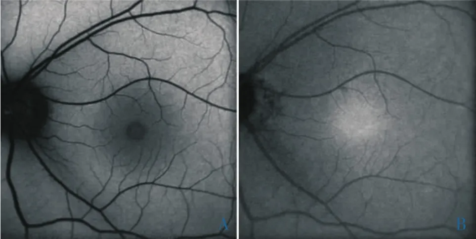
Figure 1 The fellow eye of a 26-year-old female with ICNV shows normal FAF (A)and NIA (B).

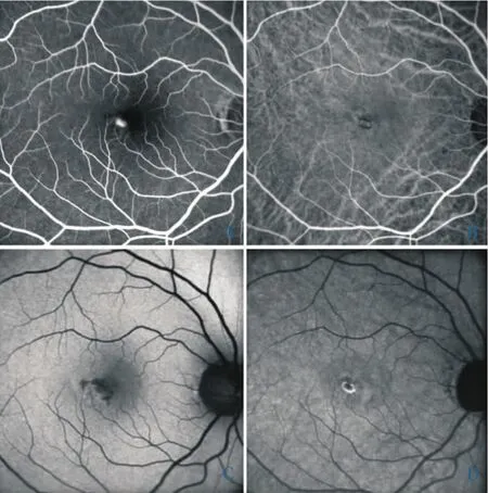
Figure 2 A 39-year-old male with ICNV.A: Early phase FFA reveals a juxtafoveal classic CNV;B:Early phase ICGA confirms a well-defined CNVM with a narrow dark rim; C:FAF shows hypoautofluorescence at the site of CNV.Around the CNV, the distribution of autofluorescence is normal.Note the hypoautofluorescent areas adjacent to CNV caused by subretinal hemorrhage;D:NIA show hypoautofluorescence at the site of CNV.A well-defined hyperautofluorescent ring around the CNV is present.
Discussion

Figure 3 A 26-year-old female with ICNV.A:Early phase FFA reveals a juxtafoveal classic CNV.B:Early phase ICGA confirms a well-defined CNVM with a dark rim;C,D:FAF and NIA show hypoautofluorescence at the site of CNV.A well-defined hyperautofluorescent ring is seen surrounding the CNV in both FAF and NIA images.
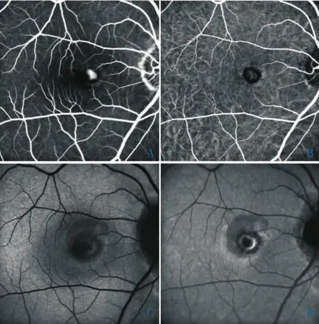
Figure 4 A 34-year-old male with ICNV.A: Early phase FFA reveals a juxtafoveal classic CNV; B: Early phase ICGA confirms a well-defined CNVM.A distinct dark rim around the CNV is present;C,D:FAF and NIA images exhibit hypoautofluorescence at the site of CNV.An ill-defined hyperautofluorescent ring is observed in FAF image(C), while a well-defined hyperautofluorescent ring is observed surrounding the CNV in NIA image (D).
- 眼科学报的其它文章
- Hydrophobic Acrylic Intraocular Lens in Both Eyes
- Different Dosages of Intravitreal Triamcinolone Acetonide Injections for Macular Edema Secondary to Central Retinal Vein Occlusion
- Clinical Analysis of Cataract Surgery Complicated by Endophthalmitis
- Efficacy of Progressive Addition Lenses in the Treatment of Ametropia after the Single Eye's IOL Implantation
- Application Value of Topical Aneasthesia in Children Strabismus Surgery
- Histological and Ultrastructural Features for Proliferation Inhibition by Delivery of Exogenous p27Kip1to Rabbit Models after Glaucoma Filtration Surgery

