Effect of arsenic trioxide on immune function and renal histopathological changes in MRL/lpr mice
WANG Xiao-bing,CHEN Dan,WANG Wu,WANG Liang-xing,ZHU Xiao-chun
(1.Department of Rheumatism,the First Affiliated Hospital;2.Laboratory of Molecular Medicine;3.Department of Respiratory Diseases,the First Affiliated Hospital,Wenzhou Medical College,Wenzhou325035,China)
Effect of arsenic trioxide on immune function and renal histopathological changes in MRL/lpr mice
WANG Xiao-bing1,CHEN Dan1,WANG Wu2,WANG Liang-xing3,ZHU Xiao-chun1
(1.Department of Rheumatism,the First Affiliated Hospital;2.Laboratory of Molecular Medicine;3.Department of Respiratory Diseases,the First Affiliated Hospital,Wenzhou Medical College,Wenzhou325035,China)
OBJECTIVETo investigate the effect of arsenic trioxide(As2O3)on immune function and renal pathology in MRL/lpr mice.METHODSForty-five MRL/lpr mice were divided into control,As2O30.8 mg·kg-1(ip,once a day)and cyclophosphamide 50 mg·kg-1(ip,once a week)groups.After continuously administration for 2 months,the serum level of anti-double stranded-DNA(dsDNA)autoantibody,interferon-γ(IFN-γ)and interleukin-12(IL-12)of mice was measured with ELISA.The subsets of the spleen lymphocytes were detected with flow cytometry.The kidney was removed for periodic acid Schiff dyeing.The expression of IgG and complement C3in the nephridial tissue was observed by immunofluorescence assay.RESULTSTwo months after therapy,compared with that of before treatment,the anti-dsDNA antibody level in normal control group increased from 1.18±0.26 to 1.80±0.26(P<0.01),while it significantly decreased from 1.14±0.58 to 0.92±0.06 in As2O3group and from 1.09±0.22 to 0.67±0.14 in cyclophosphamide group,respectively(P<0.05,P<0.01).Compared with normal control group:①the serum levels of the anti-dsDNA antibody,IFN-γ and IL-12 in As2O3and cyclophosphamide groups were lower than those of normal control group(P<0.05,P<0.01),and the anti-dsDNA antibody level was much lower in cyclophosphamide group than As2O3group(P<0.01);②percentage of CD3+,CD19+and CD3+CD4+cells in As2O3group was lower than normal control group(P<0.01),the percentage of CD3+,CD3+CD8+and CD19+cells in cyclophosphamide group was much lower than normal control group(P<0.01),CD3+CD4+cells in As2O3group were fewer than cyclophosphamide group(P<0.01);③the glomerulus cell count per glomerular cross-sections and the integral of activity in As2O3and cyclophosphamide groups were less than normal control group(P<0.05,P<0.01),while there was no significant difference between As2O3and cyclophosphamide groups;④IgG deposition along the glomerular mesangium and capillary loop in As2O3and cyclophosphamide groups was much less than normal control group(P<0.05),and there was no difference in complement C3expression among the three groups.CONCLUSIONAs2O3can decline the level of anti-dsDNA antibody and the activation and proliferation of T cells,B cells and T subsets in MRL/lpr mice.It can also decrease the serum level of IFN-γ and IL-12 hence suppress kidney lesions.
arsenic trioxide;lupus erythematosus,systemic;antibodies,antinuclear;cytokines;mice,inbred MRL lpr
Systemic lupus erythematosus(SLE)is an autoimmune disease characterized by increase in B cell activity,impairment of T lymphocyte regulation,production of autoantibodies[1-2]and a wide spectrum of organ involvement,such as lupus nephritis.SLE is currently treated with corticosteroids and cytotoxic orimmunosuppressive drugs.These therapies prolong survival but are associated with severe side effects,particularly infection,therefore discovery for a new effective drug is of great significance.
Arsenic trioxide(As2O3)is the main component of a traditional Chinese materia medicia,which is called white arsenic.It was once used to treat rheumatic disease,asthma,tumor and so on,and for the past few years it has been found to be effective in the treatment of both newly diagnosed and relapsed patients with acute promyelocytic leukemia(APL),although the molecular mechanismindetailisyetun-known[3].Wehavealreadyconfirmedthat As2O3could prolong life expectancy of lupus mice and had certain therapeutical effect on them[4-5],while the specific mechanism is still unknown.
MRL/lpr mice which can spontaneously develop hypergammaglobulinemia and high levels of autoantibodies,including anti-double stranded-DNA(dsDNA)antibody,associated with immune-complex-mediated glomerulonephritis and vasculitis[6-7].Because of the resemblance between the murine and human diseases,MRL/lpr mice have been used extensively to attempt to determine SLE etiology and to evaluate therapy.So the effect of As2O3on the level of autoantibody,lymphocyte subsets,cytokines and glomerulonephritis in MRL/lpr mice was investigated in this study.
1 MATERIALS AND METHODS
1.1 Antibodies and reagents
The following mouse cytokine specific monoclonal antibodies(mAb)and isotypematched control mAb were purchased from e Bioscience Company.PE-Cy5.5-anti-mouse CD3,RPE-anti-mouse CD19,FITC-anti-mouse CD4,APC-rat IgG1isotype control,PE-rat IgG1isotype control,anti-mouse Fc-g receptor,APC-anti-mouseinterferon-γ(IFN-γ),PE-antimouse interleukin-12(IL-12)and FITC-antimouse IgG∶sodium arsenite,Harbin Yida Pharmaceutical Co.,Ltd..Mouse IgG,Southern Biotechnology Associates.Inc..Salmon sperm DNA,Sigma.HRP-goatanti-mouseIgG,Beijing Zhong Shan Biological Technology Co.,Ltd..Mouse cytokine ELISA kits,Shenzhen Jingmei Biological Engineering Co.,Ltd..Bovine serum albumin(BSA),Shanghai Yubo Biological Technology Co.,Ltd..
1.2 Animal and treatment
Forty-five3-month-oldMRL/lprmice(weighting 37-44 g)wereboughtfrom ShanghaiS LAC Animal Laboratory(SCXK2007-0005)and bred in our pathogenfree animal facility.Forty-five MRL/lpr mice were divided into control group(ip given normal saline,once a day),As2O30.8 mg·kg-1(ip,once a day)and cyclophosphamide 50 mg·kg-1(ip,once a week)groups.After continuous administration for 2 months,the blood samples were taken at the starting point and the end point of the experiment.
1.3 ELISA for anti-ds DNA autoantibody
Ninety six-well plates were coated with 100 mg·L-1salmon-milt DNA(100 μl).After blocking with 1%BSA for 10 h,the mouse serum(1∶100)was added in triplicate for 90 min in 37℃.After washing,the bound IgG anti-DNA was detected with HRP labeled goat antimouse IgG antibody.The absorbance was determined at 450 nm(A450nm).
1.4 ELISA for cytokines
Serum levels of IFN-γ and IL-12 assayed using the ELISA kits,following the manufacturer's instructions.A450nmwas determined and the IFN-γ andIL-12contents were calculated according to the standard curve.
1.5 Flow cytometry for splenocyte subsets
The spleens were removed and gently homogenized in germ free condition,and the cells washed in PBS twice.The 1×10⁶splenocytes were incubated with PE-Cy5.5-anti-mouse CD3,FITC-anti-mouse CD4,Lyt22PE-anti-mouse CD8,APC-IgG1 isotype control,PE-Ig G1 isotype control,and RPE-anti-mouse CD19(1∶1000)antibodies for 15 min in dark at room temperature.After being washed the resuspended cells were fixed with 300 μl of 1%paraformaldehyde.Fifty thousands cells were analyzed by flow cytometry.
1.6 Periodic acid Schiff(PAS)staining for kidney tissue pathological changes
The kidneys were fixed in 10%Formalin for 3 h at 4℃,then dehydrated,and paraffin embedded.Paraffin sections(4 μm)were stained with PAS reagent.Glomerular pathological change was evaluated by assessing 20 glomerular cross-sections(GCS)per kidney and scored each glomerulus on a semiquantitative scale.0:Normal(35-40 cells per GCS);1:mild〔glomeruli with a few lesions,with slight proliferative changes,and mild hypercellularity(41-50 cells per GCS),and/or minor exudation〕;2:moderate〔glomeruli with moderate hypercellularity(50-60 cells per GCS),including segmental and/or diffuse proliferative changes,hyalinosis,and/or moderate exudates〕;and 3:severe〔glomeruli with segmental,or global sclerosis,and/or exhibiting severe hypercellularity(60 cells per GCS),necrosis,crescent formation,and/or heavy exudation〕.Damaged tubules(percentage;consisting of dilation and/or atrophy and/or necrosis)were determined in 200 randomly selected renal cortical tubules per kidney(×400).Perivascular cell accumulation was determined semiquantitatively by scoring the number of cell layers surrounding the majority of vessel walls on a 0-3 scale[8].
1.7 Immunofluorescence assay for IgG and complement C3expression in kidney tissue
Kidneycryostat cross-sections(4μm thick)were stained with FITC-conjugated goat anti-mouse IgG(1∶200)and FITC-conjugated goat IgG fraction of mouse complement C3 for 30 min at 37℃.After washing the sections were observed under fluorescent microscope.The fluorescence intensity within the peripheral glomerular capillary walls and the mesangium was scored on a scale of 0-3(0:none;1:weak;2:moderate;3:strong).At least 10 glomeruli per section were analyzed.
1.8 Statistical analysis
2 RESULTS
2.1 Effect of As2O3on serum anti-dsDNA autoantibody in MRL/lpr mice
There was no significant difference among these three groups in anti-ds DNA autoantibody levels before treatment.Two months later,the serum level of anti-dsDNA antibody in control group obviously increased(P<0.01)and those in As2O3and cyclophosphamide groups decreased(P<0.05,P<0.01)compared with before treatment.Compared with control group after treatment,As2O3and cyclophosphamide could decline anti-dsDNA autoantibody levels(P<0.01).As2O3group had higher level of anti-dsDNA antibody than cyclophosphamide group(P<0.01)(Tab.1 ).

Tab.1 Effect of arsenic trioxide(As2O3)on serum level of anti-double stranded-DNA(dsDNA)antibody in MRL/lpr mice
2.2 Effect of As2O3on serum level of IFN-γ and IL-12 in MRL/lpr mice
The level of IFN-γ and IL-12 in both As2O3and cyclophosphamide groups was dramatically lower than those in control group(P<0.05,P<0.01).There were no significant differences of IFN-γ and IL-12 levels betweenAs2O3andcyclophosphamidegroups(Tab.2 ).

Tab.2 Effect of As2O3on serum levels of interferon-γ(IFN-γ)and interleukin-12(IL-12)in MRL/lpr mice
2.3 Effect of As2O3on percentage of spleniclymphocytesubsetsinMRL/lpr mice
The percentage of CD3+,CD3+CD4+and CD19+cells in As2O3group was lower than control group(P<0.01).The percentage of CD3+,CD3+CD8+and CD19+cells in cyclo-phosphamide group was much lower than normal control group(P<0.01).CD3+CD4+cells in As2O3group were fewer than cyclophosphamide group(P<0.01)(Tab.3 ).
2.4 Effect of As2O3on renal histopathological changes in MRL/lpr mice
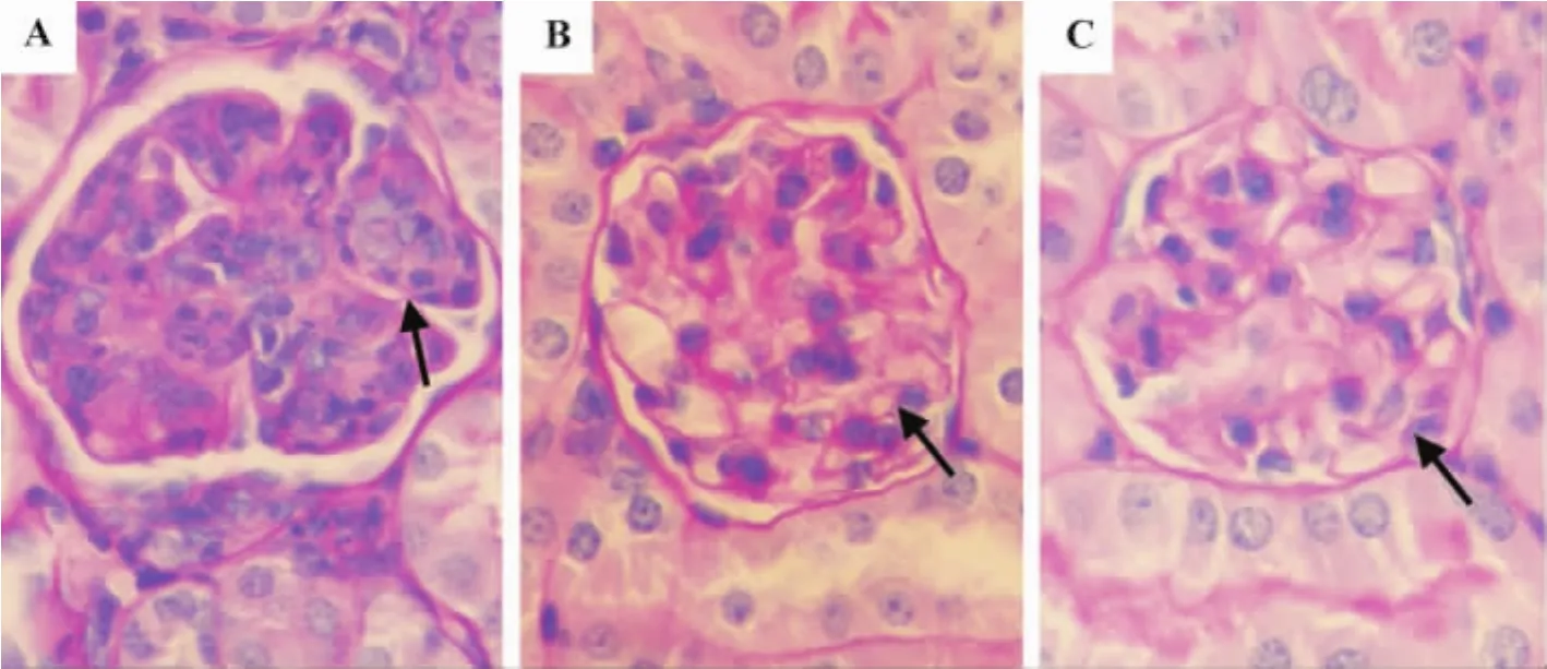
Compared with normal control group,capillary endothelial cell and mesangial cell proliferation were reduced,the membrane thickening was lesser,and perivascular infiltration with lymphocytes was decreased under microscope in glomeruliin As2O3and cyclophosphamide groups(Fig.1 ).The glomerulus cell count(numbers of cells per glomeruli)and the total activity score in As2O3and cyclophosphamide groups were less than control group(P<0.05)(Tab.4 ).However,no significant difference could be seen between As2O3and cyclophosphamide groups.
2.5 Effect of As2O3on IgG and C3 complement expression in kidney tissue in MRL/lpr mice
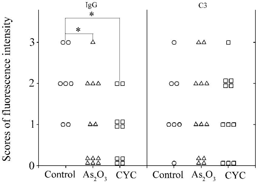
As shown in Fig.2 and Fig.3 ,the staining intensity of IgG in the kidneys of As2O3and cyclophosphamide groups was comparatively less than normal control group(P<0.05),while there was no significant difference among the three groups in complement C3deposition.The immunofluorescence staining pictures of complement C3were omitted.

Tab.3 Effect of As2O3on percentage of splenocyte subsets in MRL/lpr mice

Fig.1 Effect of As2O3on proliferation of mesangial cells and mesangial matrix in glomerulus of MRL/lpr mouse kidneys(PAS×400).See Tab.1 for the mouse treatment.Two months after treatment,the glomerulus image was detected by microscope after periodic acid-Schiff stain(PAS)staining.A:normal control group;B:As2O3group;C:cyclophosphamide group.↑:proliferation of mesangial cells and mesangial matrix.

Tab.4 Effect of As2O3on total activity score and glomerulus cell count in renal tissue in MRL/lpr mice

Fig.3 Effect of As2O3on expression of IgG and complement C3in kidney tissue in MRL/lpr mice.See Tab.1 for the mouse treatment.Two months after treatment,the histologic examination was determined by semiquantitative analysis of fluorescence spectrometry.CYC:cyclophosphamide.*P<0.05,comparedwith normal control group.
3 DISCUSSION
The pathogenesis of SLE is multifactorial and polygenic.T-helper(CD4+T)cells are considered to be significant in the immunopathogenesis of SLE,and lots of studies showed that the development of SLE is usually associated with disorder of cytokine network and imbalance of Th1/Th2[9].It demonstrated that Th1 cytokine such as IFN-γ and IL-12 may be responsible for tissue damage and severe inflammatory response both in human and mice[10-11].In MRL/lpr mice,the concentration of IFN-γ and IL-12 gradually rises over a prolonged period[12].Moreover,IFN-γ administration exacerbates the disease whereas MRL/lpr mice,with defective IFN-γ or IFN-γ receptor expression,develop less severe forms[13-14].
As2O3acts on signaling caspases and apoptosis[15],cellular redox,and cellular responses to stress[16].Although mostly focused on the APL response to As2O3,it's already been examined the therapeutic impact of As2O3on the severe autoimmune disorders manifested in lupus mice thereby predict its potential as a novel therapeutic agent for autoimmune disease[4-5,17].
This study demonstrated that anti-dsDNA autoantibody titers rose as the disease evolved,but it could be strongly inhibited by As2O3and cyclophosphamide.There was obvious hyperplasia of lymphocytes in MRL/lpr mice,but As2O3could sharply diminish the number of CD3+(T),CD19+(B)cells and CD3+CD4+(Th)cells,presenting a little advantage over cyclophosphamide in this aspect.It's supposed that these cells were eliminated by apoptosis.It was found that after treatment the serum concentrations of IFN-γ and IL-12 were markedly higher in control group compared with As2O3group.Meanwhile,control group showed obviously severer inflammation of glomerulus and tubulointerstitial lesion.Therefore,we speculate that As2O3can ameliorate lupus nephritis by suppressing the expression of anti-dsDNA antibody and inflammatory cytokines such as IFN-γ and IL-12.
As a classic cell cycle nonspecific immunosuppressive agent for SLE,cyclophosphamide has won worldwide acknowledgement for its curative effect on SLE.As2O3seemed to have a similar powerful inhibitory action on auto-antibody,lymphocytes,Th1-type cytokines and renal pathology.These suggested that As2O3be a novel therapeutic agent in changing cytokine and autoantibody production,lymphoid hyperplasia,and mononuclear-cell infiltration and immunocomplex deposition in kidneys.Thus it makes application of As2O3on human lupus or other autoimmune diseases highly promising.
It has been found that arsenic can directly inhibit JAK tyrosine kinase activity thus interfering with the Janus kinase-signal transducer and activator of transcription pathway[18].It was also reported that the methylation level of promoter,which can be regulated by arsenic,would affect the expression of IFN-γ[19].These evidences lead us to further verify whether As2O3adjust the activities and differentiation of Th cells via these pathways.Furthermore,exploring the effect of As2O3on B cells and their regulatory factor,which is not involved in this article,is our next effort to exert.
[1]Liu TF,Jones BM.Impaired production of IL-12 in systemic lupus erythematosusⅠ.Excessive production of IL-10 suppresses production of IL-12 by monocytes[J].Cytokine,1998,10(2):140-147.
[2]Mehrian R,Quismorio FP Jr,Strassmann G,Stimmler MM,Horwitz DA,Kitridou RC,et al.Synergistic effect between IL-10 and bcl-2 genotypes in determining susceptibility to systemic lupus erythematosus[J].Arthritis Rheum,1998,41(4):596-602.
[3]Leoni F,Gianfaldoni G,Annunziata M,Fanci R,Ciolli S,Nozzoli C,et al.Arsenic trioxide therapy for relapsed acute promyelocytic leukemia:a bridge to transplantation[J].Haematologica,2002,87(5):485-489.
[4]Zhu XC,Lü YQ,Xü FF,Li AL,Huang CX,Xü YL.The therapeuticall effects of sodium arsenite on lupus nephritis of BXSB spontaneous lupus mice[J].Chin J Integrated Tradit West Nephrol(中国中西医结合肾病杂志),2004,5(1):7-10.
[5]Xia XR,Lin SX,Zhou Y,Zhu XC,Gai SM,Xü FF.Effects of arsenic trioxide on the autoimmunity and survival time in BXSB lupus mice[J].Chin J Integrated Tradit West Med(中国中西医结合杂志),2007,21(2):138-141.
[6]Balomenos D,Rumold R,Theofilopoulos AN.Interferon-gamma is required for lupus-like disease and lymphoaccumulation in MRL-lpr mice[J].J Clin Invest,1998,101(2):364-371.
[7]Zhou GY,Ma CL,Bi LQ.Expression of IFN-γ and IL-4 by CD4+T cell in MRL/lpr mice[J].Chin J Rheumatol(中华风湿病学杂志),2006,10:306-308.
[8]Gamba G,Reyes E,Angeles A,Quintanilla L,Calva J,Peña JC.Observer agreement in the scoring of the activity and chronicity indexes of lupus nephritis[J].Nephron,1991,57(1):75-77.
[9]Hagiwara E.Autoimmune diseases and Th 1/Th 2 balance[J].Ryumachi,2001,41(5):888-893.
[10]Hasegawa K,Hayashi T,Maeda K.Promotion of lupus in NZB x NZWF1 mice by plasmids encoding interferon(IFN)-gamma but not by those encoding interleukin(IL)-4[J].J Comp Pathol,2002,127(1):1-6.
[11]Liu HF,Pan ZX,Tang DS,Jiang LM,Liang D,Chen XW.Excretion levels of interleukin-12 and interferongamma in whole blood cell cultures from systemic lupus erythematosus and the effect of calcineurin antagonists on them[J].Chin J Rheumatol(中华风湿病学杂志),2003,7(5):268-271.
[12]Kikawada E,Lenda DM,Kelley VR.IL-12 deficiency in MRL-Fas(lpr)mice delays nephritis and intrarenal IFN-gammaexpression,anddiminishessystemic pathology[J].J Immunol,2003,170(7):3915-3925.
[13]Sun Y,Chen HM,Subudhi SK,Chen J,Koka R,Chen L,et al.Costimulatory molecule-targeted antibody therapy of a spontaneous autoimmune disease[J].Nat Med,2002,8(12):1405-1413.
[14]Matthys P,Vermeire K,Billiau A.Mac-1(+)myelopoiesis induced by CFA:a clue to the paradoxical effects of IFN-gamma in autoimmune disease models[J].Trends Immunol,2001,22(7):367-371.
[15]Chelbi-alix MK,Bobé P,Benoit G,Canova A,Pine R.Arsenic enhances the activation of Stat1 by interferon gamma leading to synergistic expression of IRF-1[J].Oncogene,2003,22(57):9121-9130.
[16]Miller WH Jr,Schipper HM,Lee JS,Singer J,Waxman S.Mechanisms of action of arsenic trioxide[J].Cancer Res,2002,62(14):3893-3903.
[17]Bobé P,Bonardelle D,Benihoud K,Opolon P,Chelbi-Alix MK.Arsenic trioxide:a promising novel therapeutic agent for lymphoproliferative and autoimmune syndromes in MRL/lpr mice[J].Blood,2006,108(13):3967-3975.
[18]Cheng HY,Li P,David M,Smithgall TE,Feng L,Lieberman MW.Arsenic inhibition of the JAK-STAT pathway[J].Oncogene,2004,23(20):3603-3612.
[19]Yano S,Ghosh P,Kusaba H,Buchholz M,Longo DL.Effect of promoter methylation on the regulation of IFN-gamma gene during in vitro differentiation of human peripheral blood T cells into a Th2 population[J].J Immunol,2003,171(5):2510-2516.
三氧化二砷对MRL/lpr小鼠免疫功能和肾脏组织病理变化的影响
王晓冰1,陈丹1,王伍2,王良兴3,朱小春1
(温州医学院1.附属第一医院风湿免疫科,2.分子医学实验室,3.附属第一医院呼吸科,浙江温州325035)
目的研究三氧化二砷(As2O3)对MRL/lpr小鼠免疫功能和肾脏组织病理变化的影响。方法45只MRL/lpr狼疮小鼠ip给予环磷酰胺50 mg·kg-1(每周1次)和As2O30.8 mg·kg-1,每天1次,共2个月。用ELISA法检测血清抗双链DNA(dsDNA)抗体、干扰素γ(IFN-γ)和白细胞介素12(IL-12)浓度;用流式细胞术测定脾CD3+,CD19+,CD3+CD4+和CD3+CD8+细胞亚群的百分比;用PAS染色法观察肾组织病理变化;用免疫荧光方法检测肾组织IgG和补体C3的表达。结果与给药前比较,给药2个月后,正常对照组血清抗dsDNA抗体水平升高,由给药前1.18±0.26升高至1.80±0.26(P<0.01),As2O3和环磷酰胺组该抗体水平明显降低,分别由给药前1.14±0.58和1.09±0.22降低至0.92±0.06和0.67±0.14(P<0.05,P<0.01)。与正常对照组比较:①As2O3和环磷酰胺组血清抗ds-DNA抗体、IFN-γ和IL-12浓度明显降低(P<0.05),环磷酰胺组抗ds-DNA抗体比As2O3组显著降低(P<0.01);②As2O3组CD3+,CD3+CD4+和CD19+细胞百分率明显降低(P<0.01),环磷酰胺组CD3+,CD3+CD8+和CD19+细胞百分率明显降低(P<0.01);As2O3组CD3+CD4+细胞百分率明显降低(P<0.01);③As2O3和环磷酰胺组小鼠肾小球细胞计数和活动度积分明显降低(P<0.05,P<0.01),As2O3和环磷酰胺组无显著差异;④As2O3和环磷酰胺组肾IgG表达明显降低(P<0.05),补体C3表达无明显差异,As2O3和环磷酰胺组之间无显著性差异。结论As2O3能降低MRL/lpr狼疮小鼠血清抗ds-DNA抗体水平,抑制T,B和Th细胞活化和增殖,降低血清IFN-γ和IL-12水平,从而缓解狼疮肾炎的病理变化。
三氧化二砷;红斑狼疮,系统性;抗体,抗核;细胞因子类;小鼠,近交MRL Lpr
国家自然科学基金(31100576);温州市科技计划重点项目(Y20090240);温州市科技计划(Y20100287)
朱小春,E-mail:gale820907@yahoo.com.cn
2012-04-27接受日期:2012-10-01)
R994.6,R967
A
1000-3002(2012)06-0794-07
10.3867/j.issn.1000-3002.2012.06.003
(本文编辑:齐春会)

