
The application of internal suspension technique in retroperitoneal robot-assisted laparoscopic partial nephrectomy with a new robotic system KangDuo Surgical Robot-01: Initial experience
2024-01-11 11:01:58SiluChenShuboFanHuaGuanKunlinYangZhihuaLiShengweiXiongXiangWangZhenyuLiChengShenLiqunZhouXuesongLi
Asian Journal of Urology 2023年4期
Silu Chen , Shubo Fan , Hua Guan , Kunlin Yang, Zhihua Li,Shengwei Xiong, Xiang Wang, Zhenyu Li, Cheng Shen,Liqun Zhou*, Xuesong Li*
Department of Urology, Peking University First Hospital, Institute of Urology, Peking University,National Urological Cancer Center, Xicheng District, Beijing, China
KEYWORDS KangDuo Surgical Robot-01;Internal suspension;Partial nephrectomy;Retroperitoneal approach
Abstract Objective: To assess the feasibility of internal suspension technique in retroperitoneal robot-assisted laparoscopic partial nephrectomy (rRAPN) with a new robotic platform called KangDuo Surgical Robot-01(KD-SR-01)system(Suzhou KangDuo Robot Co.,Ltd.,Suzhou,China) and discuss its surgical technique.Methods: A 44-year-old male patient was admitted with a 2.5 cm tumor on dorsolateral upper pole of the left kidney.The R.E.N.A.L.nephrometry score of this patient was 4x.This patient underwent rRAPN with KD-SR-01.The perinephric fat between the tumor and Gerota’s fascia was preserved,which was used for internal suspension traction during tumor resection.Postoperative follow-up data were collected.Results: The surgery was successfully carried out with a duration of 127 min,in which the docking time was 6 min 25 s and console time was 60 min.The warm ischemia time was 19 min 53 s,and the estimated blood loss was 0 mL.The pathological histology showed a pathological tumor stage 1a clear cell renal cell carcinoma, with a negative surgical margin.The World Health Organization/International Society of Urological Pathology (WHO/ISUP) grade of this patient was Grade 2.No recurrence was observed during the 6-month follow-up.Conclusion: Internal suspension in rRAPN is feasible and effective with use of the new robotic system KD-SR-01.
1.Introduction
Renal cancer is a common genitourinary malignancy,constituting approximately 4% of adult malignancies and is on the rise [1].Partial nephrectomy (PN) has comparable oncologic outcomes and better preservation of renal function compared with radical nephrectomy (RN) in clinical tumor stage 1(cT1)renal tumors[2-4]and some cT2 renal tumors [5-7].Currently, PN has become the gold standard treatment for cT1 and even some cT2 renal tumors when technically feasible[5,8].With similar oncologic outcomes,robotic surgical systems provide a minimally invasive and sophisticated platform and could decrease the technical difficulty of intracorporeal operation for PN [9].However,the high cost of da Vinci robotic system has limited its popularization in developing areas.
In recent years,a novel robotic platform called KangDuo Surgical Robot-01 (KD-SR-01) system (Suzhou KangDuo Robot Co., Ltd., Suzhou, China) was developed in China.The prospective randomized controlled trial of robot-assisted PN (RAPN) with the KD-SR-01 versus the da Vinci Si surgical system demonstrated that KD-SR-01 system achieved noninferior outcomes regarding safety and efficacy for cT1a tumors [10].KD-SR-01 also showed promising outcomes for other urological surgeries,such as pyeloplasty[11,12] and radical prostatectomy [13].
RAPN can be performed through retroperitoneal or transperitoneal approaches.With comparable oncological outcomes versus transperitoneal approaches [14],retroperitoneal approaches have advantages of being more direct and easier to access the renal tumors and the renal vessels for posterior tumors and some lateral tumors.In addition, retroperitoneal approaches could avoid excessive intervention of abdominal organs [15,16].Nonetheless, in retroperitoneal RAPN (rRAPN), poor tumor exposure is associated with a longer warm ischemia time (WIT) and more blood loss.To further optimize the WIT and stabilize the tumor during resection in rRAPN, we present our modified technique of“internal suspension”with the use of KD-SR-01.The surgical technique and our initial experience are described in this article.
2.Patient and methods
2.1.Patient
A 44-year-old male patient with a body mass index of 28.41 kg/m2was admitted to our hospital on December 25,2020.A 2.5 cm tumor was found on the upper pole of the left kidney by three-dimensional (3D) contrast-enhanced computed tomography (CT) scan 1 week before surgery(Fig.1).The R.E.N.A.L.nephrometry score of this patient was 4x.No back pain, hematuria, or abdominal mass was observed in this patient.This patient was given an American Society of Anesthesiologists (ASA) score of II.The preoperative serum creatinine and estimated glomerular filtration rate (eGFR) was 1.03 mg/dL and 88.344 mL/min/1.73 m2,respectively.The console surgeon(Li X)has experiences with over 200 urologic robotic surgeries.The study protocol was approved by the Institutional Review Board of Peking University First Hospital (device registration number 25 [2018])and is registered at www.chictr.org.cn(ChiCTR2100045983).Written informed consent was obtained from the patient after receiving an explanation about the aim of the study.
2.2.Robotic platform
As shown in our previous researches [11-13,17,18],KD-SR-01 system is a“master-slave”platform consisting of an open surgeon control console, a suspended 3-arm patient cart, a high-definition 3D vision cart, and reusable endoscopic instruments, with compatibility of marketed laparoscopy.
2.3.Surgical procedures
The patient was securely padded and tucked on the operation bed in a 60-degree right lateral decubitus position.The trocars were placed consistent with our previous description[18](Fig.2).The retroperitoneal fat was removed, and the retroperitoneal space was expanded.The surgical robot was then docked over the patient’s head parallel to the spine.Gerota’s fascia was incised above the psoas muscle.Two renal arteries of the left kidney were exposed, and a 2.0 cm renal tumor was observed on the dorsolateral upper pole of the kidney,protruding from the renal surface.As described in our previous study [19], we took care to separate perinephric fat along the renal surface without resecting the perinephric fat atop the tumor (Fig.3A).The preserved perinephric fat was used for internal suspension traction on the tumor during later tumor resection.The renal arteries were clamped with bulldog clamps (Fig.3B), and a timer was started simultaneously to record WIT.The tumor was excised 0.5 cm beyond the margin with robot monopolar scissors (Fig.3C and D).After finishing tumor resection,the perinephric fat atop the tumor was removed(Fig.3E).Then, the resection area was continuously sutured in two layers by 2-0 absorbable barbed suture (Fig.3F).Hem-o-loks were used to clamp the thread tail to secure the stitches.Thereafter, the bulldog clamps were removed,and the timer was stopped.For this patient,the renal cyst decortication on the ventral lower pole of the left kidney was performed later.The internal suspension technique in retroperitoneal robot-assisted laparoscopic PN with a new robotic system KangDuo Surgical Robot-01(KD-SR-01) was showed in Supplementary Video 1.

Figure 1 The preoperative three-dimensional contrast-enhanced computed tomography presenting a 2.5 cm tumor on upper pole of left kidney.
Supplementary video related to this article can be found at https://doi.org/10.1016/j.ajur.2023.08.003.

Figure 2 The port placement of retroperitoneal robot-assisted laparoscopic partial nephrectomy with KangDuo Surgical Robot-01(Suzhou KangDuo Robot Co.,Ltd.,Suzhou,China).
2.4.Postoperative follow-up
Parameters of follow-up included serum creatinine levels,eGFR on postoperative Day 1, Day 3, and postoperative weeks 4-12, and urinary B-mode ultrasonography on postoperative weeks 4-12.A 3D contrast-enhanced CT scan was performed 6 months after surgery.
3.Results
The total time of this operation was 127 min, in which docking time was 6 min 25 s, and console time was 60 min.The WIT was 19 min 53 s.Estimated blood loss was 0 mL.This operation was successfully carried out, without conversion to traditional rRAPN, laparoscopic PN, or open surgery.The patient had clear liquid diet on postoperative Day 1, and resumed normal diet on postoperative Day 3.The drainage tube was removed 2 days after surgery, and this patient was discharged 4 days after surgery.No perioperative complications were recorded.
Postoperative pathology showed that the tumor size was 2.2 cm×1.8 cm×1.7 cm, with a negative surgical margin.The pathological subtype was clear cell renal cell carcinoma.The pathological tumor stage was 1a, with World Health Organization/International Society of Urological Pathology(WHO/ISUP)Grade 2.During the 6-month follow-up, no recurrence of renal cell carcinoma was observed through 3D contrast-enhanced CT (Fig.4), and the eGFR was 95.556 mL/min/1.73 m2.
4.Discussion
PN has become the first choice for the treatment of cT1 and some cT2 renal tumors when technically feasible [5,6,8].With comparable oncologic outcomes versus RN [2], the better renal function preservation of PN decreased the risk of developing metabolic or cardiovascular complications after surgery, and reduced the mortality from any cause[3,4,20].To extend the indications, PN was used in some challenging scenarios, such as cT2 renal tumors [5-7] or totally endophytic renal tumors [21].For maximal preservation of renal function, several attempts were made on off-clamp PN.The higher probability of maintaining renal function with adequate oncological outcomes has made it an attractive option for PN [22,23].In recent decades, the rapid development of surgical robotic systems has changed the way urologists performing minimally invasive PN.Compared to open procedures or laparoscopic procedures,the surgical robotic system has shown similar oncological outcomes and promising perioperative results [24,25].However, the high cost of da Vinci robotic system hindered its wider application, especially in developing areas.
Recently, a novel robotic platform called KD-SR-01 system was developed.This robotic platform consists of an open surgical console, three robotic arms suspended on the beam, and surgical instruments, with compatibility of marketed laparoscopy [17].Our preliminary experience showed that KD-SR-01 system had comparable intraoperative parameters and better ergonomics learning advantages for PN than 3D laparoscopic procedures in porcine models[17].The prospective randomized controlled trial of RAPN demonstrated that KD-SR-01 system achieved noninferior outcomes regarding safety and efficacy for cT1a tumors versus da Vinci Si surgical system [10].Subsequent clinical trials demonstrated the safety and effectiveness of KD-SR-01 system in various urological surgeries, including pyeloplasty[11,12] and radical prostatectomy [13], and this novel surgical system has been approved by National Medical Products Administration for upper urinary surgery in China (No.20223010762).
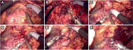
Figure 3 Surgical procedures of internal suspension technique in robot-assisted laparoscopic partial nephrectomy with KangDuo Surgical Robot-01(Suzhou KangDuo Robot Co.,Ltd.,Suzhou,China).(A)The perinephric fat atop the tumor was preserved;(B)The renal arteries were clamped with bulldog clamps; (C and D) The tumor was excised 0.5 cm beyond the tumor margin with robot monopolar scissors; (E) The perinephric fat atop the tumor was removed; (F) The resection area was continuously sutured in two layers.
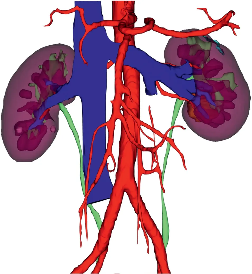
Figure 4 No recurrence of renal cell carcinoma was observed through three-dimensional contrast-enhanced computed tomography 6 months after surgery.
The transperitoneal approach was more widely used in RAPN due to its larger working space.However, bowel mobilization through the transperitoneal approach may increase the risk of iatrogenic abdominal organ injury,and it is not suitable for the patients with previous intraabdominal surgery.With similar oncological outcomes to transperitoneal approaches [14], the retroperitoneal approach may be easier to access the tumor and the renal vessels, without excessive intervention of abdominal organs [15,16].In addition, the retroperitoneal approach seemed to have advantages of reduced operative time and hospital stay for dorsal tumors or some lateral tumors[16,26].
For the retroperitoneal approach,the restriction of the working space may hinder the tumor resection and prolong the WIT.In recent years, much attention has been given to a better tumor exposure and the simplification of tumor resection in rRAPN [27,28].An additional mechanical arm could provide optimal traction on target tissue during rRAPN, which could reduce the complications and positive surgical margins.However, the introduction of an additional mechanical arm may increase surgical trauma,reduce the operation space, and demand extra medical cost [27].Jiang et al.[28] used a suspension traction suture to pull the perirenal fat to the psoas major for better tumor exposure and precise tumor excision.Nonetheless,the process of suturing perirenal fat may still be at risk of breaking tumor capsule.In our previous research[19],the preserved perinephric fat between tumor and Gerota’s fascia was used for internal suspension in laparoscopic PN.
In our previous research [18], KD-SR-01 showed the safety and effectiveness of rRAPN for the management of posterior and lateral renal tumors with R.E.N.A.L.nephrometry scores of ≤9.For this patient, the perinephric fat between the tumor and Gerota’s fascia was preserved as the internal suspension system, which could maintain tension during tumor resection.The preserved perinephric fat could also stabilize the tumor and improve the precision of the tumor excision.Thus,the risk of cutting into the tumor during resection was reduced.Unlike other reported techniques,our internal suspension system is technically simple and direct, and it does not require additional graspers,extra surgical trauma, or other procedures.With the application of this novel technique, renal tumors could be fully exposed and demand no extra medical cost.
This operation was successfully carried out within 127 min, without any conversion to other procedures.This patient had a satisfying postoperative recovery, with normal diet on postoperative Day 3, and discharged from the hospital on postoperative Day 4.In addition, no major postoperative surgical complications were observed.WIT was considered as a significant determinant of postoperative serum creatine.A shorter WIT may result in a better renal function recovery, which is recommended to be less than 25 min [29].The WIT of this patient was only 19 min 53 s, and the renal function of this patient was stable after the surgery.This patient had a negative surgical margin on histology, with no evidence of renal cell carcinoma recurrence during the 6-month follow-up.
Although the tumor of this patient was located on the dorsal side, our internal suspension technique is also suitable for ventral renal tumors, especially for those with a previous abdominal surgical history [19].With the help of our internal suspension system,we can fully expose ventral renal tumors without mobilization of the bowel, reducing the risk of iatrogenic abdominal organ injury.However,this technique has limitations in some situations.The application of this technique was limited to exophytic tumors,with the exception of hilar and anterolateral tumors.For obese patients or patients with perirenal fat adherent to the kidneys, the exposure of the tumor without resecting the perirenal fat might be challenging and time-consuming.For ventral hilar tumors, the application of this technique may make it difficult for the kidney to mobilize and rotate.With unsatisfactory exposure of the tumor and the narrow work space, subsequent excision would be difficult to perform.
The internal suspension technique is feasible and effective in rRAPN with the use of KD-SR-01.To confirm the value of the internal suspension technique, a prospective comparative study with conventional procedures with larger sample size and longer follow-up period is required.
5.Conclusion
Our initial experience demonstrates that the internal suspension technique is feasible and effective in rRAPN with the use of KD-SR-01.
Author contribution
Study concept and design: Liqun Zhou, Xuesong Li.
Data analysis: Silu Chen, Shubo Fan, Hua Guan, Kunlin Yang.
Patient follow-up: Silu Chen, Kunlin Yang, Zhihua Li,Shengwei Xiong, Xiang Wang, Zhenyu Li.
Drafting of manuscript: Silu Chen, Shubo Fan, Hua Guan.Critical revision of the manuscript:Liqun Zhou,Xuesong Li,Cheng Shen.
Conflicts of interest
The research received research grants from Suzhou Kang-Duo Robot Co., Ltd., Suzhou, China.The company did not influence the design, conduct, or publication of this research.All authors have nothing else to declare.
 Asian Journal of Urology2023年4期
- Asian Journal of Urology的其它文章
- Arterioureteral fistula: An unusual cause of haematuria 10 years after the implantation of a synthetic iliac-femoral stent
- Utility of three-dimensional virtual reconstruction for robotic-assisted partial nephrectomy using the IRIS™
- Robotic surgery in urology: Recent advances
- Right versus left fully robotic live donor nephrectomy and open kidney transplantation: Does the laterality of the donor kidney really matter?
- Contemporary outcomes of patients undergoing robotic-assisted radical cystectomy:A comparative analysis between intracorporeal ileal conduit and neobladder urinary diversions
- Unilateral post-chemotherapy robot-assisted retroperitoneal lymph node dissection in Stage II non-seminomatous germ cell tumor:A tertiary care experience