
Three-dimensional computed tomography reconstruction diagnosed digestive tract perforation and acute peritonitis caused by Monopterus albus: A case report
2023-12-05 09:27:40JinHanYangJinYingLanAnYuanLinWeiBiaoHuangJinYuanLiao
Jin-Han Yang,Jin-Ying Lan,An-Yuan Lin,Wei-Biao Huang,Jin-Yuan Liao
Abstract BACKGROUND Few reports have described living foreign bodies in the human body.The current manuscript demonstrates that computed tomography (CT) is an effective tool for accurate preoperative evaluation of living foreign bodies in clinic.The threedimensional (3D) reconstruction technology could clearly display anatomical structures,lesions and adjacent organs,improving diagnostic accuracy and guiding the surgical decision-making process.CASE SUMMARY Herein we describe a 68-year-old man diagnosed with digestive tract perforation and acute peritonitis caused by a foreign body of Monopterus albus.The patient pre-sented to the emergency department with complaints of dull abdominal pain,profuse sweating and a pale complexion during work.A Monopterus albus had entered the patient’s body through the anus two hours ago.During hospitalization,the 3D reconstruction technology revealed a perforation of the middle rectum complicated with acute peritonitis and showed a clear and complete Monopterus albus bone morphology in the abdominal and pelvic cavities,with the Monopterus albus biting the mesentery.Laparoscopic examination detected a large(diameter of about 1.5 cm) perforation in the mid-rectum.It could be seen that a Monopterus albus had completely entered the abdominal cavity and had tightly bitten the mesentery of the small intestine.During the operation,the dead Monopterus albus was taken out.CONCLUSION The current manuscript demonstrates that CT is an effective tool for accurate preoperative evaluation of living foreign bodies in clinic.
Key Words: Digestive tract perforation;Acute peritonitis;Monopterus albus;Three-dimensional computed tomography reconstruction;Case report
INTRODUCTION
Digestive tract perforation is a common acute abdominal pathology[1,2],often secondary to ulcers,trauma,inflammation,tumors,etc.Computed tomography (CT) constitutes an effective tool for accurate preoperative evaluation of foreign bodies in clinic[3].Preoperative three-dimensional (3D) CT reconstruction accurately locates perforation sites and foreign bodies,helps diagnose peritonitis and guides surgical treatment[4].In the present case,according to clinical symptoms and signs,combined with plain 3D CT reconstruction,it was determined that the patient had digestive tract perforation,and aMonopterus albushad died after entering the abdominal cavity[5].As a result,the patient’s abdominal cavity was seriously polluted,with a large amount of turbid yellow fluid and a small amount of feces attached to several intestinal areas,so it could be determined that the patient had “intestinal perforation” caused by aMonopterus albus[6].The intestinal wall is relatively weak,and may burst out afterMonopterus albusbites,which easily causes acute diffuse peritonitis[7].If not timely treated,patients may develop septic shock,which is a serious and life-threatening condition.Surgical removal of foreign bodies,e.g.,Monopterus albus,is the best treatment method,and preoperative imaging evaluation is particularly important[8].Living foreign bodies are rarely reported in the literature.
CASE PRESENTATION
Chief complaints
One patient,a 68-year-old man from China,presented to the hospital’s emergency department after suffering from dull abdominal pain,profuse sweating and a pale complexion during the two-hour workday.
History of present illness
Symptoms started 2 h before presentation with complaints of dull abdominal pain,profuse sweating and a pale complexion during work.
History of past illness
The patient didn’t have any remarkable history.
Personal and family history
The patient denied having a family history of any malignant tumors.
Physical examination
Using a physical examination,the results showed the following vital signs: Blood pressure,118/69 mmHg;body temperature,36.4 °C;heart rate,81 beats/min;respiratory frequency,18 breaths/min.Furthermore,total abdominal tenderness,plate-like abdomen,and liver dullness disappeared,with weak abdominal breathing and bowel sounds.
Laboratory examinations
Laboratory tests showed normal liver function,alpha-fetoprotein,carbohydrate antigen 19-9,and carcinoembryonic antigen.No abnormality was found in routine blood and urine analyses.Primary laboratory data upon admission are summarized in Table 1.
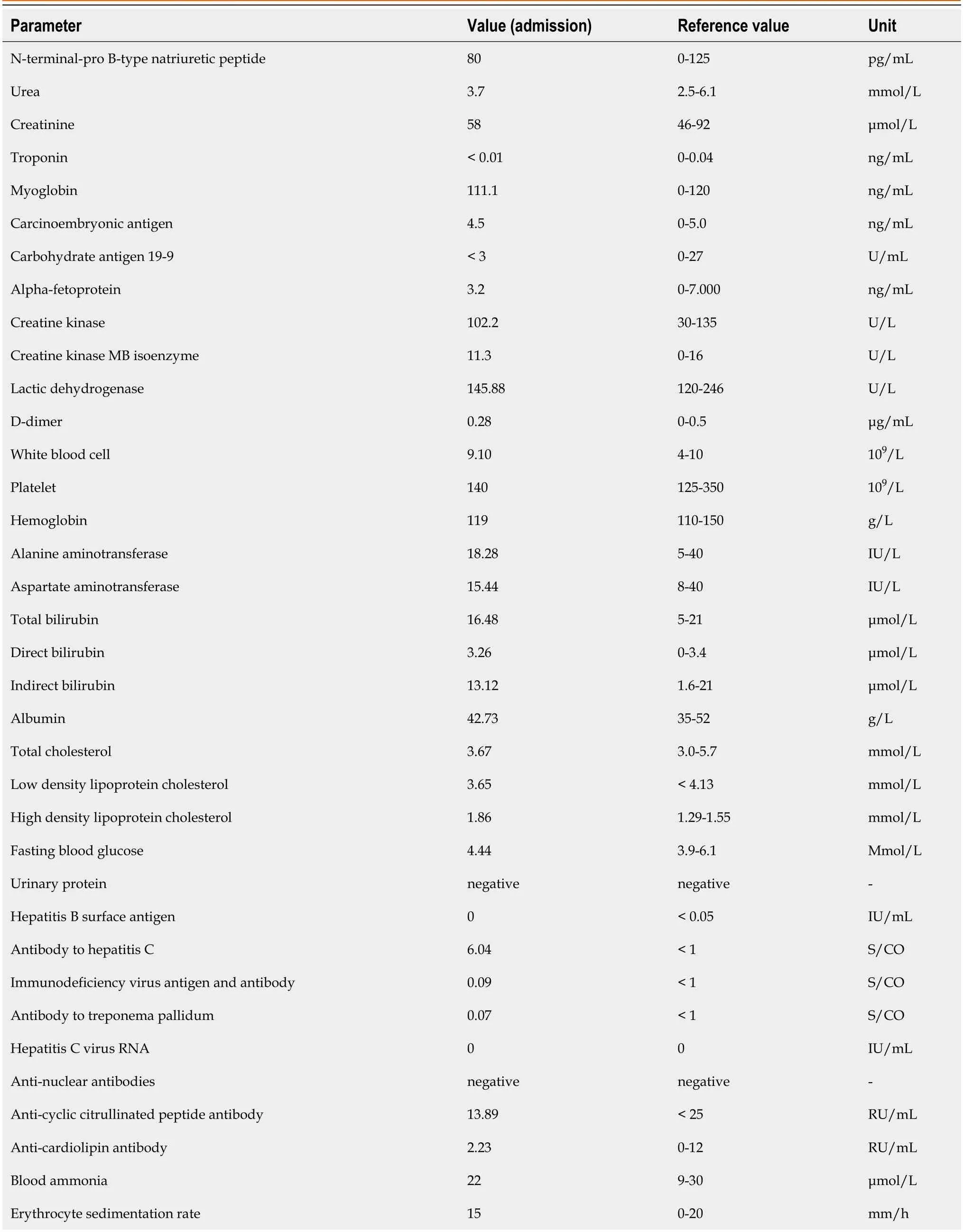
Table 1 Laboratory data at admission
Imaging examinations
CT with multi-plane reconstruction revealed scattered exudation,effusion and free gas in the abdominal cavity,indicating gastrointestinal perforation complicated with acute peritonitis (Figure 1A).Curved planar reconstruction of CT images revealed an abdominalMonopterus albusbiting the mesentery,suggesting aMonopterus albusoutside the intestinal cavity (Figure 1B).The outer margin of wall of the mid-rectum was rough and raised,and exudation and free gas were detected in the surrounding mesentery,suggesting a perforation of the mid-rectum (Figure 1C).Volume reconstruction of CT images showed clear and complete eel bone morphology in the abdominal and pelvic cavities (Figure 1D).
Further diagnostic work-up
The patient consented to laparoscopic surgery.Laparoscopic exploration revealed abundant cloudy yellow fluid and small amounts of feces-like fluid in the abdominal and pelvic cavities,and a large perforation was detected in the midrectum,with a diameter approximating 1.5 cm (Figure 1E),alongside a small amount of stool.It could be observed that theMonopterus albushas completely entered the abdominal cavity and has tightly bitten the mesentery of the small intestine (Figure 1F).During the operation,the deadMonopterus albuswas extracted,and was about 40 cm long (Figure 1G).
FINAL DIAGNOSIS
Based on the patient’s previous medical history,the patient was eventually diagnosed with digestive tract perforation and acute peritonitis.
TREATMENT
Postoperatively,the patient recovered well and was discharged on postoperative 5 d.
OUTCOME AND FOLLOW-UP
The patient recovered without complications.
DISCUSSION
Few reports have described living foreign bodies in the human body[9].CT constitutes an effective tool for accurate preoperative evaluation of living foreign bodies in clinic[3].3D CT reconstruction clearly displays anatomical structures,lesions and adjacent organs,improving diagnostic accuracy[6].In the present case,preoperative 3D CT reconstruction accurately located aMonopterus albusoutside the intestinal cavity with a perforation site,and the foreign body had damaged the mesentery in the small intestine,causing fluid and gas accumulation,as well as peritoneal thickening[5].These findings suggest preoperative 3D CT reconstruction may accurately locate perforation sites and foreign bodies,help diagnose peritonitis and guide surgical treatment[4].
CONCLUSION
Preoperative 3D CT reconstruction can accurately locate perforation sites and living foreign bodies,help diagnose peritonitis and guide surgical treatment.
FOOTNOTES
Author contributions:Yang JH and Lan JY contributed to manuscript writing and editing,as well as data collection;Lin AY and Huang WB contributed to data analysis;Liao JY contributed to conceptualization and supervision;and all authors have read and approved the final manuscript.
Informed consent statement:Written informed consent was obtained from the patient for publication of this report and any accompanying images.
Conflict-of-interest statement:All the authors report no relevant conflicts of interest for this article.
CARE Checklist (2016) statement:The authors have read the CARE Checklist (2016),and the manuscript was prepared and revised according to the CARE Checklist (2016).
Open-Access:This article is an open-access article that was selected by an in-house editor and fully peer-reviewed by external reviewers.It is distributed in accordance with the Creative Commons Attribution NonCommercial (CC BY-NC 4.0) license,which permits others to distribute,remix,adapt,build upon this work non-commercially,and license their derivative works on different terms,provided the original work is properly cited and the use is non-commercial.See: https://creativecommons.org/Licenses/by-nc/4.0/
Country/Territory of origin:China
ORCID number:Jin-Han Yang 0000-0003-4870-8799;Jin-Ying Lan 0009-0005-5568-7713;An-Yuan Lin 0009-0002-4444-7405;Wei-Biao Huang 0000-0002-9807-7668;Jin-Yuan Liao 0000-0001-9722-821X.
S-Editor:Wang JJ
L-Editor:A
P-Editor:Wang JJ
 World Journal of Gastrointestinal Surgery2023年10期
- World Journal of Gastrointestinal Surgery的其它文章
- Gastric adenosquamous carcinoma with an elevated serum level of alpha-fetoprotein: A case report
- Giant dedifferentiated liposarcoma of the gastrocolic ligament: A case report
- Hereditary hemorrhagic telangiectasia involving portal venous system: A case report and review of the literature
- Mucocutaneous ulcer positive for Epstein-Barr virus,misdiagnosed as a small bowel adenocarcinoma: A case report
- Postpolypectomy syndrome without abdominal pain led to sepsis/septic shock and gastrointestinal bleeding: A case report
- Bariatric surgery reduces colorectal cancer incidence in obese individuals: Systematic review and meta-analysis