
Construction and biological characterization of Burkholderia pseudomallei sRNA gene deletion strain
2023-09-23 09:06:58SONGXinyiLIAnyangLIYanmeiXUShuhuiXIAQianfeng
SONG Xin-yi, LI An-yang, LI Yan-mei, XU Shu-hui, XIA Qian-feng,✉
1.Hainan Medical University International School of Public Health and One Health, Haikou 571199, China
2.Laboratory of Tropical Biomedicine and Biotechnology, School of Tropical Medicine and Laboratory Medicine, Hainan Medical University, Haikou 571199, China
Keywords:
ABSTRACT Objective: Construction of Burkholderia pseudomallei (B.pseudomallei) sRNA knockout strains and observation of their biological function.Methods: Design 9sF/9sR,9xF/9xR and R1/F1 primers, which were used to amplify the homologous arm fragment upstream and downstream of the sRNA gene, through enzyme cutting, ligation, and transformation, the sRNA gene was knocked out from the B.pseudomallei by homologous recombination method.Results: The sRNA mutant was successfully constructed.In comparison with wild strain HNBP001, the growth rate, motility and biofilm formation of sRNA decreased, but the antibiotic sensitivity has no differences.Conclusion: The sRNA knockout strain of B.pseudomallei was successfully constructed, laying a foundation for further research on its mechanism of regulating B.pseudomallei.
1.Introduction
Burkholderia pseudomallei (B.pseudomallei) is a facultative intracellular gram-negative bacillus, which is the causative agent of Melioidosis[1].B.pseudomallei remains prevalent throughout tropical and subtropical regions, especially in Southeast Asia and northern Australia.The epidemic areas were Hainan, Guangdong, Guangxi,Fujian, and Taiwan, with the highest incidence in Hainan Province[2].Invade the human body mainly through contaminated water and soil and broken skin, the main symptoms were lung infection and septicemia, which is the primary risk of infection for residents or foreign tourist populations, and mortality can exceed 40% in some untreated nosocomial patients[3,4].The pathogenesis of Burkholderia pseudomallei and the immune protection mechanism of the host are not fully clarified, and there is no effective vaccine[5].
Small RNA(sRNA) is a class of non-coding RNAs with important regulatory functions, ranging in length from approximately 50 to 500 nucleotides, bacterial sRNAs act by base pairing, having complementarity with their target mRNAs, which is strongly associated with bacterial proliferation, metabolism, stress response, and pathogenicity[6].In the preliminary work, our group expressed Hfq, a molecular chaperone protein that promotes sRNA expression, and identified and characterized 9 new Burkholderia pseudomallei sRNAs by Hfq protein-based immunoprecipitation technique and RNA-seq sequencing analysis[7].Therefore, we evaluated one of the sRNA provisionally named sRNA9, which is located on chromosome 2 of HNBP001 (Sequence ID: CP038806.1,556924bp-557397bp).Through biological experiments, the important biological functions and mechanisms of this sRNA in the pathogenesis of B.pseudomallei were preliminarily explored,providing a new molecular target for the anti-B.pseudomallei infection and innovative ideas for the prevention and treatment as well as diagnosis of melioidosis.
2.Materials and Methods
2.1 Material
2.1.1 Bacteria, plasmid, and cell
Burkholderia pseudomallei wild strain HNBP001 came from the isolated and preserved strain of our research group[8].Plasmid TPR-pK18mobSacB was modified by Teacher Tian Shen of the research group to increase trimethoprim resistance fragment, which was preserved in our laboratory[9].S17-λpir Competent Cell,DH5α Competent cell from ZOMABIO.
2.1.2 Reagents and instruments
TIANamp Bacteria DNA Kit, TIANgel Purification Kit and TIANpure Mini Plasmid Kit, TIANGEN; Restriction enzyme (BamHⅠ, XbalⅠ, HindⅢ) and T4 DNA Ligase (Biolabs); Trimethoprim,Gentamicin, Kanamycin (Biosharp); PCR Instrument (Biorad);Multi-Mode Microplate Reader (Bioteck); Gel Documentation and Analysis System (JU YI).
2.1.3 Medium
Luria-Bertani (g/L): tryptone 10 g, yeast extract 5 g, NaCl 10 g,agar 16 g; 15% Sucrose solid medium (g/L): tryptone 10 g, yeast extract 5 g, saccharose 150 g, agar 16 g.
2.2 Method
2.2.1 Primers
Primers were designed for the upstream and downstream homologous arm sequences encoding sRNA9 based on the gene sequence of Burkholderia cepacia HNBP001 uploaded to NCBI in our laboratory: primers 9sF/9sR, 9xF/9xR, to amplify the homologous arm of the target sRNA, R1/F1 primers were applied to validate the knockout strain.All primers were synthesized by Sangon Biotech.(Table 1)
Tab 1 PCR primer sequence
2.2.2 Vector Construction
Extract the genome of HNBP001, primers 9sF/9sR and 9xF/9xR were used for upstream and downstream homologous arm PCR amplification, and ligated after enzyme digestion.The plasmid TPR-pK18mobSacB was used as the template.It was purified by double digestion using HindⅢ and BamHⅠ, with the enzyme digestion system shown in Table 2.The upstream and downstream homologous arms are ligated to the plasmid cleavage product at 16℃ for several hours using T4 DNA ligase.The post-ligation product was added to DH5α competent cell, heat-stimulated transformed by standard procedures, and antibiotic-containing plates were coated.After 12 h incubation, single colonies were picked in an LB liquid medium containing antibiotics and incubated overnight for plasmid extraction using the Plasmid min kit, followed by enzymatic verification of the plasmids.
Tab 2 System of enzyme digestion
2.2.3 Construction and identification ofΔsRNA9 knockout strain
The appropriate amount of verified plasmid was diluted and transformed into S17-1 receptive cells.After recovery, the receptive cells were coated with antibiotic LB solid plates and cultured for 12 h.Single colonies from the plates were selected and added to an LB liquid medium containing antibiotics.At the same time, single colonies of HNBP001 were selected and cultured in a non-antibiotic LB medium overnight.1 mL of transformed S17-1 and HNBP001 bacterial solution was taken, and the supernatant was obtained by centrifugation.The precipitate was resuspended in 500 μL of the medium, and the resuspended bacterial solution was mixed and dripadded to the filter membrane and incubated for several hours.The bacteria were washed with LB liquid medium, diluted with different concentration gradients (10-1, 10-2), coated with plates containing antibiotics, and cultured for 36-48 h.
Single bacteria were selected from LB solid plates and cultured in an LB liquid medium containing antibiotics overnight.Extract the bacteriophage genome, verify by PCR, and obtain the bacteriophage with successful binding.Take 500 μL of the successfully combined bacteriophage and add it into LB medium without antibodies and incubate it overnight.Dilute the bacterial solution and spread it on LB solid screening medium.Single colonies on the plates were picked and primer R1/F1 was used to verify whether HNBP001 was successfully knocked out.HNBP001 was used as a positive control,amplified, purified and sent for sequencing, and the sequenced sequences were compared, and the successful knockout strain was tentatively named as sRNA9.
2.2.4 Growth curve
Single colonies of wild strain HNBP001 and knockout strain sRNA9 were selected and added into fresh LB medium one night in advance, and the bacteria were cultured to logarithmic growth stage.The bacterial solution in the logarithmic phase of growth was diluted to OD600=0.1, and added into the 96-well plate according to the liquid volume of 200 μL.Each sample was set up for 5 replicates.The multifunctional microplate reader program was set at 37℃, double orbital vibration, and the interval time was 2 h.Growth curves were drawn using GraphPad 8.0.2 software.
2.2.5 Mobility
The single colony plates of fresh wild strain HNBP001 and knocked out strain sRNA9 were prepared one night in advance, and the single colony was selected and cultured in LB medium until the logarithmic growth stage.Dilute the bacterial solution to OD600=0.1,aspirate 2.5 μL diluted bacterial solution suspension drop to 3% LB soft agar medium, set 3 replicates, air dry for 10-15 min, put into 37℃ incubator and incubate for 24 h, measure the diameter of bacterial inhibition circle.
2.2.6 Biofilm
The bacterial culture method was the same as before, the concentration of a bacterial solution of wild strain HNBP001 and knocked strain sRNA9 was adjusted to OD600= 0.1, and 5 mL glass test tubes were taken to add 3 mL diluted bacterial solution of HNBP001 and sRNA9, respectively.Three repeats were set, and the glass test tubes were placed in 37℃ incubator for 48 h.Obvious differences were observed.The bacterial liquid in the glass tube was sucked out with a pipette, the residual impurities were cleaned with 1 × PBS, and 4mL 1% crystal violet was added to the stain for 30 min.After the crystal violet solution was sucked out, the test tube was cleaned again with 1 × PBS, and 3 mL 30% acetic acid was added for 20 min to measure the absorbance value of OD595.
2.2.7 Drug sensitivity test
Use the MIC plates strictly according to the instructions.Prepare a fresh single colony plate in advance, pick a suitable size single colony and add it to the dilution bottle, grind and mix to prepare bacterial suspension, aspirate 50 μL of bacterial suspension, add M-H broth medium, mix well and add to the antimicrobial drug wells, 100 μL/well, and read the results of antimicrobial drug MIC determination after 18 h.
2.2.8 Statistical analysis
GraphPad 8.0.2 software was used for drawing and statistical analysis.Wilcoxon signed-rank test was used to analyze the difference between paired samples, and the Mann-Whitney U test was used to analyze the difference between unpaired samples.
3.Results
3.1 Construction and validation of knockout strain ΔsRNA9
3.1.1 PCR amplification results of the upstream and downstream arms of the target gene
Using HNBP001 as a template, the upper and lower arm fragments were amplified by PCR using primers 9sF/R and 9xF/R, respectively,to obtain bands with lengths of 682bp and 793bp.The PCR amplification results were shown in Figure 1.
Fig 1 PCR amplification results of the up and downstream sequences of the target gene.M: 2000bp DNA Marker; 1: upstream fragment of sRNA9; 2:downstream fragment of sRNA9.
3.1.2 Verification of recombinant plasmid
The upper and lower arm fragments of sRNA9 were ligated to the plasmid TPR-pK18mobSacB, the ligation product was transformed to DH5ɑ, coated to the plate containing methotrexate, and the plasmid was extracted by picking a single colony, and verified by double digestion with HindⅢ and BamHⅠ.
3.1.3 Validation of knockout strains
The constructed recombinant plasmid TPR-pK18mobSacB-sRNA9 was transformed into S17-λpir Competent Cell, which was doubleexchanged with HNBP001, and screened by trimethoprim and gentamicin double-resistant plates and 15% sucrose plates.If the double-resistant plate did not grow and 15% sucrose plate grew, a single colony was selected for PCR verification.The knockout strain of Burkholderia meliorate sRNA9 was obtained, and the genome was extracted and sent to a sequencing company for further verification.Validation primer R1/F1 was used for PCR amplification of wild strain HNBP001 and knockout strain △sRNA9.HNBP001 contained a sRNA9 knockout target fragment with an amplification size of 2227bp, and △sRNA9 did not contain a sRNA9 knockout target fragment with amplification product size of 1753bp.The gel electrophoresis results were shown in Figure 3.The knockout plant was successfully constructed.
3.2 Growth curve
Fig 2 Verification of recombinant plasmid TPR-pK18mobSacB-sRNA9 by double enzyme digestion.M: 2000bp DNA Marker; BP: HNBP001; 9:sRNA9 enzyme digestion product.
Prepare the single colony plate in advance, select the single colony,and culture overnight to the logarithmic growth stage, dilute the bacterial solution to OD600=0.1, the multifunctional enzyme marker Fig 3 Validation of gene sRNA9 knock-out mutant.M: 2000bp DNA Marker; BP: HNBP001; 9: sRNA9 knockout product validation.is programmed to measure OD600absorbance at 4h intervals.The growth curve was repeated 3 times in different time periods, and 3 replicates were set in each group.HNBP001 strain was used as the control, and the growth curve was drawn.The results were shown in Figure 4.Both HNBP001 and △sRNA9 entered the logarithmic growth stage at 8h, and the growth rate of knocked-out strain △sRNA9 was significantly lower than that of HNBP001 after logarithmic growth stage.The results showed that the deletion of sRNA9 fragment affected the growth of B.pseudomallei.
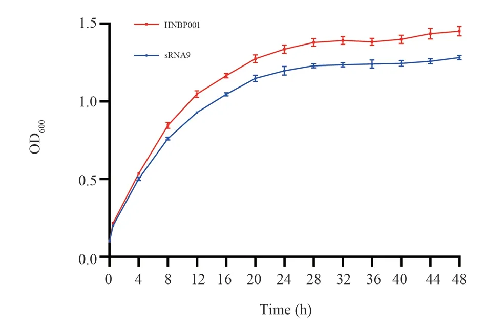
Fig 4 Growth curve of B.pseudomallei HNBP001 WT and △sRNA9.Growth curve comparison of SRNA9 and HNBP001, paired sample Wilcoxon signed rank test, P<0.0001.
3.3 Mobility experiment of bacteria
The mobility ability of Burkholderia melioides plays an important role in nutrient acquisition and invasion of host cells.In this experiment, the mobility ring diameter of bacteria in 3% soft medium was observed to determine the mobility ability of bacteria after sRNA9 gene knockout.Taking wild strain HNBP001 as the reference object, it was found that the diameter of △sRNA9 mobility ring decreased, indicating that the mobility ability was weakened after sRNA9 gene knocked out, as shown in Figure 5.
3.4 Biofilm formation experiment
Biofilm formation plays an important role in protecting bacteria from extreme external environments.The B.pseudomallei HNBP001 and △sRNA9 were incubated in cuvette, for 48 h, and the biofilm thickness was observed.After PBS washing, crystalline violet staining, acetic acid dissolution and other steps 200 μL of liquid was aspirated and added to a 96-well plate for OD595absorbance detection, and the biofilm results were obtained.The biofilm of △sRNA9 was sparse compared with that of HNBP001.After staining, the absorbance of OD595 was detected, and △sRNA9 of the knocked out strain was significantly lower than that of the wild strain HNBP001, as shown in Figure 6.The results showed that the deletion of sRNA9 affected the biofilm formation.
3.5 Drug sensitivity test
The sensitivity of wild strain HNBP001 and knocked out strain△sRNA9 to 20 kinds of antibiotics was analyzed through the MIC test, and △sRNA9 had no change in MIC of all antibiotics.The results were shown in Table 3.The results showed that knockout sRNA9 had no significant change in B.pseudomallei resistance.
Fig 5 Mobility of B.pseudomallei HNBP001 WT and △sRNA9.A: Mobility of HNBP001; B: Mobility of △sRNA9; C: Comparison between △sRNA9 and HNBP001, Mann-Whitney U test, P= 0.0286, P<0.05
Fig 6 Biofilm of B.pseudomallei HNBP001 WT and △sRNA9.A: 48h biofilm; B: crystalline violet staining; C: Comparative results of biofilms; Mann-Whitney U test, P=0.0286, P < 0.05.
Tab 3 MIC of B.pseudomallei HNBP001 WT and △sRNA9
4.Discussion
Bacterial gene expression is regulated by multiple molecules,with sRNAs mediating the diversity of regulatory functions by modifying the stability of ribosomes and mRNAs to act as repressors or promoters[10].Numerous researches proved that sRNA is closely related to bacterial virulence, host-pathogen messaging, antibacterial ability and drug resistance[11].Includes proteins compositions regulating cell membranes[12], biofilm formation[12], metabolism of nitrogenous compounds[13], glucose metabolism[14], regulation of extra-chromosomal DNA elements[15], control of transcription and RNA degradation[16], together with the regulation of bacterial virulence and host organism interactions during infection[17],sRNA has been shown to be involved in the regulation of posttranscriptional bacterial virulence levels and environmental stress adaptation through binding to mRNAs of target genes.
sRNAs have been identified in a variety of bacteria and have been found to play key regulatory roles in many mechanisms.French researchers found sRNA called SprD plays a virulence-regulating role in Staphylococcus aureus, and found its molecular target Sbi protein, indicating that sRNA plays an important role in bacterial pathogenicity[18].In Escherichia coli, SgrS participate in the stress process of phosphoglucose metabolism by down-regulating PtsG transporters and resist glucose phosphorylation accumulation in bacteria[19].There are fewer studies on the mechanism of sRNA in Burkholderia pseudomallei, Researchers in Thailand predicted 1306 B.pseudomallei sRNA genes and selected 8 novel sRNA through experiments, but their functions were not further verified[20].Malaysian scholars predicted the expression profiles of 12 differentially expressed cis-coding sRNAs that may be associated with the adaptation of B.pseudomallei to nutrient-deficient environments by simulating different nutrient environments [21].
Previous studies have confirmed the regulatory role of sRNA in other bacteria, as SmsR4 promotes the growth of Streptococcus pyogenes in a sorbitol-containing medium[22].ArcZ, CsrB,and OxyS sRNAs affect biofilm formation by regulating the expression of flagellar regulators[23], DicF regulates the virulence of Escherichia coli through the regulation of enterocyte abscission site genes and Shiga toxin expression[24].In this study, based on the sRNA chaperone protein Hfq protein expressed and purified by the group, nine sRNAs interacting with Hfq were screened by immunoprecipitation, and the TPR-pK18mobSacB suicide plasmid vector system was used to construct the sRNA9 knockout strain.After knocking out the sRNA9 gene fragment, the growth rate of bacterial plateau phase was lower than that of HNBP001, and the mobility ability and biofilm formation ability were reduced, but the drug resistance was not different compared with the wild strain.These results suggest that sRNA9 may play an important role in the growth and virulence of HNBP001.
In summary, the construction of an sRNA knockout strain of Burkholderia pseudomallei and the preliminary evaluation of its biological function provides an important basis for the subsequent study of the molecular mechanism of sRNA in the pathogenesis of B.pseudomallei.
 Journal of Hainan Medical College2023年9期
- Journal of Hainan Medical College的其它文章
- Advances in the efficacy and safety of nano-silver dressings for diabetic foot ulcers (DFU) healing
- Research progress of extracorporeal shock wave therapy for plantar fasciitis
- Pathogenesis and drug resistance mechanism of Burkholderia pseudomallei
- Screening and verification of the lncRNA-miRNA-mRNA regulatory network in muscle atrophy after spinal cord injury
- Clinical efficacy of dapagliflozin in the treatment of type 2 diabetes mellitus with heart failure with mildly reduced ejection fraction
- Application of ARFI technology to explore the clinical study of Gandou Tang in treating liver fibrosis of wilson's disease with damp-heat accumulation