SPHK1 expression in gastric cancer and clinical value based on bioinformatics analysis
LING Qian-long, GUAN Jia-jia, JI Kai, ZHU Shuang-qiu, ZHU Bing✉
1. Department of Gastrointestinal Surgery, the First Affiliated Hospital of Bengbu Medical College, Bengbu 233000, China
2. Department of Radiation Oncology, the First Affiliated Hospital of Bengbu Medical College, Bengbu 233000, China
Keywords:
ABSTRACT Objective: To elucidate the expression of SPHK1 in gastric cancer tissues and to analyse its clinical value in gastric cancer using bioinformatics.Methods: The expression of SPHK1 in gastric cancer tissues was analysed using HPA and TCGA databases.The association between the expression of both SPHK1 and MKI67 in gastric cancer was analysed using GEPIA database.Based on Kaplan-Meier Plotter database to analyse the relationship between SPHK1 mRNA expression level and survival of gastric cancer patients.Gene enrichment pathway predicted the potential biological function of SPHK1 on gastric cancer.The TIMER database was used to predict the expression of SPHK1 in immune-infiltrating cells.The expression of SPHK1 in gastric cancer tissues was analysed by IHC and Western Blot.Results: Bioinformatics analysis showed that SPHK1 was highly expressed in gastric cancer tissues compared to paracancerous tissues (P < 0.001).The analysis from the GEPIA database reveals a significant increase in the expression of SPHK1 and MKI67 in gastric cancer tissues(P<0.05).Furthermore, there is a positive correlation between the two proteins (P=0.00049).Kaplan-Meier Plotter survival analysis showed that high expression of SPHK1 predicted worse overall survival tine (logrankP=0.002) and post-progression generation time (logrankP=0.0019)in gastric cancer.Gene enrichment pathway results showed that SPHK1 may be involved in multiple gastric cancer progression pathways.TIMER database analysis showed that in macrophages, gastric cancer patients with low SPHK1 expression group had a better postprogression period than those with high SPHK1 expression group (P=0.002).IHC showed that the staining intensity and staining positive area of SPHK1 in gastric cancer tissues were higher than that of adjacent non-tumour tissues.And the staining intensity of TNMI and II stages was weaker than that of III and IV stages.Western blot analysis revealed higher levels of SPHK1 protein in gastric cancer tissue compared to paired adjacent non-cancerous tissue in 9 cases(P=0.0 265).Conclusion: SPHK1 gene is highly expressed in gastric cancer tissues and is associated with poor prognosis and may reduce tumour immunosuppression, which may serve as a potential marker for evaluating the prognosis of gastric cancer patients.
1.Introduction
Gastriccancer (GC) is one of the most common gastrointestinal malignancies and has been an important cause of cancer-related death in South America, Eastern Europe, and China[1-3].Although screening in some high-prevalence areas has reduced gastric cancerrelated mortality and morbidity, the overall 5-year survival rate remains low[4].Despite a series of advances in laparoscopic surgery and drug therapy, patients with advanced GC still have a poor prognosis[5-6].Therefore, in order to identify the main molecular mechanism of GC progression, the search for prognostic biomarkers and gene regulatory targets will enable individualized therapy.
Sphingosinekinase1 (SPHK1), as a lipid active kinase in vivo,can phosphorylate sphingosinekinase1 to produce sphingosine-1-phosphate (S1P)[7-8], which has been shown to play various functions in different stages of cancer progression[9-10].SPHK1 is involved in the disease process by up-regulating the expression of S1P receptor in cancer cells[11].More and more studies have found that SPHK1 plays a crucial role in gastric cancer[12].Now, many studies have shown that SPHK1 may be associated with biomarkers for the diagnosis and prognosis of gastric cancer[13].However, the role and specific mechanism of SPHK1 in gastric cancer progression remain unclear.This study used bioinformatics analysis to predict SPHK1 expression in gastric cancer.By analyzing the effect of SPHK1 on immune cell infiltration in gastric cancer tissues, the relationship between SPHK1 and clinicopathological features and prognosis, we hope to provide basis for diagnosis and treatment of patients with gastric cancer.
2.Materials and Methods
2.1 Main materials of the experiment
The clinical samples were collected from the required tissue samples and clinicopathological data of the Gastrointestinal Surgery Department of the First Affiliated Hospital of Bengbu Medical College from January 2020 to December 2022.Both the protein concentration BCA kit and gel PAGE kit were purchased from Biyuntian Biotechnology.Omni-ECLTM development kit was purchased from Shanghai Yase Biotechnology Co., LTD.Mouse polyclonal antibodies SPHK1 and GAPDH were purchased from Wuhan Sanying (Proteintech) company.The PVDF membrane was purchased from Millipore.Other reagents are imported.
2.2 Database Analysis
From the HPA (https://www.proteinatlas.org) download the gastric cancer and tissue adjacent to carcinoma data.Use TIMER2.0 SPHK1mRNA (http://timer.cistrome.org) database analysis in different expression level of malignant tumor.The expression of SPHK1 and tumor growth factor MKI67 in gastric cancer tissues was analyzed by GEPIA(http://gepia.cancer-pku.cn) database.Kaplan-Meier Plotter(http//: Kmplot.com) database was used to analyze the relationship between the expression level of SPHK1mRNA and the survival prognosis of patients with gastric cancer.CBioPortal database download and SPHK1 in the stomach of the communist party of China (https://www.cbioportal.org) table of potential target genes.DAVID (https://david.ncifcrf.gov) database download enrichment related data analysis.Use of microscopic letter web site(http://www.bioinformatics.com.cn) online editing download data processing.Based on the data provided by TISCH (http://tisch.compgenomics.org/home), the correlation between SPHK1 and TME was discussed.The gastric cancer chip provided by the website was GSE134520.
2.3 Immunohistochemistry
Fifty gastric cancer tissues and 34 matched adjacent normal tissues were implanted according to standard procedures.Refer to Deng et al[14] for specific operations.The scores were independently evaluated blind by 2 pathologists.Staining percentage score :0(<5%positive cells);1 (5~24% positive cells);2 (25~49% positive cells);3(50~74%) and 4 ( 75%).The staining intensity scores were 0(unstained), 1(weak), 2(medium), 3(strong).Combined staining scores were equivalent to percentage score×staining intensity scores, with a combined score 4 indicating positive staining and a combined score <4 indicating negative staining.
2.4 Western Blot
The protein was extracted from gastric cancer and adjacent tissues and quantified by BCA method.Appropriate amount of protein samples were taken for SDS-PAGE electrophoresis, and the protein gel was transferred to 0.45 um PVDF film for membrane transfer after the electrophoresis test was completed.Then the membrane was immersed in the sealing liquid and sealed for 60 min.After three times of film washing, the PVDF film was diluted and incubated in the refrigerator at 4 ℃ overnight.The film was washed the next day and incubated with a secondary antibody room temperature shaker for 90 min.ECL-plus detects protein signals on the PVDF membrane, which is then exposed and developed.
2.5 Statistical Processing
Use the default statistical methods of the database.Person correlation was used to analyze the relationship between SPHK1 and related gene expression.Kaplan-MeierPlotter was used to calculate the survival rate of patients, and Log-rank test was used to analyze the relationship between SPHK1 expression and survival prognosis of patients.χ2test was used to analyze the relationship between positive and negative expression of SPHK1 and clinicopathological features of patients.WB results of gastric cancer and adjacent tissues were determined by wilcoxon test.Bilateral P < 0.05 was considered statistically significant.
3.Result
3.1 Location detection of SPHK1 in cells
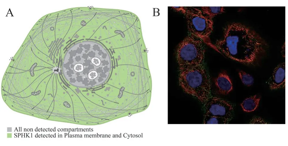
Based on HPA database retrieval, SPHK1 may be expressed in the cytoplasm of the cell model (Figure 1A).For further mapping,immunofluorescence of cells expressing SPHK1 was analyzed using the HPA database, indicating that SPHK1 in human high expression in the cytoplasm ( Figure 1B).

Fig 1 Localization detection of SPHK1 in cells
3.2 Expression of SPHK1 in normal human stomach tissue
The analysis of HPA database showed that SPHK1 was widely distributed in various tissues of the body.The expression of SPHK1 is low in various human tissues and low in normal human stomach tissues ( Figure 2A).In addition, SPHK1 is also widely expressed in normal gastric tissue cells, such as stromal cells, fibrocytes, and T cells ( Figure 2B).

Fig 2 Expression of SPHK1 in normal human tissues and cells
3.3 Expression of SPHK1 in different tumor types
TCGA database data showed that SPHK1 showed a high expression trend in a variety of cancers, such as breast invasive carcinoma(BRCA), cholangiocarcinoma (CHOL), colon cancer (COAD),hepatocellular carcinoma (LIHC), lung adenocarcinoma (LUAD),etc.(P < 0.05).The expression level of SPHK1 in gastric cancer tissues was much higher than that in adjacent tissues (P < 0.001), and the difference was statistically significant (Figure 3A).Meanwhile,immunohistochemical staining results of SPHK1 expression in gastric cancer tissues and normal tissues were downloaded according to the HPA database, indicating that SPHK1 was highly expressed in gastric cancer tissues (Figure 3B).
3.4 Relationship between SPHK1 expression level and prognosis of patients
To explore the correlation between SPHK1 and prognosis of patients with gastric cancer.Firstly, GEPIA database was used to analyze the expression of SPHK1 and tumor growth factor MKI67[15] in gastric cancer tissues.The results showed that the expression level of SPHK1 in gastric cancer tissues was higher than that in adjacent tissues (P < 0.05), and the difference was statistically significant (Figure 4A).Secondly, the expression level of MKI67 in gastric cancer tissues was also higher than that in adjacent tissues (P< 0.05), and the difference was statistically significant (Figure 4B).In addition, the expressions of the two were positively correlated, and the difference was statistically significant (P=0.00049) (Figure 4C).It may be suggested that high expression of SPHK1 is associated with poor prognosis of gastric cancer patients.In addition, Kaplan-MeierPlotter database was used to evaluate the relationship between SPHK1mRNA expression and survival prognosis of gastric cancer patients online.The results showed that high expression of SPHK1 predicted poor overall survival in gastric cancer, logrankP=0.002(Figure 4D).At the same time, it also predicted a worse postprogressive generation period, logrankP=0.0019, as shown in Figure 4E, and the difference was statistically significant.
3.5 Enrichment of gene pathways in gastric cancer
In this study, the top 500 potential target genes co-expressed with SPHK1 in gastric cancer were first downloaded using the cBioportal database, and the data related to SPHK1mRNA enrichment were analyzed and downloaded using the DAVID database.It includes BiologicalProcess (BP), CellComponent (CC), MolecularFunction(MF), and KEGG pathway.The data were analyzed and the results were obtained by using Weisheng website.As shown in Figure 5A-C,it is found that in the GO analysis composed of BP, CC and MF.The cells were enriched in cancer-related signaling pathways including Intracellular receptors, ATP enzymes activation, and Growth binding factor (P < 0.05).In the KEGG enrichment pathway, SPHK1 potential target gene also enriched the information pathway that has been widely studied and confirmed to be related to the development of gastric cancer.For example, the Hedgehog signaling pathway,Relaxin signaling pathway and cAMP signaling pathway are shown in Figure 5D (P < 0.05), and the differences are statistically significant.

Fig 3 Expression of SPHK1 in various tumor tissues

Fig 4 Relationship between SPHK1 expression levels and prognosis of gastric cancer patients.
3.6 Effect of SPHK1 on tumor microenvironment
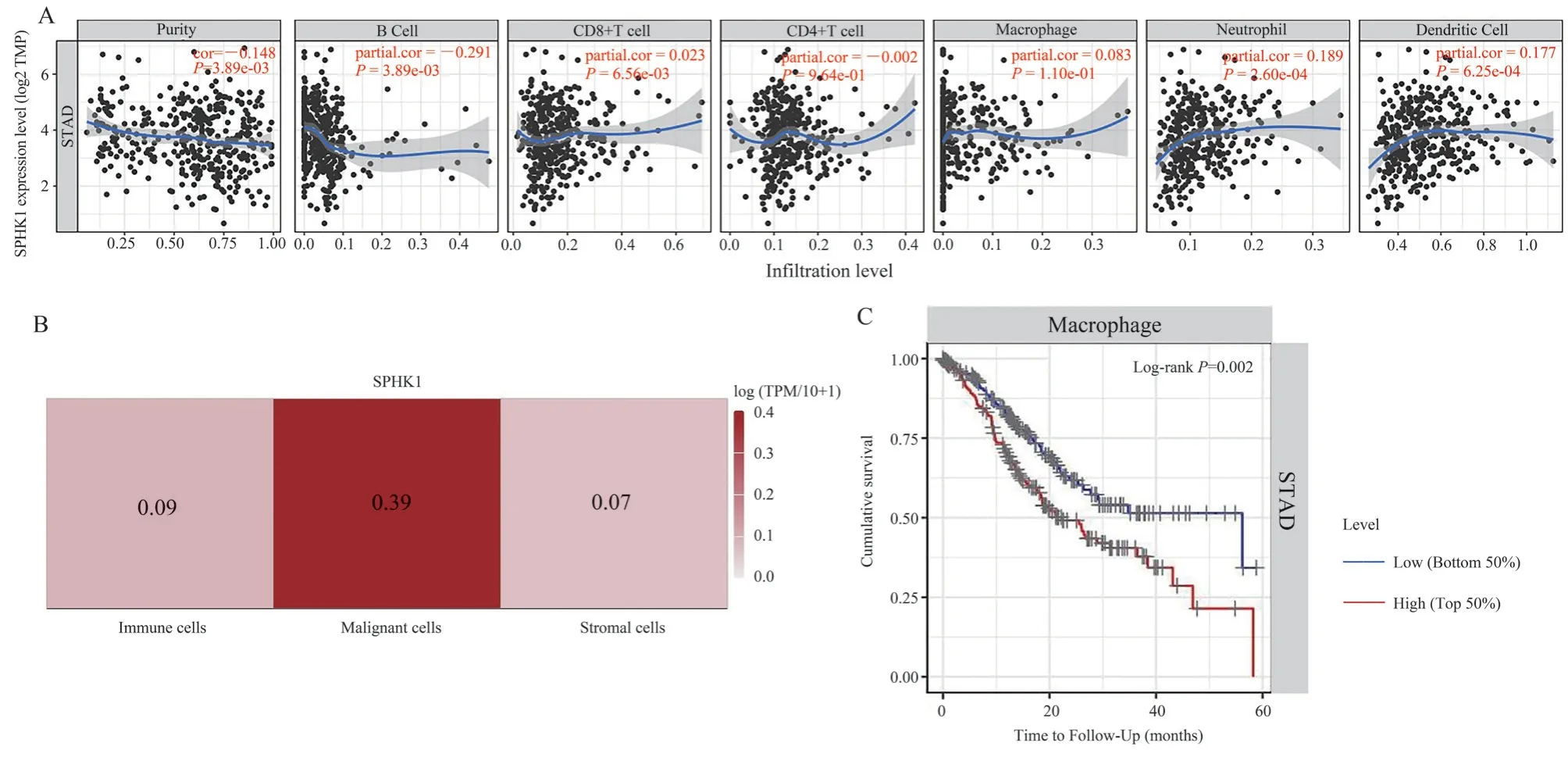
The tumor microenvironment (TME ) is very complicated.Various immune stromal cells in TME interact with cancer cells to regulate the proliferation and invasion of tumor cells[16-18].Therefore, it is necessary to further study the relationship between SPHK1 and TME.Using the TIMER database, it was found that in the immune microenvironment of gastric cancer, the expression level of SPHK1mRNA was negatively correlated with tumor purity[partial correlation coefficient (Partialcor) = -0.148], P < 0.001.On the other hand, SPHK1 was also negatively correlated with B cells in the immune microenvironment [partial correlation coefficient(Partialcor) = -0.291] (P < 0.001), showing statistical significance.In addition, in the immune microenvironment, SPHK1 was positively correlated with neutrophils and dendritic cells (partial correlation coefficients were all positive), but not definitively correlated with the expression of CD8+T cells, CD4+T cells and macrophages (P> 0.05), as shown in Figure 6A.Based on the data given by TISCH,the correlation between SPHK1 and TME was discussed.The gastric cancer chip provided by the website is GSE134520, which contains TME-related cells including tumor cells, immune cells(such as CD8+T cells, plasma cells, etc.), and interstitial cells (such as mucous cells, fibroblasts, etc.).The correlation analysis results between SPHK1 gene and TME are as follows:The expression level of SPHK1 gene was the highest in Malignant cells (log(TPM/10+1)=0.39), as shown in Figure 6B.In addition, in macrophages, the postoperative survival of gastric cancer patients in the low SPHK1 expression group was better than that in the high SPHK1 expression group, with logrankP=0.002, and the difference was statistically significant ( Figure 6C).In neutrophils, dendritic cells, CD8+T cells,CD4+T cells and B cells, low expression of SPHKmRNA had no significant difference in survival of gastric cancer patients (P > 0.05).
3.7 Expression of SPHK1 in gastric cancer tissues
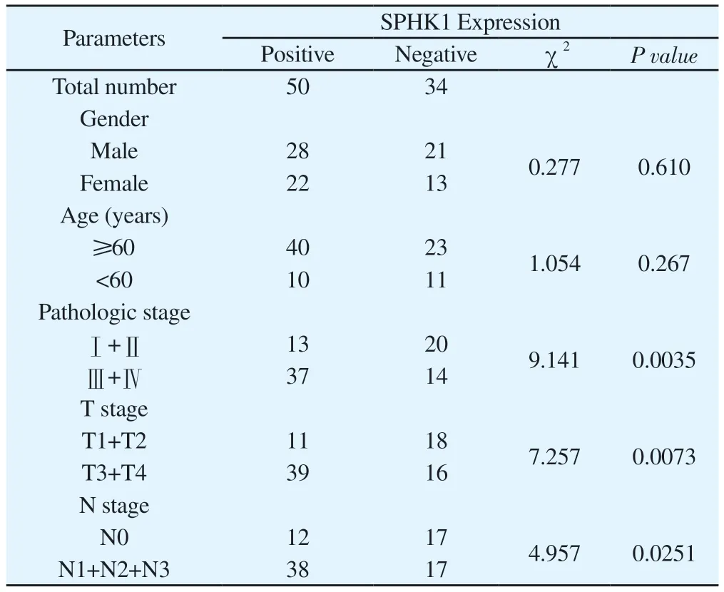
Clinical gastric cancer tissue samples were collected from 50 cases and adjacent non-tumor tissue samples from 34 cases.Statistical analysis showed that SPHK1 expression was not correlated with gender (χ2=0.277, P=0.610) and age (χ2=1.054, P=0.267), but correlated with pathological stage (χ2=9.141, P=0.0035) and TNM stage (χ2=7.257, P=0.0073) and N stages (χ2=4.957, P=0.0251).And there was statistical significance, as shown in Table 1.

Tab 1 Relationship between SPHK1 expression and clinicopathological features
To further investigate the expression of SPHK1 in gastric cancer tissues, immunohistochemical staining of clinical tissue samples will be performed.IHC results showed that, as shown in Figure 7A,the staining intensity and positive area of SPHK1 in gastric cancer tissues were higher than those in adjacent non-tumor tissues, and the staining intensity of gastric cancer tissues in TNM stage TNMI, II was weaker than that in stage III, IV, suggesting that the expression of SPHK1 might be related to the progression of gastric cancer.The IHC staining score showed that SPHK1 was highly expressed in gastric cancer tissues (P < 0.001), as shown in Figure 7C, with statistical significance.In addition, WesternBlot showed that in 9 gastric cancer tissues and paired para-cancerous tissues, the median expression of SPHK1 in gastric cancer tissues and para-cancerous tissues was 0.9030 and 0.6150, respectively, and its protein level was higher in gastric cancer tissues than in para-cancerous tissues(P=0.0265), as shown in FIG.7B and D, showing statistical significance.At the same time, some results of Shengxin data analysis are verified.Based on this, the above data further suggest that the high expression of SPHK1 in gastric cancer tissues may be related to the poor prognosis of gastric cancer progression.

Fig 6 Impact of SPHK1 on the immune microenvironment of gastric cancer
4.Discussion
Generally, the onset of gastric cancer is relatively insidious, and most cases have reached the stage of local development at the time of initial diagnosis, resulting in a high mortality[19].Traditional surgery and drug therapy still have poor prognosis for patients with gastric cancer[20].The SPHK1 gene plays a crucial role in cell growth and signal transduction[21].Peritoneal dissemination of gastric cancer (GCPD) has been recognized as the most common form of metastasis in advanced gastric cancer, and SPHK1-driven autophagy of human peritoneal mesoepithelial cells (HPMC) accelerates the occurrence of GCPD in vitro and in vivo[13].Xia et al[22] found that SPHK1 expression was elevated in gastric cancer, and downregulation of SPHK1 could inhibit the proliferation of gastric cancer cells and shorten the survival time of patients.In human MGC-803 gastric cancer cells by UV irradiation, SPHK1 conferred apoptotic resistance to apoptosis via the Akt / FoxO3a / Bim pathway[23].The ERK1-dependent up-regulation of SPHK1 transcription stimulates the proliferation of gastric cancer cells[24].Based on this, it is predicted that SPHK1 will play an important role in the diagnosis,treatment and prognosis prediction of gastric cancer, and then point out a new trend for the accurate diagnosis and treatment of gastric cancer.Qin et al[25] found in their study that SPHK1 promoted cisplatin resistance in bladder cancer cells through the NONO/STAT3 axis, thereby enabling tumor cells to generate immune escape.In a clinicopathological study of hepatocellular carcinoma(HCC) associated with non-alcoholic steatohepatitis (NASH),immunohistochemical staining showed that SPHK1 was expressed at a higher level than normal tissues[26].In addition, Peng et al[27]showed that high level expression of SPHK1 played an important role in the development of lung adenocarcinoma (LUAD).SPHK1,which is associated with drug resistance to targeted therapy, is also overexpressed in predicting poor prognosis of renal cell carcinoma[28].Therefore, the study of target genes in the progression of gastric cancer has more important clinical significance[29].This also provides the research basis and direction for this study.
In this study, based on TCGA and HPA databases, there were significant differences in the expression of SPHK1 in gastric and paracancer tissues.The results of IHC and WesternBlot showed that SPHK1 was highly expressed in gastric cancer tissues, which was consistent with the results of biological information analysis.Through further biological information retrieval, SPHK1 showed a positive correlation with the expression of tumor growth factor MKI67.The Kaplan-MeierPlotter database also suggested that high expression of SPHK1 predicted worse survival in gastric cancer.The results of rich pathway analysis also suggest that SPHK1 is involved in multiple pathways to affect the occurrence and development of gastric cancer.The exact role of various immune cells in the tumor microenvironment in cancer is very complicated[30-31].Lymphocyte infiltration can be induced by dendritic cell immunotherapy, which may have a positive impact on clinical outcomes[32].In this study,the expression of SPHK1 may be significantly correlated with the infiltration of three immune cell types, suggesting that SPHK1 can reflect not only the prognosis of cancer patients, but also the immune status.In addition, the expression of SPHK1 gene was the highest in tumor cells, which in turn regulated tumor progression.
In summary, SPHK1 has a significant role in gastric cancer, and it may be a potential prognostic marker for gastric cancer.However,this study still has some limitations, so we will further collect clinical data of gastric cancer patients and follow up to analyze the 5-year survival rate.At the same time, the expression of SPHK1 gene was interfered, and the influence of SPHK1 on the biological behavior of gastric cancer cells was discussed from the cellular level, and then the molecular mechanism of the occurrence and development of gastric cancer was further studied.
Conflict of Interest
The owners of this study declare that there is no conflict of interest.Authors’ Contribution
Ling Qianlong was mainly involved in the execution of experiment design, database search and paper writing.Guan Jiajia participated in some database searches.Ji Kai and Zhu Shuangqiu participated in some basic experiments.Zhu Bing participated in experimental design and paper writing guidance.
Ethics: Bengbu Medical College Human Ethics Approval number[2023] No.311, No.405.
 Journal of Hainan Medical College2023年23期
- Journal of Hainan Medical College的其它文章
- Advances in molecular mechanisms of oral submucosal fibrogenic carcinogenesis
- Research progress of ICOSL/ICOS pathway in maternal-fetal immune tolerance
- Effect and drug sensitivity analysis of PRPF18 on HCC
- Research progress of sphingosine 1-phosphate and its signal transduction in central nervous system diseases
- Meta-analysis of the effects of high-intensity intermittent exercise on cardiopulmonary function rehabilitation in patients with stroke
- Predictive value of controlling nutritional status score for progression to chronic critical illness in elderly patients with sepsis
