Shunxin decoction (顺心组方) improves diastolic function in rats with heart failure with preserved ejection fraction induced by abdominal aorta constriction through cyclic guanosine monophosphatedependent protein kinase Signaling Pathway
ZHANG Jiaying,WEI Xiangxiang,LI Xuefeng,YUAN Yang,DOU Yinghuan,SHI Yanbin,XIE Ping,ZHOU Mengru,ZHAO Junnan,LI Miao,ZHANG Shuwen,ZHU Rui,TIAN Ying,TAN Hao,TIAN Feifei
ZHANG Jiaying,WEI Xiangxiang,LI Xuefeng,YUAN Yang,DOU Yinghuan,SHI Yanbin,ZHOU Mengru,ZHAO Junnan,LI Miao,ZHANG Shuwen,ZHU Rui,TIAN Ying,TAN Hao,TIAN Feifei,School of basic medical sciences,Institute of Integrated Chinese and Western Medicine,Lanzhou University,Lanzhou 730000,China
XIE Ping,Department of Cardiology,Gansu Provincial hospital,Lanzhou 730000,China
Abstract OBJECTIVE: To determine whether Shunxin decoction(顺心组方) improves diastolic function in rats with heart failure with preserved ejection fraction (HFpEF) by regulating the cyclic guanosine monophosphatedependent protein kinase (cGMP-PKG) signaling pathway.METHODS: Except for control group 8 and sham surgery group 8,the remaining 32 male Sprague-Dawlay rats were developed into HFpEF rat models using the abdominal aorta constriction method.These rats in the HFpEF model were randomly divided into the model group,the Shunxin high-dose group,the Shunxin lowdose group,and the Qiliqiangxin capsule group.The three groups received high-dose Shunxin decoction,lowdose Shunxin decoction,and Qiliqiangxin capsule by gavage,respectively,for 14 d.After the intervention,the diastolic function of each rat was evaluated by testing E/A,heart index,hematoxylin-eosin staining,Masson,myocardial ultrastructure,and N-terminal pro-brain natriuretic peptide (NT-proBNP).The Bioinformatics Analysis Tool for Molecular Mechanism of Traditional Chinese Medicine (BATMAN-TCM) software was used to predict targets for which Shunxin decoction acts on the cGMP-PKG pathway.Natriuretic peptide receptor A(NPRA) and guanylate cyclase (GC) were detected by immunohistochemistry,and eNOS,phosphodiesterase 5A (PDE5A),and cGMP-dependent protein kinase 1(PKG I) were determined by Western blotting.RESULTS: Compared to the model group,the thickness of the interventricular septum at the end of diastole (IVSd)and the thickness of the posterior wall at the end of diastole (PWd) of the Shunxin decoction high-dose group,Shunxin decoction low-dose group,and Qiliqiangxin capsule group were all significantly reduced (P < 0.01).Furthermore,Shunxin decoction high-dose group E/A value was decreased (P < 0.01).Compared to the model group,the expression of NPRA and GC increased in the Shunxin decoction low-dose group and the Qiliqiangxin capsule group (P < 0.01).Compared to the model group,the expressions of eNOS and PKG I increased (P < 0.05)in the Shunxin decoction high-dose group.The expression of PDE5A expression decreased in the myocardium of the Shunxin decoction high-dose group,Shunxin decoction low-dose group,and Qiliqiangxin capsule group compared to the model group (P < 0.01).CONCLUSIONS: Shunxin decoction can improve diastolic function in rats with HFpEF.It increases the expression of NPRA,GC,and eNOS in the myocardial cell cGMP-PKG signaling pathway,upregulates cGMP expression,decreases PDE5A expression to reduce the cGMP degradation.Thus,the cGMP continually stimulates PKG I,reversing myocardial hypertrophy and improving myocardial compliance in HFpEF rats.
Keywords: heart failure;preserved ejection fraction;diastole;cyclic GMP-dependent protein kinases;signal transduction;Shunxin decoction
1.INTRODUCTION
The incidence of heart failure with preserved ejection fraction (HFpEF) has increased in recent years,accounting for about 50% of all patients with heart failure.1,2The 2013 American heart failure guidelines define HFpEF as having clinical symptoms of heart failure with EF ≥ 50%,excluding symptoms caused by other noncardiac diseases.3The 2016 European guidelines for heart failure provide a more precise definition of HFpEF.This definition includes the presence of typical signs and symptoms of heart failure,left ventricular ejection fraction (LVEF) ≥ 50%,increased levels of natriuretic peptides (brain natriuretic peptide (BNP) > 35 pg/mL or N-terminal pro-BNP (NTproBNP) > 125 pg/mL),left ventricular hypertrophy and left atrial dilatation,or left ventricular diastolic dysfunction.1The primary pathological mechanisms of HFpEF are left ventricular centripetal hypertrophy and diastolic dysfunction.4Recent studies have shown that the cardiac cyclic guanosine monophosphate/cGMPdependent protein kinase (cGMP-PKG) signaling pathway regulates the expression of downstream proteins through cGMP-dependent protein kinase 1 (PKG I) to inhibit pathological cardiac hypertrophy and enhances cardiac cell compliance.2Except for diuretics,drugs that can effectively treat patients with heart failure with a reduced ejection fraction are ineffective in improving HFpEF in patients.2
Recent studies have demonstrated that traditional Chinese medicine is effective in the treatment of HFpEF.5,6In clinical practice,we found that the primary pathogenesis of HFpEF is the deficiency ofQiand blood stasis,mutual obstruction of phlegm and blood stasis,and internal stagnation of water retention.Therefore,for the last 5 years,Shunxin decoction (顺心组方) was used to treat patients with HFpEF by invigoratingQi,improving blood circulation,and eliminating phlegm and blood stasis.It has shown promising clinical efficacy for HFpEF patients.It is also available as Shunxin sustainedrelease granules for patients with HFpEF.7
We designed this study to explore whether Shunxin decoction cures HFpEF through the cGMP-PKG signaling pathway.In this study,the natriuretic peptide receptor A (NPRA),guanylate cyclase (GC),eNOS,PKG I,and phosphodiesterase 5A (PDE5A) in the cGMP-PKG signaling pathway were selected as observation targets to explore the mechanism of Shunxin decoction intervention in HFpEF rats.
2.MATERIALS AND METHODS
2.1.Constructing a Rat Model of HFpEF
A total of 48 specific pathogen free healthy adult male Sprague Dawley rats [weight (250 ± 20) g] were purchased from the Lanzhou University Experimental Animal Center [license number SCXK (gan) 2013-0002].The experimental procedures adhered to the ethical regulations of animal experiments (The written approval of the agreement for all experimental procedures was awarded by Lanzhou University School of Basic Medicine Ethics Committee).Before the experiment,Esaote B MyLab Five was used to examine the hearts of rats.The cardiac structure and function of the rats’ hearts were normal.Except for control group 8 and sham operation group 8,the remaining 32 male Sprague-Dawlay rats were used as the HFpEF rat models.The rat model of HFpEF was created by constricting the abdominal aorta.These rats were anaesthetized with 0.4%pentobarbital administered intraperitoneally,and then the abdominal aorta was dissected by laparotomy.The abdominal aorta above the left renal artery was measured with a Vernier calliper.The abdominal aorta was narrowed to about 50% of its original diameter.A needle(the needle tip was flat) was placed parallel to and near the abdominal aorta.The abdominal aorta and the needle were ligated with a 2-0 suture,the needle was gently withdrawn,and the abdomen closed.8,9In the sham surgery group,the abdominal aorta was dissected but not ligated.After 3 months,echocardiography was performed to detect cardiac function in rats and evaluate the success of the HFpEF rat model.Interventricular septal thickness at end-diastole (IVSd),left ventricular end-diastolic dimension,the posterior wall thickness at end-diastole (PWd),left ventricular end-diastolic dimensions (LVDs),interventricular septal thickness at end-systole (IVSs),posterior wall thickness at endsystole,EF,and heart rate (HR) were identified using the left ventricular long-axis section and M-mode ultrasound.The peak blood flow in the early mitral diastolic period(E value) and late mitral diastolic period (A value),as well as E/A,were measured on the apical four-chamber surface with pulse-Doppler ultrasound.After the HFpEF rats’ model was created successfully,32 HFpEF model rats were randomly divided into the Model group (n=8),Shunxin decoction high-dose group (n=8),Shunxin decoction low-dose group (n=8),and Qiliqiangxin capsule group (n=8).
2.2.Drug intervention
Shunxin decoction consisted mainly of 12 herbs:Hongshen (Radix Ginseng Rubra),Huangqi (Radix Astragali Mongolici),Baizhu (Rhizoma Atractylodis Macrocephalae),Fuling (Poria),Chuanxiong (Rhizoma Chuanxiong),Chishao (Radix Paeoniae Rubra),Tinglizi(Semen Lepidii Apetali),Danshen (Radix Salviae Miltiorrhizae),and Gancao (Radix Glycyrrhizae).These herbs were purchased from the Hui Ren Tang Pharmaceutical Company and identified by Li Jianyin,a lecturer at the School of Pharmacy at Lanzhou University.The voucher specimen numbers of the stored herbarium specimens are 620031220010918321LY for Hongshen(Radix Ginseng Rubra),6203212018080-7063LY for Huangqi (Radix Astragali Mongolici),6201232001-0921355LY for Baizhu (Rhizoma Atractylodis Macrocephalae),620321199008101001LY for Fuling(Poria),62032120090828301LY for Chuanxiong(Rhizoma Chuanxiong),62032120180811156LY for Chishao (Radix Paeoniae Rubra),620321201708-10122LY for Tinglizi (Semen Lepidii Apetali),62012320100602101LY for Danshen (Radix Salviae Miltiorrhizae),and 62032120180812164LY for Gancao(Radix Glycyrrhizae).All of these herbs were evaluated for the prescription requirements and then decocted,concentrated,and put in the refrigerator at 4 ℃ for use.Qiliqiangxin capsules were purchased from Hui Ren Tang.The rats of each group were given the corresponding drug by intragastric gavage for 14 d:Shunxin decoction high-dose (42.81 g/kg),Shunxin decoction low-dose (14.27 g/kg),and Qiliqiangxin capsule intervention (0.375 g/kg).The control,sham operation,and model groups received normal saline.After drug intervention,cardiac function was assessed using echocardiography.
2.3.Weighing and histopathological examination of organs
After being weighed,the rats were anaesthetized by intraperitoneal injection of 0.4% pentobarbital.They were sacrificed by collecting blood from the abdominal aorta without causing any pain.The rats' hearts were excised and weighed.Furthermore,the heart index (heart mass/body weight) was calculated.The parts of each heart were put in the sample tube in a cryopreserved refrigerator at-80 ℃.The rest of the samples were placed in a 4% paraformaldehyde solution for a paraffin section.The morphological changes of myocardial cells stained with hematoxylin-eosin were observed under the light microscope.The myocardial collagen fibres were evaluated using the Masson staining kit (G1340,Solebao,Beijing,China).Moreover,these collagen fibers were quantitatively analyzed using Image Pro-plus 6.0 (Media Cybernetics,Rockville,MD,USA) under the light microscope according to the operation instructions.In each group,a section of each rat heart's left ventricular posterior wall was fixed in 2.5% glutaraldehyde solution for examination under an electron microscope.
2.4.Transmission electron microscopy
Take out cardiac tissue from 2.5% glutaraldehyde solution,phosphate buffer saline (PBS) rinsing and osmium fixation for 1.5 h,PBS rinse,Gradient ethanol dehydration,100% acetone dehydration,Acetone,and embedding solution were prepared in a 1∶1 ratio and placed for 2 h,35 ℃ for 24 h,45 ℃ for 24 h,65 ℃ for 24 h.Ultra-thin slicer cut 50-60 nm slices;Staining with lead citrate,transmission observed with the electron microscope,film.
2.5.Network pharmacological analysis
The BATMAN-TCM (Beijing Proteome Research Center,Beijing,China) was used to search the targets of each component herb of Shunxin decoction in the cGMPPKG signaling pathway.The score was 80,theP-value was 0.01,and three herbs were used as a single group for retrieval,and targets of each herb of Shunxin decoction on the cGMP-PKG signaling pathway were collected.At present,there are few studies on HFpEF,and it has not been included in the database.Therefore,the targets of treating heart failure are selected for each herb of Shunxin decoction.According to the multi-target prediction results of network pharmacology,nine of the 12 herbs of Shunxin decoction affected the cGMP-PKG pathway.In combination with related references,the final confirmed targets were NPRA,GC,eNOS,PDE5A,and PKG I.
2.6.Immunohistochemistry for detection of NPRA and GC
Paraffin sections of the myocardium were dewaxed using water.After antigen repair,NPRA and GC were detected with the rabbit SP kit (SP-9001,Zhongshanjinqiao,Beijing,China),and the images were observed and photographed using a microscope.Image-pro plus 6.0 software was used for image analysis.
2.7.Western blotting detection of eNOS,PDE5A,and PKG I
Protein was extracted from the rats' homogenate heart tissue,and its concentration was measured using BCA.Primary anti-dilution: antibodies PDE5A,eNOS,and PKGI (Beijing Bioss Antibodies,Beijing,China) were diluted with 5% skimmed milk powder at a ratio of 1∶500.Glyceraldehyde-3-phosphate dehydrogenase was diluted with 3% skimmed milk powder at a ratio of 1∶5000.
Second anti-dilution: sheep anti-rabbit with 5% skim milk powder to 1∶5000 ratio of dilution;Sheep anti-rat with 5% skim milk powder to 1∶2500 ratio of dilution.Electrophoresis (MP-300v Major science,CA,USA)was used to transfer the film (Beijing Liuyi Biological Technology Co.Ltd.,Beijing,China),and exposure was analyzed using MINICHEMI.
2.8.Statistical analysis
SPSS 21 statistical software (IBM Corp.,Armonk,NY,USA) was used to process the data,and all measurement data were expressed as mean ± standard deviation (±s).One-way analysis of variance was used for multigroup comparison.Pairwise comparisons were made using the Bonferroni method.P< 0.05 was considered statistically significant.
3.RESULTS
3.1.Assessment of heart function in rats
Three months after the surgery,the cardiac function tests of the rats in each group revealed that IVSd,PWd,and E/A increased in the model group compared with the control group (P< 0.01),and the differences were statistically significant.IVSd,PWd,and E/A were significantly higher in the model group than the sham group (P< 0.01) (Figure 1).There were no statistically significant differences between the control and the sham groups.LVEF of rats in each group did not differ significantly.
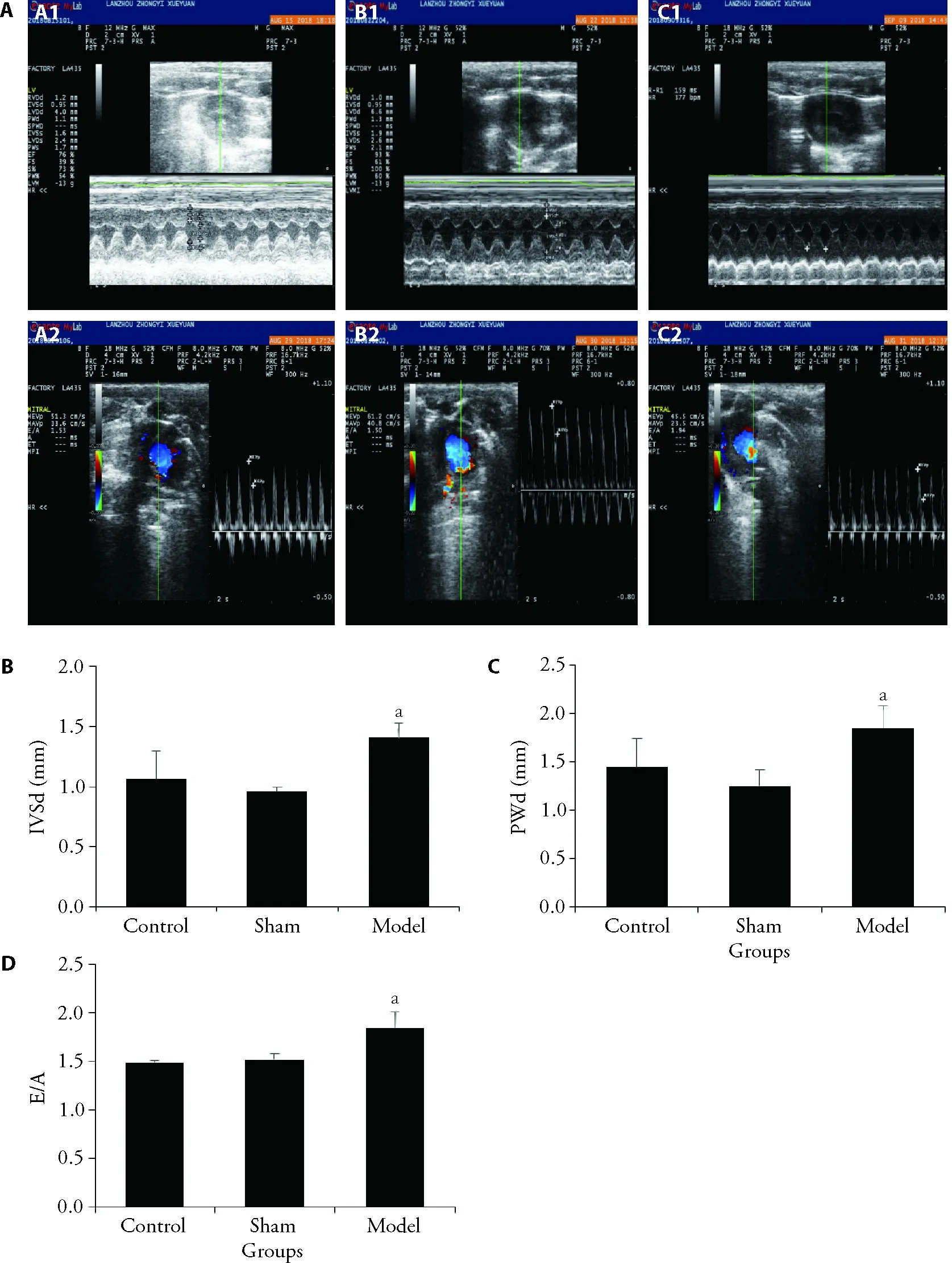
Figure 1 Cardiac function in rats in each group after three months of surgery
After the intervention,compared with the model group,IVSd and PWd were reduced in Shunxin decoction highdose group,Shunxin decoction low-dose group,and Qiliqiangxin capsule group (P< 0.01).E/A was decreased in Shunxin decoction high-dose group (P<0.01) (Figure 2).
3.2.Cardiac index
After the intervention,the cardiac index of the model group increased significantly (P< 0.05) compared with the control group (Figure 3 B).
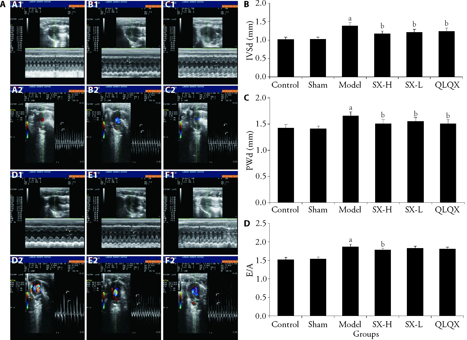
Figure 2 Cardiac function in rats in each group after drug intervention
3.3.Hematoxylin-eosin (HE) staining
HE staining of rat myocardium revealed that the myocardium cells in the control group were wellorganized.The arrangement of myocardial cells in the model group was partially disordered,the myocardial intercellular space was widened,the myocardial cells were enlarged,and some myocardial nuclei were enlarged and fused.The degree of damage in myocardial cells was less in Shunxin decoction high-dose group,Shunxin decoction low-dose group,and Qiliqiangxin capsule group than in the model group (Figure 3 A).
3.4.Masson staining
Myocardial cells were stained red,and collagen fibres blue after Masson staining of rat myocardium.The changes in myocardial collagen fibers in each group were observed,and resulting images were analyzed using Image-Pro Plus software.The cardiac collagen volume fraction (CVF)=myocardial collagen area/measured field area was calculated.Compared with the control group,CVF was higher in the model group (P< 0.05).Furthermore,CVF decreased in Shunxin decoction highdose group,Shunxin decoction low-dose group,and Qiliqiangxin capsule group compared with the model group;however,the difference was not statistically significant.The results indicated that the myocardial collagen fibres of the HFpEF rat model were increased(Figure 3 C,D).
3.5.Ultrastructure of myocardial cells
In the control group,the myofibrils were arranged in order and straight in direction,the thick and thin myofilaments were evenly distributed,tidy,and compact,and the bands within the sarcomere were visible.The mitochondria were round or oval with a clear bilayer membrane and a smooth outer membrane and were arranged in a regular pattern.In the model group,some myofibrils were ruptured,some sarcomeres were dissolved,some myofibrils were loose,the intercalated disc of the myocardium was dissected,and some mitochondria were enlarged and ruptured.Slightly enlarged mitochondria were observed in both the Shunxin decoction high-dose group and the Shunxin decoction low-dose group.The visible muscle-silk arrangement was slightly loose in the Qiliqiangxin capsule group,part of the sarcomere was lysed,and mitochondria were slightly swollen.However,the degree of damage to the ultrastructure of myocardial cells of the Shunxin decoction high-dose group,Shunxin decoction low-dose group,and Qiliqiangxin capsule group was smaller than that in the model group (Figure 3E).
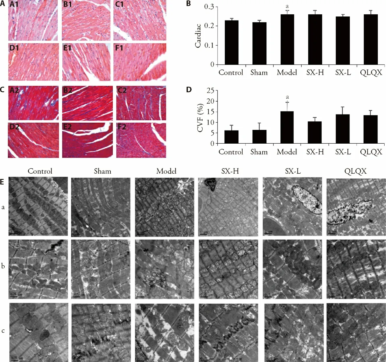
Figure 3 Morphological observation of rat myocardium in each group
3.6.Serum NT-proBNP levels in rats in each group
Serum NT-proBNP levels significantly increased in the model group compared with the control group (P< 0.05).Compared with the model group,NT-proBNP decreased in Shunxin decoction high-dose group,Shunxin decoction low-dose group,and Qiliqiangxin capsule group,but there was no statistically significant difference.
3.7.Expressions of NPRA and GC in rats’ myocardium in each group
The results of the NPRA assay in rat myocardium rats showed that compared with the control group,the expression of NPRA in the model group was decreased(P< 0.01).In comparison to the model group,myocardial NPRA expression increased in Shunxin decoction low-dose group and the Qiliqiangxin capsule group (P< 0.01) (Figure 4A,4B).
The expression of GC in the myocardium of the model group was reduced compared with the control group (P< 0.01).Compared with the model group,the Shunxin decoction low-dose group and the Qiliqiangxin capsule group showed increased expression of cardiac GC (P<0.01) (Figure 4C,4D).
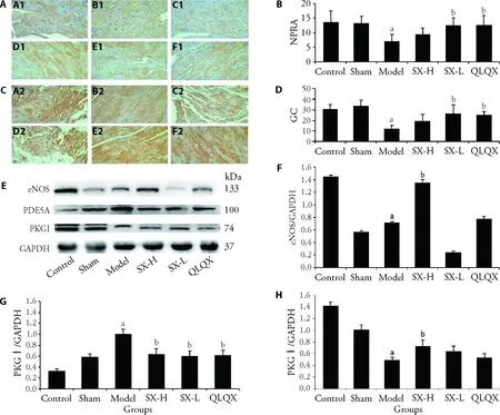
Figure 4 Expression of major target moleculars of cGMP-PKG pathway in each group
3.8.Expressions of eNOS,PDE5A,and PKG I in the myocardium of rats in each group
The results of the Western blotting test showed that the expression of eNOS in the model group was lower than that in the control group (P< 0.01).Compared with the model group,the expression of eNOS increased in the Shunxin decoction high-dose group (P< 0.01).
PDE5A assay revealed that the expression of PDE5A in the myocardium of the model group was increased compared with the control group (P< 0.01).Compared with the model group,myocardial PDE5A expression was significantly reduced in Shunxin decoction highdose group,Shunxin decoction low-dose group,and Qiliqiangxin capsule group (P< 0.01).
Compared with the control group,myocardial PKGI expression decreased in the model group (P< 0.01).Moreover,the PKG I expression increased in Shunxin decoction high-dose group compared with the model group (P< 0.05) (Figure 4E-4H).
4.DISCUSSION
The myocardial tissues of HFpEF patients have characteristics that include varying degrees of cardiomyocyte hypertrophy,interstitial fibrosis and microvascular reduction,10and downregulation of cardiac cGMP-PKG signaling pathway.11,12The cGMPPKG signaling pathway is activated when NO activates soluble guanylate cyclase acid,and natriuretic peptides activate particles GC at the same time.This further converts GTP into cGMP,and then this cGMP activates PKG I.The activated PKG I regulates the phosphorylation of many target proteins and then starts the downstream effects.For example,the PKG I acts on N2B of muscle protein to decrease myocardial cell stiffness,on phosphorous protein to enhance the reuptake of sarcoplasmic reticulum for calcium,and on G protein signal transduction regulators 2 and 4 to inhibit hypertrophy signal.13-15cGMP can be selectively hydrolyzed by PDE5A.16
This study showed that Shunxin decoction could effectively reverse cardiac hypertrophy in rats with HFpEF.It can improve the diastolic function and myocardial cells morphology and can decrease the collagen fibre distribution and ultrastructural damage of the myocardium.The mechanism behind these effects includes not only upregulation of NPRA,GC,and eNOS of cGMP-PKG signaling pathways to increase the cGMP expression but also downregulation of PDE5A to decrease cGMP degradation.These two effects keep cGMP expression at a high level in myocardial cells.cGMP continually activated PKGI,causing it to regulate phosphorylation of target proteins and subsequently cause a wide range of downstream effects.Similarly,it reduces myocardial cell stiffness,enhances calcium reuptake by the sarcoplasmic reticulum,and inhibits hypertrophic signaling.11These effects can reverse pathological cardiac hypertrophy.17
Shunxin decoction has effects such as warmingYang Qi,inducing diuresis to alleviate oedema,and activating blood and removing blood stasis.Its herb composition mainly includes Hongshen (Radix Ginseng Rubra),Huangqi (Radix Astragali Mongolici),Baizhu (Rhizoma AtractylodisMacrocephalae),Fuling (Poria),Chuanxiong (Rhizoma Chuanxiong),Chishao (Radix Paeoniae Rubra),Tinglizi (Semen Lepidii Apetali),Danshen (Radix Salviae Miltiorrhizae),and Gancao(Radix Glycyrrhizae).Among these herbs,Hongshen(Radix Ginseng Rubra) is warm,restores vital energy,and is especially beneficial for weak YangQi,whereas Huangqi (Radix Astragali Mongolici) boostsQiand ascends Yang,inducing diuresis to reduce swelling.Both are the Monarch drugs.Baizhu (Rhizoma Atractylodis Macrocephalae) heals spleen andQi,eliminates moisture,and induces diuresis.With flat in nature,sweet taste,and light Poria(Fuling),together,they can strengthen the spleen,benefit the function of water infiltration and accumulation,make the spleen healthy,remove water accumulation and alleviate oedema.These two are the Minister drug.HFpEF patients with longtermQideficiency have blood stasis;therefore,their treatment medication must include drugs that increase blood circulation to remove blood stasis.Chuanxiong(Rhizoma Chuanxiong),Danshen (Radix Salviae Miltiorrhizae),Chishao (Radix Paeoniae Rubra) are used together to remove blood stasis and restore heart’s function and blood circulation.Tinglizi (Semen Lepidii Apetali) has diuretic and anti-inflammatory properties.These two herbs can assist in curing oedema in HFpEF patients.Gancao (Radix Glycyrrhizae) works synergistically with various herbs,restoresYangQi,removes congestion,eliminates oedema,removes HFpEF symptoms,and promotes gradual recovery.18
Modern pharmacological studies have proved that Huangqi (Radix Astragali Mongolici) can effectively improve cardiac dysfunction and abnormal metabolic alterations in rats with heart failure.19Astragaloside has been shown to increase the release of eNOS,which leads to vasodilation.19Chishao (Radix Paeoniae Rubra)’s main active ingredient is paeoniflorin,which has antiinflammatory,antioxidant,and anti-thrombotic effects.Studies have proved that paeoniflorin can promote angiogenesis.20,21Ligustrazine,the main active ingredient of Chuanxiong (Rhizoma Chuanxiong),has anti-oxidant,anti-platelet aggregation,anti-inflammatory properties and regulates calcium homeostasis.22,23Danshen (Radix Salviae Miltiorrhizae) has a protective effect on rat cardiac myocytes.24,25Danshen(Radix Salviae Miltiorrhizae),Huangqi (Radix Astragali Mongolici),and Chuanxiong (Rhizoma Chuanxiong) can resist myocardial fibrosis.26Glycyrrhizin and glycyrrhizic acid are active ingredients of Gancao (Radix Glycyrrhizae),which can treat heart failure.27-30To various degrees,many studies on the properties of these drugs support the findings of this study.
Pathological myocardial hypertrophy can be reversed.The researchers used the pressure load to induce pathological myocardial hypertrophy and heart failure model.20,31,32Taking PDE5A inhibitors (sildenafil) orally for stopping the degradation of cGMP then increase the cGMP can activate the PKG I,33,34thereby inhibiting myocardial cell hypertrophy and ventricular hypertrophy.These actions could improve cardiac function in rats with chronic pressure load induced by constricted aorta.30,32,33However,to reverse pathological cardiac hypertrophy,activating the PKG I signaling pathway is more important than increasing the activity of PKG I itself.In hypertrophic cardiomyocytes,the activity of PKG I was increased,but the cardiac cells were significantly enlarged.Therefore,activating the PKGI signaling pathway may be a potential therapeutic target for reversing pathological myocardial hypertrophy.34These study results are consistent with the findings of this study.Therefore,Shunxin decoction can improve myocardial blood supply,protect myocardial cells,alleviate pathological myocardial fibrosis,reverse myocardial hypertrophy,and improve the diastolic function in rats with HFpEF by restoringQi,promoting blood circulation,and removing blood stasis.In a study,the Qiliqiangxin capsule was clinically used to treat heart failure patients.35In this study,Shunxin decoction as Qiliqiangxin capsule had a similar effect in rats with HFpEF.
In conclusion,Shunxin decoction can effectively reverse myocardial hypertrophy in HFpEF rats and improve their diastolic function.It increases cGMP expression in cardiomyocytes by upregulating the NPRA,GC,eNOS of the cGMP-PKG signaling pathway.Furthermore,it also downregulates PDE5A to reduce the degradation of cGMP.These effects make cGMP continue to act on the target of PKG I,reversing myocardial hypertrophy and effectively improving the myocardial compliance of HFpEF rats.
 Journal of Traditional Chinese Medicine2022年5期
- Journal of Traditional Chinese Medicine的其它文章
- Shugan Jieyu capsule (舒肝解郁胶囊) improve sleep and emotional disorder in coronavirus disease 2019 convalescence patients: a randomized,double-blind,placebo-controlled trial
- Application value of Qisexingtai hand diagnostic method in diagnosis of coronary artery disease
- An infrared thermographic analysis of the sensitization acupoints of women with primary dysmenorrhea
- Preliminary single-arm study of brain effects during transcutaneous auricular vagus nerve stimulation treatment of recurrent depression by resting-state functional magnetic resonance imaging
- Effect of treatment with Fufang Huangqi decoction (复方黄杞汤剂)on dose reductions and discontinuation of pyridostigmine bromide tablets,prednisone,and tacrolimus in patients with type I or II myasthenia gravis
- Wenshen Jianpi recipe (温肾健脾方) induced immune reconstruction and redistribution of natural killer cell subsets in immunological nonresponders of human immunodeficiency virus/acquired immune deficiency syndrome: a randomized controlled trial
