First report of polymicrobial necrotizing fasciitis caused by Eggerthia catenaformis and Finegoldia magna
Claudius Illg, Jonas Kolbenschlag, Ruth Christine Schäfer, Adrien Daigeler, Sabrina Krauss
Department of Hand, Plastic and Reconstructive Surgery, BG Unfallklinik Tuebingen, Eberhard Karls University, Tuebingen 72076, Germany
Corresponding Author: Claudius Illg, Email: Claudius.Illg@gmail.com
Dear editor,
, formerly knownas, was first described in 1933 and is a gram-positive anaerobic spherical-shaped bacterium.It belongs to the class Clostridia and is part of the flora of the gastrointestinal and genitourinary tract, while it is presumably the most pathogenic bacterium among the gram-positive anaerobic cocci.is a gram-positive anaerobic rod-shaped bacterium that is a part of the human intestinal flora and was first isolated from human stools in 1935.To our knowledge, this is the first report of necrotizing fasciitis involving.
CASE
A 56-year-old woman with a medical history of diabetes mellitus type 2 (hemoglobin A1c [HbA1c] 12.6%), chronic renal failure, nicotine abuse, and slight overweight (body mass index 26.3 kg/m²) was assigned with suspicion of necrotizing fasciitis. A Bartholin’s cyst had been incised in a municipal hospital’s department of gynaecology six days before presentation. A second look with local debridement and drain insertion was performed two days before transfer to our clinic, as the patient’s condition deteriorated and inflammation values increased. The initial operation’s wound swabs showedand, and an antiinfective therapy with cefuroxime, metronidazole, meropenem and anidulafungin was gradually escalated during the primary hospital stay.
At presentation, she was awake and afebrile, with a blood pressure of 96/52 mmHg (1 mmHg=0.133 kPa) and a heart rate of 107 beats/min. Clinical examination was remarkable for an open wound of the left labia majora draining foul-smelling pus and a surrounding erythema, blistering, warmth and tenderness reaching from the left anterior superior iliac spine to the medial and dorsal left thigh (Figure 1A). Contrast-enhanced computed tomography (CT) showed marked suprafascial gas entrapment (Figure 2).
A complete blood count showed a leukocytosis (18,200 cells/mm³), a heavily elevated C reaction protein (265.1 mg/L), a slightly elevated procalcitonin (0.63 ng/mL) and a blood glucose level of 196 mg/dL, indicative of a severe systemic inflammation. In addition, the calculation of the Laboratory Risk Indicator for Necrotizing Fasciitis (LRINEC) score yielded a score of 8, which was strongly predictive of necrotizing fasciitis.
Considering all findings, we strongly suspected necrotizing fasciitis and instantly took the appropriate measures.
Preoperative preparation took place at our burn intensive care unit, and an emergency operation was immediately initiated. Intraoperative findings included typical dishwater pus and lytic abdominal external oblique muscle fascia (Figures 1B-D). An extensive fascial debridement of the groin, abdomen (including partial resection of the abdominal external oblique muscle), left thigh and left gluteal area resulted in a 12% total body surface area tissue defect with exposed right and left iliac crest and symphysis pubis. The wound was covered in moist dressings to allow monitoring of the wound several times daily. Multiple smears and tissue samples were taken, and aerobic and anaerobic bacterial blood cultures were obtained. The calculated systemic antibiotic treatment regimen immediately initiated included 20 ×10U penicillin G three times, 900 mg clindamycin three times, 1 g metronidazole three times and 1 g vancomycin twice a day.
Her blood cultures were negative, yet surgical wounds yielded Eggerthia catenaformis and Finegoldia magna. Candida glabrata could no longer be cultivated at this stage of disease anymore, in combination with the gas formation, so we concluded that Candida glabrata was not a leading pathogen in the development of necrotizing fasciitis in this case. The histological findings showed an infiltration of subcutaneous fat and skin with neutrophil granulocytes, focal necrosis zones and superficial ulceration confirming necrotizing fasciitis.
Simultaneously, a diabetes treatment, including insulin replacement therapy with basal insulin and regular insulin, was initiated. The therapy was complemented by metformin later on, and eventually the insulin therapy could be terminated. With oral antidiabetic medication, the HbA1c was reduced to 6.2% at the time of discharge. The creation of a transversostomy was necessary to avoid wound contamination with stool and facilitate wound management. The necrotizing fasciitis did not progress, allowing wound conditioning with negative-pressure wound therapy to promote granulation tissue building up. Plastic reconstruction followed in two subsequent surgeries until complete coverage of the tissue defect was achieved. The covering of the tissue defect was accomplished with a local abdominal advancement flap to cover the right lateral abdominal region and the right iliac crest, a pedicled rectus femoris muscle flap from the right thigh to cover the symphysis pubis, a pedicled rectus femoris muscle flap from the left thigh to cover the left iliac crest and a right labia majora flap to cover the right groin region. Split-thickness skin grafts, 0.2 mm thick, from both thighs and the right, lower leg were meshed and expanded to a ratio of 1:1.5 and were transplanted to cover the remaining epifascial tissue defects and the pedicled rectus femoris muscle flaps (Figure 3). Multiple smaller wound healing disorders were treated conservatively, and the prolonged remobilization process was physiotherapeutically supported.

Figure 1. Clinical presentation at admission (A). Intraoperative findings with typical dishwater pus (B) and tissue defect after initial debridement (C-D).

Figure 2. Computed tomographic scan shows gas inclusions superficial to the abdominal muscle fascia and in the subcutaneous tissue (A-C).

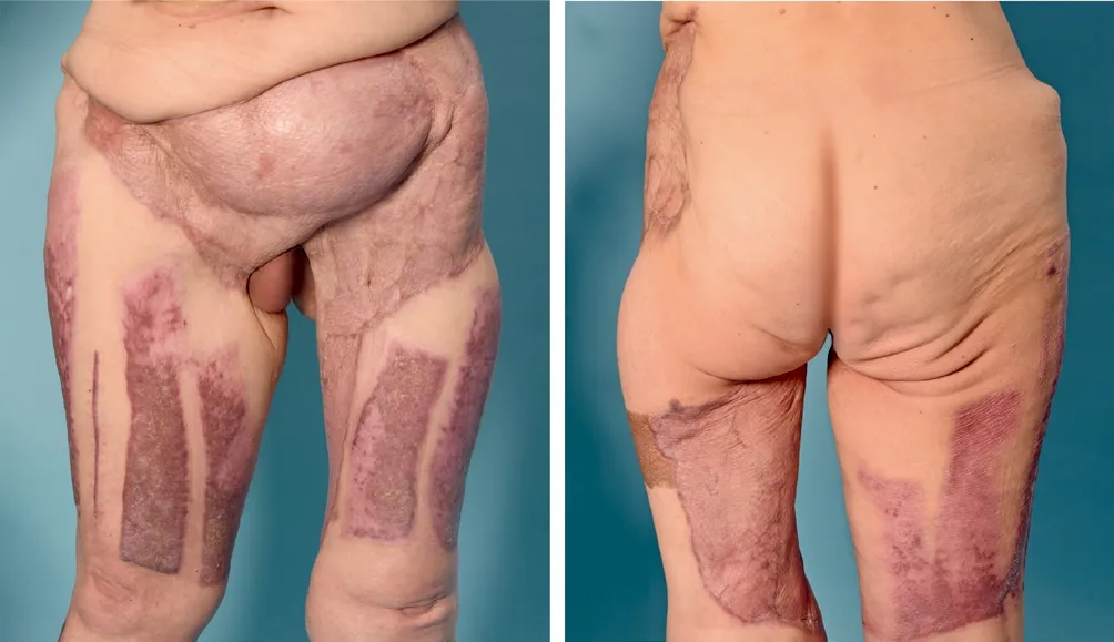
Figure 3. Outcome after four months of discharge (A-B).
After admission, the patient recovered and was discharged from the hospital 113 d later (including 31 d in the intensive care unit).
●While sowing highland barley, mix its seeds with husks. The interpretation of locals is, “ask pests to eat husks instead of the seeds of highland barley”.
DISCUSSION
We report the first case in the literature of polymicrobial necrotizing fasciitis due toand.
Necrotizing fasciitis is a rapidly evolving softtissue infection, characterized by spread along the fascial planes and fascial ischemia.Despite its rarity, and not least because of its mortality, physicians need to consider the diagnosis in case of disproportionate pain to clinical findings, blistering, and symptoms of systemic impairment. The paths for microbial invasion include blunt and penetrating trauma, postoperative complications and insect bite, but the infection may also occur idiopathically.
Early diagnosis and immediate therapeutic measures, namely, intravenous broad-spectrum antibiotics, extensive debridement and further treatment in a specialized burn intensive care unit, are key to increasing the odds of survival, as the time to surgical intervention was shown to be the crucial predictor for survival.Frequent monitoring of all wound margins should initially be performed several times daily to notice disease progression early and allow undelayed countermeasures. Even extensive soft tissue defects with exposed functional or bony structures can be covered when a steady state is achieved in subsequent plastic reconstructive surgeries.
Two cases of monomicrobialnecrotizing fasciitisand one case of polymicrobialnecrotizing fasciitis associated withandhave been reported before in adults. In accordance with our case, all of the affected patients suffered from diabetes mellitus type 2 or type 1 (one case) as a predisposing immunocompromising condition. Diabetes limits micro- and macrovascular circulation, and results in immunodeficiency by impairing of polymorphonuclear leukocyte and monocyte function, predisposing for soft tissue infections and necrotizing fasciitis.In concordance with all known descriptions in literature, the involvement ofin necrotizing fasciitis seems to be linked to diabetes. This assumption is supported by the characterization of the diabetic foot ulcer microbiome by Smith et al, verifying the frequent presence ofin diabetic foot ulcers.The CT scan (Figure 2) showed massive subcutaneous gas collections, pathognomonic for the synergistic toxicity in polymicrobial type I necrotizing fasciitis, as seen innecrotizing fasciitis before.
has been rarely described in human disease; only a few cases of severe systemic infections have been reported, originating most often from a dental abscess.In three out of these five cases, as well as in our case, comorbidities such as chronic alcoholism, smoking, obesity or active malignancy preceded the infection, while in two cases the previous clinical history was unremarkable for immunodeficiency disorders.
The spectrum of pathogens has been shown to influence the course and mortality of necrotizing fasciitis; therefore, the commonly used classification is derived from the bacterial culture results.The disease progression in this case was rather slowly advancing, compared to the usually rapid progression in type II necrotizing fasciitis, resembling the clinical progression of type I necrotizing fasciitis.Type I necrotizing fasciitis often originates from the trunk and may result from a surgical complication.The clinical course and the predisposing immunocompromising diabetes matched the microbiological classification of type I necrotizing fasciitis in this case.
CONCLUSIONS
We report the first case of necrotizing fasciitis involving. Necrotizing fasciitis is a rapidly advancing soft-tissue infection with high mortality, requiring early diagnosis and immediate aggressive antibiotic and surgical intervention.
Funding: None.
Ethic approval: Written informed consent for the publication of the case and the photographs was obtained from the patient before submission.
Conflicts of interests: The authors have no conflict of interest.
Contributors: CI and SK designed the study and collected the patient’s clinical data. All authors contributed substantially to writing and revision of this manuscript. All authors read and approved its contents.
