Bioinformatics prediction of potential mechanisms and biomarkers underlying dilated cardiomyopathy
Zhou Liu,Ying-Nan Song,Kai-Yuan Chen,Wei-Long Gao,Hong-Jin Chen,Gui-You Liang
Zhou Liu,Gui-You Liang,School of Basic Medical Sciences,Guizhou Medical University,Guiyang 550025,Guizhou Province,China
Zhou Liu,Ying-Nan Song,Kai-Yuan Chen,Wei-Long Gao,Hong-Jin Chen,Gui-You Liang,Translational Medicine Research Center,Guizhou Medical University,Guiyang 550025,Guizhou Province,China
Ying-Nan Song,Hong-Jin Chen,Gui-You Liang,Department of Cardiovascular Surgery,the Affiliated Hospital of Guizhou Medical University,Guiyang 510000,Guizhou Province,China
Abstract BACKGROUND Heart failure is a health burden responsible for high morbidity and mortality worldwide,and dilated cardiomyopathy (DCM) is one of the most common causes of heart failure.DCM is a disease of the heart muscle and is characterized by enlargement and dilation of at least one ventricle alongside impaired contractility with left ventricular ejection fraction < 40%.It is also associated with abnormalities in cytoskeletal proteins,mitochondrial ATP transporter,microvasculature,and fibrosis.However,the pathogenesis and potential biomarkers of DCM remain to be investigated.AIM To investigate the candidate genes and pathways involved in DCM patients.METHODS Two expression datasets (GSE3585 and GSE5406) were downloaded from the Gene Expression Omnibus database.The differentially expressed genes (DEGs)between the DCM patients and healthy individuals were identified using the R package “linear models for microarray data.” The pathways with common DEGs were analyzed via Gene Ontology (GO),Kyoto Encyclopedia of Genes and Genomes (KEGG),and gene set enrichment analyses.Moreover,a protein-protein interaction network (PPI) was constructed to identify the hub genes and modules.The MicroRNA Database was applied to predict the microRNAs (miRNAs)targeting the hub genes.Additionally,immune cell infiltration in DCM was analyzed using CIBERSORT.RESULTS In total,97 DEGs (47 upregulated and 50 downregulated) were identified.GO analysis showed that the DEGs were mainly enriched in “response to growth factor,” “extracellular matrix,” and“extracellular matrix structural constituent.” KEGG pathway analysis indicated that the DEGs were mainly enriched in “protein digestion and absorption” and “interleukin 17 (IL-17) signaling pathway.” The PPI network suggested that collagen type III alpha 1 chain (COL3A1) and COL1A2 contribute to the pathogenesis of DCM.Additionally,visualization of the interactions between miRNAs and the hub genes revealed that hsa-miR-5682 and hsa-miR-4500 interacted with both COL3A1 and COL1A2,and thus these miRNAs might play roles in DCM.Immune cell infiltration analysis revealed that DCM patients had more infiltrated plasma cells and fewer infiltrated B memory cells,T follicular helper cells,and resting dendritic cells.CONCLUSION COL1A2 and COL3A1 and their targeting miRNAs,hsa-miR-5682 and hsa-miR-4500,may play critical roles in the pathogenesis of DCM,which are closely related to the IL-17 signaling pathway and acute inflammatory response.These results may provide useful clues for the diagnosis and treatment of DCM.
Key Words: Dilated cardiomyopathy; Bioinformatics; Differentially expressed genes; Function enrichment analysis; Protein-protein interaction network; Immune cell infiltration
lNTRODUCTlON
Dilated cardiomyopathy (DCM) is a progressive myocardial disease.It accounts for 30%–40% of heart failure cases and leads to high mortality worldwide[1].DCM is characterized by biventricular dilatation,cardiac systolic dysfunction,and ventricular remodeling[2].Recently,several studies have reported that mutations,myocarditis,hypertension,and ischemia are the induction factors of DCM[3,4].Increasing evidence shows that various gene mutations and biomarkers are associated with DCM[5-7].Mutations in cytoskeletal proteins,including dystrophin[8] and desmin[9],impair muscular force transmission and thereby contribute to the development of DCM.Mutations in lamin A/C,a nuclear membrane protein,usually cause DCM with atrioventricular block and atrial fibrillation[10].Liet al[11] reported that mutation of aryl hydrocarbon receptor nuclear translocator-like protein 1 (known as BMAL1) plays a critical role in the development of DCM through the regulation of mitochondrial fission and mitophagyviamitochondrial protein B cell leukemia/lymphoma 2 interacting protein 3.Mutations in thin filament regulatory proteins including cardiac troponin T,cardiac troponin I,and α-tropomyosin can cause DCM with systolic dysfunction by reducing fractional shortening and systolic calcium level[12].Moreover,some biomarkers associated with the development of DCM have been reported.For example,syndecan-1 and syndecan-4 may serve as useful biomarkers for predicting adverse cardiovascular events in DCM patients[13,14].Carbonic anhydrase 2 and 3 are associated with heart failure and are potential risk biomarkers for DCM[15],and serum fibroblast growth factor 21 level is linked to the prognosis of DCM[16].
Several studies have sought DCM-related genes and mechanismsviabioinformatic methods and found some meaningful results.Huanget al[17] found that Fos proto-oncogene,AP-1 transcription factor subunit,tissue inhibitor of metalloprotease-1,and serpin family E member 1 may serve as therapeutic targets in DCM.Zhaoet al[18] identified 89 differentially expressed genes (DEGs),mainly enriched in the extracellular matrix and biological adhesion signaling pathways,which may play significant roles in the development of DCM.However,the main cause(s) and pathogenic mechanism(s)underlying DCM are still unknown; thus,DCM is mostly diagnosed late,which causes a poor prognosis in turn.More studies are urgently needed to improve the diagnostic and therapeutic efficiency in DCM.The Gene Expression Omnibus (GEO) database includes many DCM-related microarray data,which have not been fully utilized.These data can be used to identify additional candidate biomarkers and pathways to further explore the cause(s) of DCM.To investigate the candidate genes and pathways involved in DCM patients,we analyzed the two gene expression data sets GSE3585 and GSE5406.Using the “linear models for microarray data” (limma) method,we identified 97 DEGs between healthy individuals and DCM patients.In addition,we identified the mechanisms commonly regulated by the DEGsviaGene Ontology (GO),Kyoto Encyclopedia of Genes and Genomes (KEGG) analyses,and gene set enrichment analyses (GSEA).Moreover,a protein-protein interaction network (PPI) network was applied to identify the hub genes that may contribute to the pathogenesis of DCM and predict the microRNAs (miRNAs) targeting the hub genes.Furthermore,we investigated the pattern of immune cell infiltration in DCM.
MATERlALS AND METHODS
Microarray data extraction from the GEO database
The mRNA expression profiles GSE3585 and GSE5406 in the GEO database (http://www.ncbi.nlm.nih.gov/geo/),which is a shared platform in which researchers deposit their microarray data related to various diseases,were downloaded.The GSE3585 dataset,generated by Barthet al[19],and the GSE5406 dataset,generated by Hannenhalliet al[20],consisted of 7 DCM patients and 5 healthy individuals,and 86 DCM patients and 16 healthy individuals,respectively.In total,114 samples of the left ventricular myocardium,consisting of 93 DCM and 21 healthy samples (control group),were included in this study.
Data processing and DEGs identification
The two datasets GSE3585 and GSE5406 were loaded onto the GPL96 platform (Affymetrix Human Genome U133A Array [HG-U133A]).Additionally,the series matrix and platform annotation for the two databases were downloaded from the GEO database.The probe identity documents were transformed into gene symbols.Then,viathe R package “Surrogate Variable Analysis,” the two databases were merged,and any batch effect was removed using the “Empirical Bayes” method[21].The R package “limma” was applied to identify the DEGs between the DCM and healthy myocardium tissues[22].The screening criteria were set asP< 0.05 and |log fold change (FC)| > 0.589 (FC > 1.5).Volcano and heat maps were generated using R software.
Gene expression enrichment analysis
The gene expression enrichment analysis in this study included GO analysis (https://www.geneontology.org)[23],KEGG (https://www.genome.jp/kegg)[24] pathway analysis,and GSEA (https://www.gsea-msigdb.org/gsea) analysis[25].The DEGs were inputted into Metascape (https://metascape.org)[26]: The species was selected asHomo sapiens; The screening standard was set asP< 0.05; and The GO terms of biological process (BP),cellular component (CC),and molecular function(MF) were analyzed and KEGG pathway analysis was performed with the criteria ofP< 0.05.GSEA interprets the biological function of the expression dataset.The expression dataset in the DCM casesvshealthy tissues was loaded into GSEA 4.0.3 software,set gene sets database as GO gene set(c5.all.v7.1.symbols.gmt),set number of permutations as 1000,set phenotype labels as controlvsDCM,set collapse/remap to gene symbols as no collapse,set permutation type as phenotype,and the other parameters were set at default parameters.Then the GSEA software was used to obtain the enrichment results.The enriched terms were defined as significant with nominalP< 0.05.
PPI network construction and hub gene identification
The PPI network was constructed with the online website Search Tool for the Retrieval of Interacting Genes/Proteins (STRING,https://stringdb.org/cgi/input.pl)[27] to contribute to the understanding of the interactive relationship among DEGs.The DEGs were inputted into this website,species ofHomo sapienswas selected,and the identification criterion was set as combined score > 0.4 (medium confidence).Then the profile of interacting node pairs was imported into Cytoscape (https://cytoscape.org)[28] to visualize the PPI network.The top 10 hub genes were identified with the standard of connectivity degree by using the CytoHubba plugin.The plugin Molecular Complex
Detection (MCODE) was applied to identify the essential module within the PPI network in Cytoscape with the default parameters (degree cutoff,2; node score cutoff,0.2; kcore,2; and maximum depth,100).
Construction of the miRNA-mRNA interaction network
miRNAs,a class of small non-coding RNAs,regulate the expression of various genes by binding to their transcripts and play critical roles in DCM progression[29].By using the MicroRNA Database (miRDB)[30] (http://mirdb.org/),we predicted miRNAs targeting any of the top 10 hub genes.Then,we sorted these miRNAs according to their prediction scores and selected the top 10 miRNAs.The mRNA-miRNA pairs were imported into Cytoscape to visualize the miRNA-mRNA network.
Immune cell infiltration analysis
The CIBERSORT (cibersort.stanford.edu) algorithm was applied to analyze the normalized gene expression data,and the proportions of 22 types of immune cells in each sample were analyzed[31].The gene expression data were normalizedvia“limma” and transformed into the 22 types of immune cell expression data through the source of CIBERSORT[32]viaR.Then the results were filtered outviaPerl (https://www.perl.org) withP< 0.05,and the immune cell infiltration matrix was obtained.Next,the“vioplot” package was used to draw violin diagrams to visualize the difference in immune cell infiltration between the DCM and healthy groups in detail.The “ggplot2”[33] package was applied to perform principal component analysis (PCA) and draw a PCA clustering map.The “corrplot” package was used to analyze the correlation among immune cell infiltration and draw a correlation heatmap.
RESULTS
Identification of DEGs
After merging the two datasets,97 DEGs,including 47 upregulated and 50 downregulated genes,were obtained in the DCM group compared with the control group.Figure 1 shows the volcano map and heatmap of the 97 DEGs.The details of the top 10 upregulated or downregulated genes are shown in Table 1.
GO and KEGG enrichment analyses
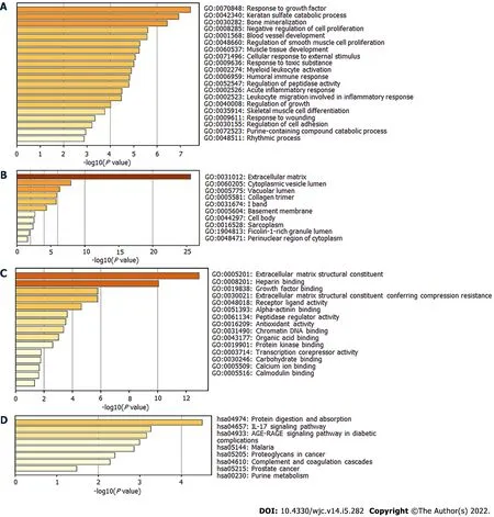
Next,the DEGs were used to perform enrichment analysis for BP,CC,MF,and KEGG pathways.By using the Metascape website,the BP of GO was found to be significantly enriched in “response to growth factor,” “blood vessel development,” “regulation of smooth muscle cell proliferation,” “muscle tissue development,” and “acute inflammatory response” (Figure 2A).The DEGs in CC were mainly enriched in “extracellular matrix,” “cytoplasmic vesicle lumen,” “collagen trimer,” and “sarcoplasm”(Figure 2B).Regarding MF,the DEGs were mainly enriched in “extracellular matrix structural constituent,” “growth factor binding,” “alpha-actinin binding,” and “calcium ion binding” (Figure 2C).KEGG pathway analysis revealed significant pathway enrichment of DEGs in “protein digestion and absorption,” “interleukin 17 (IL-17) signaling pathway,” “advanced glycation end products-receptor for advanced glycation end products signaling pathway in diabetic complications,” “complement,” and“coagulation cascades” (Figure 2D).

Figure 2 Gene Ontology and Kyoto Encyclopedia of Genes and Genomes analysis of enrichment for differentially expressed genes.
GSEA analysis
GSEA analysis results revealed that,compared with the control group,the DCM group was significantly enriched in GO terms “heart development,” “response to ischemia,” “vascular smooth muscle cell differentiation,” “response to transforming growth factor beta,” “stem cell proliferation,” and“regulation of mitochondrial fission” (Figure 3).

Figure 3 Gene set enrichment analyses of enrichment of differentially expressed genes.
PPI network and identification of hub genes
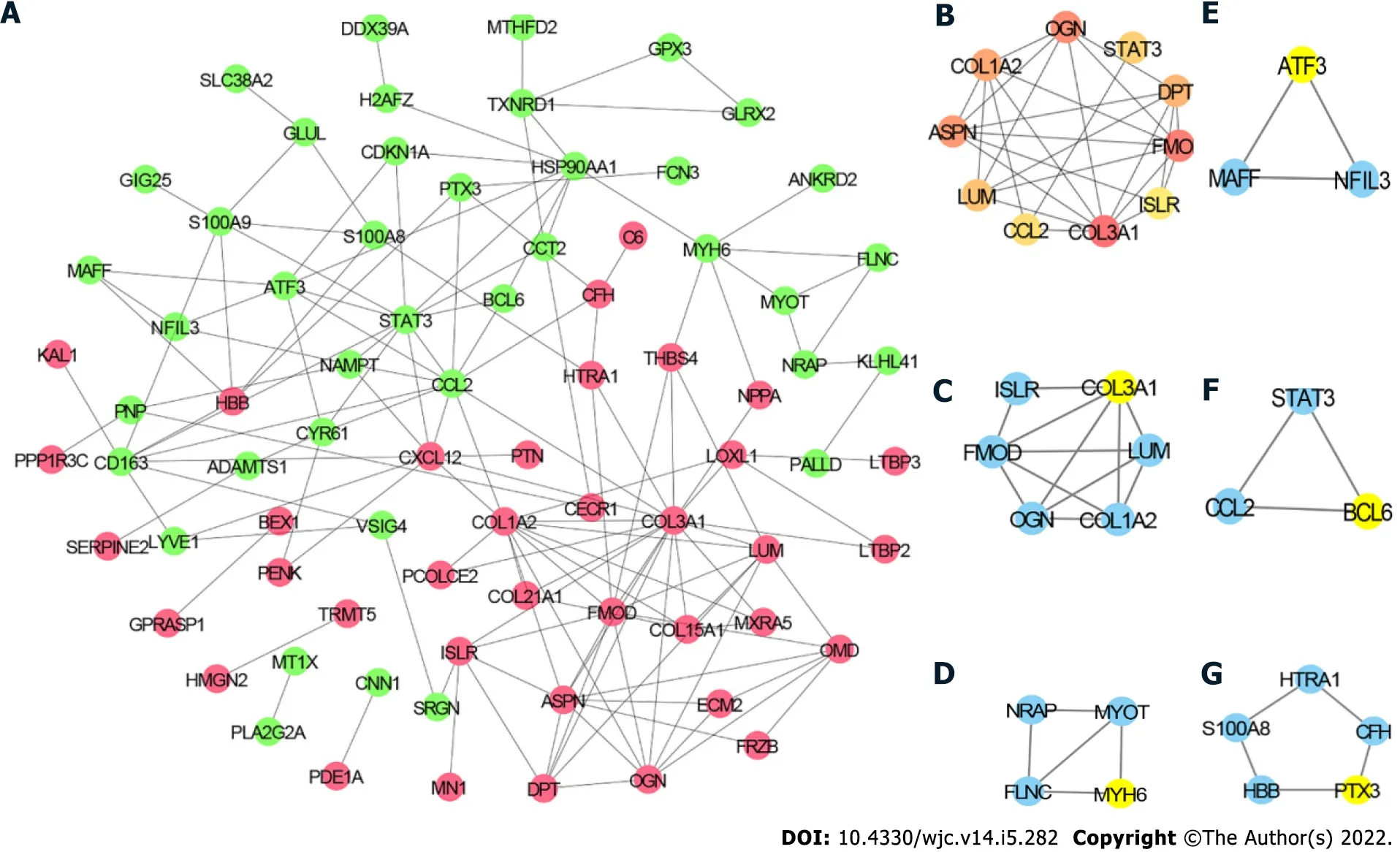
To further explore the relationship among the DEGs at the protein level,the PPI network of the 97 DEGs was constructed using STRING with the criterion of combined score > 0.4 and visualized using Cytoscape.The PPI network consisted of 77 nodes and 145 edges (Figure 4A).The top 10 hub genes included collagen type III alpha 1 chain (COL3A1),COL1A2,signal transducer and activator of transcription 3 (STAT3),C-C motif chemokine ligand 2 (CCL2),fibromodulin (FMOD),asporin (ASPN),C-X-C motif chemokine ligand 12 (CXCL12),lumican (LUM),heat shock protein 90 alpha family class A member 1 (HSP90AA1),and osteoglycin (OGN) (Figure 4B).The detailed information of these hub genes is provided in Table 2.MCODE analysis identified five essential modules,and COL3A1,myosin heavy chain 6,activating transcription factor 3,B cell leukemia/lymphoma 6 transcription repressor,and pentraxin 3 were the seeds of clusters 1,2,3,4,and 5,respectively (Figure 4C–G).

Table 2 Top 10 hub genes in the protein-protein interaction network

Figure 4 Protein-protein interaction network.
miRNA–mRNA interaction network
Increasing evidence shows that miRNAs play important roles in the development and progression of DCM.By using the miRDB database,we predicted miRNAs targeting any of the top 10 hub genes.Then we sorted these miRNAs according to their prediction scores and selected the top 10 miRNAs.Additionally,the top 100 miRNA–mRNA pairs were visualized using Cytoscape (Figure 5).Consequently,hsa-miR-5682,hsa-miR-4500,hsa-miR-32-3p,and hsa-miR-374a-3p were each found to target ≥ 2 hub genes.

Figure 5 MicroRNA-mRNA network.
Immune cell infiltration in DCM
Immune cells infiltrate into the myocardium upon myocardial injury[28].Thus,a violin plot was constructed to investigate the difference in immune-cell infiltration between the DCM and control groups (Figure 6A).Compared with the control group,the DCM group had more infiltrated plasma cells and fewer infiltrated B memory cells,T follicular helper cells,and resting dendritic cells (DCs),whereas there was no significant difference in the remaining 18 types of immune cells.However,the PCA results showed that the control and DCM groups could not be well distinguished according to the infiltration patterns of the 22 types of immune cells (Figure 6B).We generated a correlation heatmap to assess the correlation among the 19 immune cells that were found infiltrated in the DCM or control group.As shown in Figure 6C,the number of infiltrated B memory cells was positively correlated with that of the infiltrated resting DCs,activated natural killer (NK) cells and T follicular helper cells,the number of infiltrated activated NK cells was negatively correlated with that of infiltrated resting NK cells,and the number of infiltrated resting memory CD4 T cells was negatively associated with that of infiltrated B memory cells and regulatory T cells.

Figure 6 lmmune cell infiltration analyses of differentially expressed genes.
DlSCUSSlON
DCM is one of the main reasons of sudden cardiac death and heart failure.It is a heterogeneous disease caused by various types of pathogenic factors including genetic,infectious,hormonal,and environmental factors[34].The causes of DCM should be explored in depth to improve the diagnosis,treatment,and prognosis of DCM patients.Therefore,it is of great significance to elucidate the genetic mechanisms involved in DCM.
In this study,97 DEGs,consisting of 47 upregulated and 50 downregulated genes,were identified between the DCM and control groups.GO of BP and GSEA analysis revealed that the DEGs were not only enriched in the development of the cardiovascular system,such as in the development of the muscle tissue,heart,and blood vessels,but also in the etiology of DCM,such as in acute inflammatory response and mitochondrial fission (Figures 2 and 3).Growing evidence shows that infiltration of inflammatory cells is associated with the pathogenesis of DCM[35-37],and prelamin A accumulation[38] and myosin binding protein C3 mutation[39] can promote DCM pathogenesisviaregulation of inflammation.Xiaet al[40] reported that dynamin-related protein 1 (Drp1,myocardial fission protein) is significantly upregulated in DCM patients.Moreover,Sacubitril (known as LCZ696),a novel inhibitor of the angiotensin receptor neprilysin,can alleviate the cardiac dysfunction in doxorubicin-induced DCM and reduce apoptosis by inhibiting mitochondrial fissionviathe Drp1-mediated pathway.
Regarding CC,the DEGs were enriched in sarcoplasm.Previous studies have reported that mutations in phospholamban (related to abnormal contractility)[41] and B-cell lymphoma 2-associated athanogene 3 (alter the cardiac response)[42] are closely associated with DCM in the sarcoplasmic reticulum.GO analysis of MF indicated that the DEGs were enriched in alpha-actinin binding and calcium ion binding.Alpha-actinin and calcium ion are critical for myocardial contraction[43,44].Other studies have demonstrated that most DCM patients exhibit abnormalities related to calcium ion and α-actinin,which cause decreased heart contractility[45-47].Cardiac troponin contributes to myocardial contraction[48].Mutations in cardiac troponin T,troponin C,and troponin I are mainly related to DCM pathogenesis[49,50]
KEGG pathway analysis showed that the DEGs were significantly enriched in the IL-17 signaling pathway.DCM is induced by viral myocarditis,accompanying autoimmune dysfunction,affecting the secretion of IL-17 cytokine by T helper 17 cells,and IL-17 itself promotes myocardial cell injury[51].Wanget al[52] reported that elevated IL-17 levels are significantly associated with DCM incidence and progression.Additionally,the serum levels of other inflammatory factors such as IL-6,tumor necrosis factor-α,and IL-21 are significantly increased in DCM patients[53].Thus,the IL-17 signaling pathway may be one of the major signaling pathways involved in the development of DCM.
Through construction of PPI and miRNA-mRNA interaction networks,we identified the hub genes and the miRNAs targeting them.The hub genes,COL1A2 and COL3A1,encode the pro-alpha2/1 chains of type I and III collagens,respectively.Collagens I and III,the main collagens of cardiac extracellular matrix,are classical biomarkers of cardiac fibrosis in DCM[54].Mihailoviciet al[55] reported that collagens I and III are upregulated in DCM patients compared with matched healthy controls.Additionally,Zhaoet al[18] reported that COL1A2 may participate in DCM pathogenesis by regulating the cardiac remodeling characterized by collagen deposition in the extracellular matrix[56].Consistent with our results,Liuet al[53] identified STAT3 as a hub gene in DCMviabioinformatic analysis.Other studies have also indicated a role of STAT3 in DCM.Podewskiet al[57] showed that STAT3 protein level is significantly decreased in the cardiomyocytes of patients with end-stage DCM.Moreover,inhibition of the IL-6–mediated STAT3 signaling pathway can improve myocardial remodeling through reducing myocardial apoptosis in a mouse model of DCM[58].Thus,the roles of COL1A2,COL3A1,and STAT3 in DCM should be further investigated.Moreover,the identified miRNAs hsa-miR-5682,hsa-miR-4500,hsa-miR-32-3p,and hsa-miR-374a-3p may participate in DCM pathogenesis through their interaction with ≥ two hub genes.Previous studies have also suggested that miRNAs play significant roles in DCM.It has been found that miR-21,miR-29a,and miR-133b are differentially expressed in DCM patients[59].miR-133a expression is associated with fibrosis,myocyte necrosis,left ventricular function,and clinical outcome in patients with inflammatory DCM[60].Moreover,Satohet al[61] showed that a low let-7i level can serve as an independent predictor of cardiac death and heart failure (relative risk = 3.76).
Immune cells commonly infiltrate into the myocardium upon various types of cardiac damage[49,62].Overactivation of immune cells could be investigated in pathological examination about cardiac biopsy specimens in DCM patients.Noutsiaset al[63] reported that the upregulation of genes associated with T cells exacerbates DCM progression.Therefore,our study also assessed the correlation between DEGs and immune cell infiltration.The results indicated infiltration of 19 types of immune cells in DCM pathogenesis.Notably,compared with the control group,the DCM group had more infiltrated plasma cells and fewer infiltrated B memory cells,T follicular helper cells,and resting DCs.However,Liuet al[53] have demonstrated that,compared with healthy controls,DCM patients have more infiltrated T follicular helper cells and fewer T follicular regulatory cells,and infiltration of T follicular regulatory cells is positively correlated with left ventricular ejection fraction.
Our study had some limitations.First,the gene expression data were acquired from a public database.Moreover,we did not experimentally verify the relevance of the identified DEGs with DCM and their enriched functions or hub genes.Likewise,we did not verify the predicted miRNA-mRNA interactions and their relevance.
CONCLUSlON
In summary,we first identified that COL1A2 and COL3A1 may be both presumably regulated by hsamiR-5682 and hsa-miR-4500,and play significant roles in the pathogenesis of DCM through acute inflammatory response and IL-17 signaling pathway.These results may provide useful biomarkers for the diagnosis and treatment of DCM,but further research is needed to clarify the roles of the predicted genes and pathways.
ARTlCLE HlGHLlGHTS
Research background
Dilated cardiomyopathy (DCM),a disease of the heart muscle,is one of the most common causes of heart failure.However,the original cause and pathogenesis in development of DCM are still remain elusive.
Research motivation
The early diagnosis and prognosis of DCM patients are unsatisfactory because of DCM main cause and pathogenesis are still unclear.Increasing DCM datasets were provided online but little was been explored.Bioinformatics could further investigate the DCM mechanism and biomarkers for improving the diagnostic and therapeutic efficiency.
Research objectives
This study investigated the candidate genes and pathways involved in DCM patients.
Research methods
Expression datasets were downloaded from the Gene Expression Omnibus database.Gene Ontology,Kyoto Encyclopedia of Genes and Genomes,and gene set enrichment analyses investigated the key pathway in differentially expressed genes (DEGs) between the DCM patients and healthy individuals.Protein-protein interaction network identified the hub genes and modules in DCM.MicroRNA Database predicted the microRNAs which targeting the hub genes.CIBERSORT analyzed the immune- ell infiltration in DCM.
Research results
Ninety-seven DEGs mainly enriched in “response to growth factor,” “extracellular matrix,” and“extracellular matrix structural constituent.” Moreover,the top two pathways were “protein digestion and absorption” and “interleukin 17 signaling pathway.” Collagen type III alpha 1 chain (COL3A1) and COL1A2,whose were regulated by hsa-miR-5682 and hsa-miR-4500,mainly contributed to the pathogenesis of DCM.Compared with the control group,DCM patients had more infiltrated plasma cells and fewer infiltrated B memory cells,T follicular helper cells,and resting dendritic cells.
Research conclusions
DCM progression closely related to IL-17 signaling pathway and acute inflammatory response.COL1A2 and COL3A1 and their targeting miRNAs,hsa-miR-5682 and hsa-miR-4500,are the potential biomarkers of DCM.
Research perspectives
This study may provide valuable pathways and biomarkers for the diagnosis or treatment of DCM.Further studies should investigate the functions of the predicted genes and pathways.
FOOTNOTES
Author contributions:Liu Z and Song YN designed this study; Chen KY collected the relevant data; Liu Z analyzed the data; Liu Z and Gao WL drafted the manuscript; Chen HJ and Liang GY reviewed and supervised this manuscript; All authors approved the final version of the article.
Supported byNational Nature Science Foundation of China,No.81960051,No.8217021743,and No.82160060;Project of High–Level Innovative Talents of Guizhou Province,No.[2016]4034; and Construction Funding from Characteristic Key Laboratory of Guizhou Province,No.[2021]313.
Conflict-of-interest statement:The authors have no conflicts of interest to declare.
Data sharing statement:No additional data are available.
Open-Access:This article is an open-access article that was selected by an in-house editor and fully peer-reviewed by external reviewers.It is distributed in accordance with the Creative Commons Attribution NonCommercial (CC BYNC 4.0) license,which permits others to distribute,remix,adapt,build upon this work non-commercially,and license their derivative works on different terms,provided the original work is properly cited and the use is noncommercial.See: https://creativecommons.org/Licenses/by-nc/4.0/
Country/Territory of origin:China
ORClD number:Zhou Liu 0000-0002-6773-1073; Ying-Nan Song 0000-0003-4464-0512; Kai-Yuan Chen 0000-0002-0375-1091; Wei-Long Gao 0000-0002-0464-970X; Hong-Jin Chen 0000-0002-0513-9584; Gui-You Liang 0000-0002-4555-9102.
S-Editor:Ma YJ
L-Editor:Filipodia
P-Editor:Ma YJ
 World Journal of Cardiology2022年5期
- World Journal of Cardiology的其它文章
- Comparative efficacy and safety of adenosine and regadenoson for assessment of fractional flow reserve: A systematic review and meta-analysis
- Day-to-day blood pressure variability predicts poor outcomes following percutaneous coronary intervention: A retrospective study
- Pledget-assisted hemostasis to fix residual access-site bleedings after double pre-closure technique
- Same day discharge after structural heart disease interventions in the era of the coronavirus-19 pandemic and beyond
