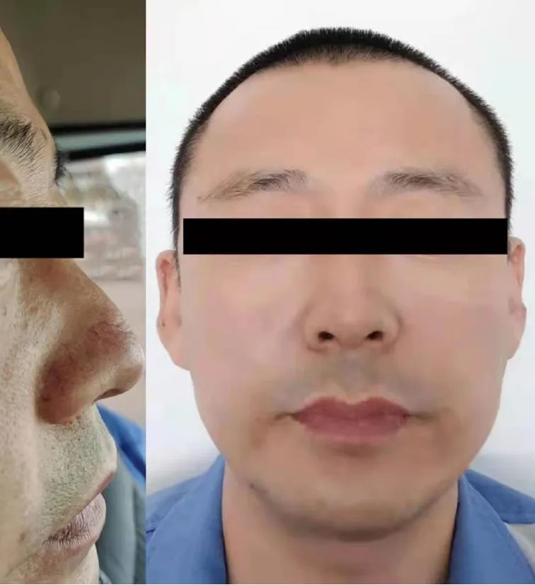
Multiple gouty tophi in the head and neck with normal serum uric acid:A case report and review of literatures
INTRODUCTION
Gout is a clinical syndrome caused by an increase in uric acid production or a weakening of renal excretion function,resulting in a continuous increase in serum uric acid (SUA) level and the deposition of urate in joints,synovium,or other tissues and organs[1,2].Consequently,complications including gouty arthritis,gouty tophus,uric acid renal stones,and gouty nephropathy may develop.Gouty tophus is a sign of chronic gout,and its formation can lead to bone destruction,joint deformity,joint dysfunction,and even fracture and infection of the tophus[3],which can seriously affect the quality of life of patients.The formation of gouty tophus is a complex process,and many factors can lead to and accelerate its formation.The gold standard for the diagnosis of gout is the detection of birefringent acicular urate crystals in joint fluid or gouty tophus under a polarised light microscope[4].
And that day the slaves in their black robes, and each having a large bat perched upon his head, marched in slow procession with the Prince in their midst, chanting a melancholy62 song, to the iron gate that led into a kind of Temple
An increased SUA level is considered as the basis for gout.When a patient presents with an SUA level>7 mg/dL[5,6],characteristic arthritis,urinary calculi,or renal colic,the clinical diagnosis of gout can be considered.However,most patients with hyperuricemia (HUA) do not develop gout[7].Further,several patients have normal SUA levels at gout onset and some gout patients have gouty tophus with no acutegout attack or gouty tophus before the attack[7,8],potentially leading to clinical misdiagnosis and mistreatment.Herein,we report a case of multiple gouty tophi in the head and neck,specifically in the auricle and bridge of the nose,with normal SUA level.
CASE PRESENTATION
Chief complaints
A 48-year-old male patient was admitted to the hospital with a chief complaint of ‘recurrent nasal swelling and pain for 3 years’.
History of present illness
Since 2016,the nasal swelling and pain were mild,without nasal congestion,runny nose,epistaxis,fever,headache,trauma,or mosquito bites.In recent years,regular outpatient reviews of SUA had been normal.Drinking alcohol and high purine diet could occasionally aggravate nasal symptoms.
History of past illness
Thirteen years ago,the patient was admitted to another hospital due to swelling and tingling of ankle joints and was diagnosed with gout and hyperuricemia.He regularly received febuxostat.The patient’s SUA level had been regularly reviewed in the outpatient department,and was well controlled for several years (Figure 1).The patient had no history of diabetes mellitus,hypertension,cystic fibrosis and metabolic disorders.
With the new environment, the girl learnt sign language and started a new life. She told herself every day that she must forget the guy. One day, her friend visited her and told her that he was back. She asked her friend not to let him know what happened to her. Since then, there wasn t any more news of him.

Personal and family history
The patient was a male,48 years old,reported no history of dental surgery,facial trauma,or previous sinus surgery.
Physical examination
The gold standard for the diagnosis of gout is the detection of birefringent acicular urate crystals in joint fluid or gouty tophus under a polarised light microscope.In China,HUA is defined as the condition of a normal purine diet,in which the fasting SUA level is higher than 420 μmol/L (7 mg/dL) in men and 360 μmol/L (6 mg/dL) in women.Nonetheless,the definition of HUA varies widely across published studies in different countries,ranging from 6 to 7 mg/dL[13],and the clinical diagnosis of gout should be considered with the onset of characteristic arthritis,urinary calculi,or renal colic occurrence.In the chronic stage of gout,X-ray and conventional CT examinations can better reveal bone and joint destruction in patients,and can show the characteristic chisel-like and worm-like changes with high specificity;however,they are only suitable for the evaluation of bone destruction in patients with late gout.Magnetic resonance imaging (MRI) has shown that in the early stages of gout,though crystal deposition cannot be observed with the naked eye,the display level is clear,diagnostic sensitivity is high,and the diagnostic value is favourable on MRI.However,because of the high cost and long appointment period,MRI is rarely used in clinical practice.In recent years,the application of dual-energy CT (DECT) in the diagnosis and treatment of gout has become a popular research topic[14-16].It can identify the chemical composition of urate crystals and the deposition of urate in deep tissues,which can be applied to a patient’s whole body.With the improvement in ultrasound resolution,ultrasound can be used to observe urate crystals,gouty tophus,and bone erosion damage as well as evaluate joint inflammation[17,18].It has become an effective means of diagnosing gout and monitoring the effect of reducing uric acid levels.

Laboratory examinations
There was no recurrence in nearly 2 years after operation (Figure 5).
The essence of gout inflammation is the deposition of monosodium urate (MSU) in bone,joint,kidney,and subcutaneous tissue,which leads to tissue damage and inflammation[9].The development of inflammation depends on changes in the surface protein for MSU crystals.Repeated inflammation depends on the innate immune response mediated by the MSU crystal.Gouty tophus is a granulomatous substance formed by urate crystals encapsulated by monocytes and multinucleated giant cells.Gouty tophus is mainly composed of three layers[3]:(1) MSU crystals forming the centre of the gouty tophus;(2) Monocytes and multinucleated macrophages that are wrapped around the MSU crystal;and (3) Dense connective tissue constituting the outermost layer.Age,sex,genetic susceptibility,SUA level,disease duration,metabolic syndrome,lifestyle,drugs,high purine diet,and drinking are risk factors for gout[10-12].Gouty tophus is a sign of chronic gout.The formation of gouty tophus can lead to bone destruction,joint deformity,joint dysfunction,fracture,and infection of gouty tophus,seriously affects the quality of life of patients.
Imaging examinations
And that is the life drama that passes before the old maid whileshe looks out upon the rampart, the green, sunny rampart, where thechildren, with their red cheeks and bare shoeless feet, arerejoicing merrily, like the other free little birds.

FINAL DIAGNOSIS
The patient underwent nasal lumpectomy and autologous cartilage graft repair under general anaesthesia.During the operation,the skin and subcutaneous tissues were incised.The deep capsule of the mass was incomplete,the left nasal bone compressed and absorbed,the bone defect obvious,and the surface of the right bone not smooth.The tumour was completely removed along the residual bone.Its contents showed yellowish silt-like changes,and 0.5 mL of yellowish viscous caseous secretions were extracted from the tumour.Nasal endoscopy detected no fistula in the nasal cavity.The operative region was flushed with normal saline,and no liquid flowed out of the nose.Cartilage from the left tragus was taken to repair the nasal bone defect,and the skin was continuously sutured subcutaneously with 4-0 absorbable protein thread and bandaged under pressure.Postoperatively,the patient received anti-infection treatment and was advised to relinquish drinking,pay attention to a gout-suitable

TREATMENT
The accurate diagnosis of nasal gouty tophus was confirmed by postoperative histopathological examination (urate crystallized and granulation tissue formed around the tumour) (Figure 4).
diet,and engage in active follow-up at the internal medicine department for the regulation of SUA levels.
OUTCOME AND FOLLOW-UP
Laboratory examination revealed a uric acid level of 384 μmol/L (reference value range 208-428 μmol/L).The water sample secretion of rice swill was punctured from the local uplift,and general bacterial and fungal cultures showed no abnormal flora.The patient declined invasive cytological examination.
“Now, how would you like a chance to earn a few easy straws like the rest of us? I still have the name I picked tonight in my pocket, and I haven’t looked at it yet. Why don’t we switch, just for the last day? It will be our secret.”

DISCUSSION
A quiet peace filled Trint s heart. He was lucky guy. He had a job he loved, Melinda s phone number in his pocket, clear weather and miles of open road ahead.
Skin swelling with a diameter of 2 cm on the bridge of the nose,obvious tenderness,and nasal deformity.Under the nasal endoscope,the nasal cavity was unobstructed,the nasal septum was in the centre,no obvious bulge or neoplasm was observed in the top wall of the nasal cavity (Figure 2).A new greyish-white creature with a diameter of 5 mm was observed on the outer upper edge of the right auricle.
The current patient's SUA level was 384 μmol/L (in China,the reference value is 208–428 μmol/L).He took febuxostat regularly for teen years,and his SUA level was regularly reviewed in the outpatient department.The uric acid level was well under control for several years.When he was diagnosed with gout 13 years ago,gouty tophi appeared in his lower limbs,feet,and ankles.Although his SUA level was reduced to normal by drug treatment,he drank alcohol for a prolonged period and enjoyed consuming seafood with high purine.The patient was convinced that the gout condition was well under control,in recent years,the symptoms of foot and ankle gouty tophus had not been aggravated,and because the occurrence of gouty tophus in the head and neck is extremely rare,the nasal gouty tophus was not considered the main diagnosis before the operation.Nasal ultrasound examination was not performed before the operation,and the patient refused the puncture cytology examination;therefore,it was impossible to make a definite diagnosis before the operation.A CT scan of the nose revealed bone erosion of the tumour,and considering the possibility of a malignant tumour of the nose[19],we did not perform an external inverted-V incision along the columella,in case of residual tumour.Subsequent to complete exposure of the tumour during the operation,the capsule could be observed surrounding the tumour.There was sand-like filling and rice swill water liquid in the tumour;we considered it a gouty tophus infection that caused exudate formation,although the bacterial and fungal cultures were negative[10].The diagnosis of gouty tophus lesions was confirmed during the operation and by postoperative pathology.The auricle lesions were mild,and surgery was not performed.Therefore,attention should be directed to the formation of tumours in atypical parts of the body and atypical chondritis in gout patients without HUA as well as to the establishment of a clinical understanding of gouty tophus to prevent misdiagnosis[20].
Enhanced computed tomography (CT) imaging revealed a mixed density mass shadow on the left side of the nose (Figure 3).
Most certainly, said the little tailor: just you take the trunk on your shoulder; I ll bear the top and branches, which is certainly the heaviest part
Gouty tophus can be deposited in different parts of the human body,which can be categorised into typical and atypical parts[21].Gouty tophus in the head and neck is atypical and rarely observed in the bridge of the nose[22].In addition,gout often mimics the process of malignant tumours,infections,or other unrelated diseases.A few reports have described the deposition of gout in unusual body parts[23-26].Therefore,a more systematic,scientific,and comprehensive diagnosis of gout is necessary.In 2015,the American College of Rheumatology/European Alliance Against Rheumatology developed new classification criteria for gout[27];ultrasound and DECT were included in the gout classification for the first time.If patients met the diagnostic criteria of clinical,laboratory,and imaging examinations,the sensitivity and specificity of diagnosis could be as high as 92%,which also stratified the level of SUA,taking into account that SUA level may not be high during a gout attack.After a definite diagnosis,we further emphasise that drug treatment of patients with gout is particularly important[28-30].The recommended serum uric acid level is below 6 mg/dL in all gouty patients or 5 mg/dL in severe gout patients to allow more rapid dissolution of the crystals[23].
Why a multiple gouty tophi in the head and neck with normal serum uric acid have been developed in this patient? The possible reason is that although the blood uric acid decreased to normal through drug treatment,the gouty tophus symptoms did not stop developing with the normal blood uric acid due to long-term drinking and eating seafood high purine diet,and gradually occurred in the auricle and nasal,which did not attract enough attention from the patient.Another important reason is that although the patient's blood uric acid is in the normal range,it is still not low enough.The blood uric acid of patients with gout stone should be controlled at 5 mg/dL.
CONCLUSION
This report describes a case of gouty tophus with normal SUA in an atypical location and atypical symptoms.In clinical settings,especially for patients with normal SUA,the possibility of atypical symptoms and atypical parts of gouty tophus should be considered.Strict control of diet,drinking habits,and SUA levels are needed to avoid the progression of gouty tophus and the development of more serious complications.Surgery is an effective treatment method for gouty tophus.
We would like to thank all the participating departments for their cooperation.
 World Journal of Clinical Cases2022年4期
- World Journal of Clinical Cases的其它文章
- Surgical treatment of acute cholecystitis in patients with confirmed COVID-19:Ten case reports and review of literature
- Rituximab as a treatment for human immunodeficiency virusassociated nemaline myopathy:What does the literature have to tell us?
- Eustachian tube involvement in a patient with relapsing polychondritis detected by magnetic resonance imaging:A case report
- Endoscopic clipping for the secondary prophylaxis of bleeding gastric varices in a patient with cirrhosis:A case report
- Inflammatory myofibroblastic tumor after breast prosthesis:A case report and literature review
- Langerhans cell histiocytosis presenting as an isolated brain tumour:A case report
