Gut microbiota and immune system in liver cancer:Promising therapeutic implication from development to treatment
Ilenia Bartolini,Matteo Risaliti,Rosaria Tucci,Paolo Muiesan,Maria Novella Ringressi,Antonio Taddei,Amedeo Amedei
Ilenia Bartolini,Matteo Risaliti,Rosaria Tucci,Paolo Muiesan,Maria Novella Ringressi,Antonio Taddei,Department of Experimental and Clinical Medicine,University of Florence,Azienda Ospedaliero Universitaria Careggi(AOUC),Florence 50134,Italy
Amedeo Amedei,Department of Experimental and Clinical Medicine,SOD of Interdisciplinary Internal Medicine,Azienda Ospedaliero Universitaria Careggi(AOUC),Florence 50134,Italy
Abstract Liver cancer is a leading cause of death worldwide,and hepatocellular carcinoma(HCC)is the most frequent primary liver tumour,followed by cholangiocarcinoma.Notably,secondary tumours represent up to 90% of liver tumours.Chronic liver disease is a recognised risk factor for liver cancer development.Up to 90% of the patients with HCC and about 20% of those with cholangiocarcinoma have an underlying liver alteration.The gut microbiota-liver axis represents the bidirectional relationship between gut microbiota,its metabolites and the liver through the portal flow.The interplay between the immune system and gut microbiota is also well-known.Although primarily resulting from experiments in animal models and on HCC,growing evidence suggests a causal role for the gut microbiota in the development and progression of chronic liver pathologies and liver tumours.Despite the curative intent of “traditional” treatments,tumour recurrence remains high.Therefore,microbiota modulation is an appealing therapeutic target for liver cancer prevention and treatment.Furthermore,microbiota could represent a non-invasive biomarker for early liver cancer diagnosis.This review summarises the potential role of the microbiota and immune system in primary and secondary liver cancer development,focusing on the potential therapeutic implications.
Key Words:Gut microbiota;Immune system;Liver cancer;Primary liver cancer;Colorectal liver metastasis;Liver cancer treatment
INTRODUCTION
Primary liver cancer is a leading cause of death worldwide.Hepatocellular carcinoma(HCC)is the most common primary liver tumour,accounting for about 80% of the cases.Cholangiocarcinoma(CCA)is the second primary liver tumour,representing approximately 15% of the malignancies[1].Finally,the secondary tumours represent up to 90% of liver tumours,and the liver is the most frequent metastatic site[2].
Chronic liver disease is a recognised risk factor for HCC.Up to 90% of the patients with HCC have an underlying liver alteration,and about 30% of the patients with cirrhosis will suffer from HCC[3].Several pathologies may cause liver cirrhosis,including viral hepatitis,alcohol abuse,diabetes and non-alcoholic fatty liver disease(NAFLD)[4].Although most CCAs occur without any specific predisposing factors,about 20% of the patients harbour some of the same causal pathologies as HCC[5].
The gut microbiota consists of the biological community of bacteria,Archaea,fungi and viruses harboured within a host showing a commensal,symbiotic or pathogenetic attitude[6,7].The liver is exposed to both microbiota and microbial metabolites through the portal flow.There is a bidirectional relationship between gut microbiota and liver through signalling-sensing pathways,known as the “gut microbiota-liver axis”[3,7,8].
The intestinal barrier has a crucial role in preserving the host from the environment.Intestinal barrier and gut microbiota can influence each other in a dynamic process,and alterations in each of them may impair this balance[9].
The interplay between the immune system and gut microbiota is also well-known.The liver immune system is a dynamic microenvironment subjected to changes related to the received stimuli[10].In the liver,there are cells of the innate immune system,including Kupffer cells,natural killer cells,natural killer T(NKT)cells and cells belonging to the adaptive immune system,including T lymphocytes[11].The γδT cells are unconventional T lymphocytes and act as a bridge between innate and adaptive immunity.γδT cells are getting attention due to their pleiotropy and their potential causal or beneficial role in the different aspects of tumour progression[12,13].
Metagenomic analysis,polymerase chain reaction and 16S ribosomal RNA sequencing can identify bacteria and their products[14].Although primarily resulting from experiments in animal models and on HCC,growing data suggest a causal role for the gut microbiota in the development and progression of both chronic liver pathologies and tumours[3].
Several guidelines propose the best treatment strategies for liver cancer.Traditional treatments include surgery,transplantation,locoregional therapies,chemotherapy or chemoradiotherapy[15].Despite curative treatments,tumour recurrence remains high.Therefore,gut microbiota modulation may represent a promising therapeutic target for liver cancer prevention and therapy[7].Since complete prevention is not achievable,as with any other cancer,an early diagnosis may allow better patient outcomes,and the gut microbiota may represent a novel,non-invasive biomarker[16-18].
This review aims to provide the actual state of the art of the potential role of microbiota-immunity in every step of both primary and secondary liver cancer,focusing on the potential therapeutic implications.
CANCER DEVELOPMENT
Different microbial composition in health and disease
Several studies reported the progressive increase in the number of pathogenic bacteria with the decrease of those showing a healthy behaviour and the different stages of chronic liver disease and HCC development[7,19].Furthermore,faecal biodiversity seems to decrease along with the cirrhosis progression,but it seems to increase again along with early HCC progression[20].Tables 1 and 2 summarise the most important changes in chronic liver disease and HCC,respectively.

Table 1 Different microbial composition in healthy and disease — chronic liver disease

Table 2 Different microbial composition in healthy and disease — hepatocellular carcinoma
Studies in humans reported a significant difference between the gut microbiota of patients with chronic viral hepatitis and healthy volunteers.In particular,a significant decrease in the number ofAlistipes,Bacteroides,Asaccharobacter,Butyricimonas,Ruminococcus,Clostridium cluster IV,ParabacteroidesandEscherichia/Shigellawas found together with a significant increase in the number ofMegamonas,unclassifiedLachnospiraceae,Clostridium sensu strictoandActinomycesin patients with chronic hepatitis B[21].
Patients with chronic hepatitis C presented in their stool samples a lower bacterial diversity,a higher presence ofStreptococcus,LactobacillusandBacteroidetes,and a lower presence of Clostridiales andBifidobacterium[22].
Alcoholic patients showed an increased number of gram-negative bacteria[6],but also the contributing role of theEnterococcus faecalisin alcoholic hepatitis has been described[23].
Dysbiosis has also been found in patients with NAFLD,though different studies reported a different relative abundance of bacteria in the gut of this subgroup of patients[3].For example,compared with healthy controls,NAFLD patients’ microbiota presented enriched inProteobacteriaandFusobacteriawith higher representations of the bacteria belonging to the familyErysipelotrichaceae,Enterobacteriaceae,LachnospiraceaeandStreptococcaceae.An increased number ofEscherichiaandShigellawere also found together with a reduced number ofPrevotella[24].
Independently from the aetiology,patients with cirrhosis presented with a progressively lower number of bacteria from the family ofLachnospiraceae,together with increased levels of the bacteria belonging to the familiesEnterobacteriaceae,VeillonellaceaeandStreptococcaceae.The last two families are typically found in healthy people’s oral microbiota and can colonise cirrhotic patients’ guts[25,26].In particular,the number ofStreptococcusseems to correlate with the Child-Pugh score directly.In contrast,the number of bacteria belonging to the family ofLachnospiraceaeappears to correlate inversely with the Child-Pugh score[26].
Patients with hepatitis B-related HCC resulted in having a higher level of proinflammatory bacteria,includingEscherichia,Shigella(Enterobacteriaceae)andEnterococcus,with a reduced amount ofFaecalibacterium,RuminococcusandRuminoclostridiumcompared with healthy subjects[27].
The presence ofVeillonella parvulaandBacteroides caecimurisseems to allow the differentiation of patients with NAFLD-related HCC from those with NAFLD-related cirrhosis only[28].
In a murine model of non-alcoholic steatohepatitis-induced HCC,Clostridium,Corynebacterium,Bacillus,Desulfovibrio,andRhodococcuswere highly represented in male mice and were associated with a higher risk of HCC development[29].
A particular abundance of bacteria,includingClostridiumand CF231,were uniquely observed in HCC patients,independently from the cirrhosis stage or other environmental factors[30].
Interestingly,Western and Eastern people showed different gut microbiota,but they shared similar pathogenic microbial signatures[31].Luet al[17]analyzed the tongue coating microbiota in cirrhosis-related HCC patients.They found significantly higher biodiversity and dysbiosis in patients compared with healthy controls.Epsilonproteobacteria,Actinobacteria,Clostridia andFusobacteriawere increased in the patients,while there was a higher presence of Gammaproteobacteria andBacteroidetesin the volunteers.In particular,the number ofFusobacteriumandOribacteriumseems to differentiate HCC patients from healthy people[17].
Data about CCAs have been rarely reported.Lactobacillus,Actinomyces,AlloscardoviaandPeptostreptococcaceaeincreased in stool samples from patients with intrahepatic CCA compared to those with HCC or healthy people.Furthermore,the overgrowth of bacteria belonging to the family of theRuminococcaceae,together with higher levels of interleukin-4(IL-4)and lower levels of IL-6,correlated with vascular invasion and,thus,with patients’ prognosis.Furthermore,LactobacillusandAlloscardoviaare directly related to the tauroursodeoxycholic acid levels,and tauroursodeoxycholic acid levels showed a negative association with survival[1].
In a small study on bile samples taken during endoscopic retrograde cholangiopancreatography of patients with extrahepatic CCA,first episodes of bile duct stones and recurrent bile duct stones,Chenet al[32]showed a significant increase in the presence of Gemmatimonadetes,Latescibacteria,Planctomycetes and Nitrospirae in the patients with extrahepatic CCA.At the same time,they were absent in patients with the first episode of bile duct stones.
Similarly,in another different study on bile samples of patients with gallbladder cancer and gallbladder lithiasis,Tsuchiyaet al[33]found the predominance ofFusobacterium nucleatum,Escherichia coliandEnterobacterspecies in cancer patients and a predominance ofEscherichia coli,Salmonellaspecies andEnterococcus gallinarumin the patients with gallbladder lithiasis.
Despite these data,it remains challenging to assess whether these modifications in microbiota composition are related to liver disease rather than the medications used in these patients[3].Furthermore,some results may appear conflicting.Many reasons could explain these differences,including:(1)The different study models(in vitro,animal,human);(2)The influence of the environment,diet,lifestyle,and,eventually,other comorbidities;(3)The different methods to take and manage the samples;(4)The potential confounding effects of known or unknown factors;and(5)The relationship between testable and untestable microbiota.
Further large-scale human studies are needed.Complete knowledge of the relationship between progressive microbiota modifications and tumour initiation/progression may represent the basis for attractive therapeutic options,mostly for tumour prevention or,at least,for early diagnosis[7].
Pathogenetic pathways
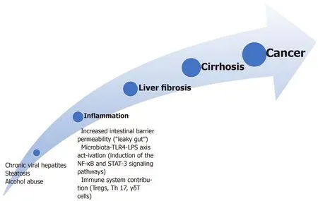
Inflammation-liver fibrosis-cirrhosis-cancer:The pathway comprehending inflammation-liver fibrosis-cirrhosis-cancer is one of the most commonly recognised for HCC development[7].Figure 1 summarises this pathway.On the contrary,most studies reported these alterations as a protective factor for liver metastasis development.However,some papers showed similar pathogenesis for both primary and secondary liver cancers[34].

Figure 1 The inflammation-liver fibrosis-cirrhosis-cancer pathway.Dysbiosis,alcohol abuse and high-fat diet cause the alteration of the intestinal barrier permeability.With increased gut permeability,both microbiota and toxins(e.g.,endotoxins or flagellin)may reach the liver through the portal vein stimulating an inflammatory reaction.The toll-like receptor(TLR)4 is expressed in the Kupffer,hepatic stellate,endothelial cells and hepatocytes.TLR4 activation causes the upregulation of the epidermal growth factor epiregulin that shows a mitogenic effect on hepatocytes causing hepatocellular carcinoma promotion.The lipopolysaccharide,a component of the gram-negative bacteria wall,binds to the transmembrane TLR4 causing the expression of the hepcidin showing an antiapoptotic effect on the hepatocytes via the activation of the nuclear factor-κB and signal transducer and activator of transcription 3 signalling and the production of interleukin(IL)-17,IL-6,IL-1β,and tumour necrosis factor-α.Regulatory T cells can suppress the host antitumor immunity and cause tumour progression worsening CD8+ T cells function.T helper 17 cells showed pro-inflammatory effects through the secretion of IL-17A and IL-22 while γδT cells show pleiotropic activities.TLR:Toll-like receptor;LPS:lipopolysaccharide;NF:nuclear factor;Tregs:Regulatory T cells;Th:T helper;STAT-3:Signal transducer and activator of transcription 3.
In addition,bacterial dysbiosis causes a higher release of inflammatory cytokines and an increased intestinal barrier permeability[35].
Finally,there are growing data about the role of the microbiota toll-like receptor(TLR)4 axis and of the lipopolysaccharide(LPS)-TRL4 axis in the development of inflammation and liver fibrosis from experimental and in clinical settings[36-38].
The TLR4 is expressed in the Kupffer,hepatic stellate,endothelial cells and hepatocytes[3].TLR4 activation causes the upregulation of the epidermal growth factor epiregulin that shows a mitogenic effect on hepatocytes causing HCC promotion[39].
LPS,a component of the gram-negative bacteria wall,is a well-recognised inflammation inducer.It binds to the transmembrane TLR4 causing the expression of the Hepcidin(an inflammatory molecule),showing an anti-apoptotic effect on the hepatocytesviathe activation of the nuclear factor-κB and signal transducer and activator of transcription 3 signalling and the production of IL-17,IL-6,IL-1β,and tumour necrosis factor(TNF)-α[3,40].Furthermore,the binding between LPS and TLR4 in the Kupffer cell causes hepatocyte proliferation due to reducing TNF and IL-6 release[41].
Higher levels of LPS,together with a higher presence of bacterial unmethylated CpG DNA that binds to the TLR9,have been found in peripheral blood of patients with chronic liver disease and liver metastasis[42,43].While there is little specific data about modification of the microbiota in the subgroup of viral hepatitis-related cirrhosis[44],a synergic action between TLR4 signalling pathway and hepatitis C infection in promoting HCC has been reported[45].
Alcohol intake may also cause increased blood LPS levels by increasing gramnegative bacteria numbers[6].Furthermore,alcohol abuse may interfere with the tight junctions enabling intestinal translocation[46].Similarly,a high-fat diet can increase LPS levels up to three-fold and increase intestinal barrier permeability[47].
On the other hand,mouse models of HCC demonstrated that the overexpression of the granulocyte-macrophage colony-stimulating factor(GM-CSF)promoted by the microbiota might help reduce the inflammatory status through the modulation of the immune system.In particular,GM-CSF downregulated the pro-inflammatory cytokines IL-1β and IL-2 and TLR4 expression while increasing levels of the antiinflammatory cytokines IL-4 and IL-10.Furthermore,mice with HCC and the overexpression of GM-CSF showed a different microbiota composition,with an increased anti-inflammatory generaRoseburia,BlautiaandButyricimonassand a significantly reduced presence ofPrevotella,Parabacteroides,Anaerotruncus,Streptococcus,ClostridiumandMucispirillum,together with modification in microbial metabolites.In particular,mice with HCC and GM-CSF overexpression showed higher biotin levels,reduced level of IL-2,and a low level of succinic acid levels together with an increased level of IL-4 and IL-10,thus showing a decreased intestinal barrier function and dysbiosis[48].
Alteration of the intestinal barrier has a role in the inflammation-cirrhosis-cancer pathway.The intestinal barrier is composed of a high turnover epithelium;a double layer mucus covers the epithelium and allows the microbes not to be carried away by the peristaltic movements;immunoglobulin A and defensins are secreted within the mucus layer;Paneth cells can produce antibacterial peptides;lastly,there is mucosaassociated lymphoid tissue.At the apical side of the cells,there are tight junctions that harbour signalling molecules[6,9,49].
The status of increased intestinal barrier permeability is known as “leaky gut”[8].With increased gut permeability,microbiota and toxins,including endotoxins or flagellin,may reach the liver through the portal vein stimulating an inflammatory reaction[50].Although the exact pathogenetic mechanism under this alteration is not yet wholly explained,both acute and chronic liver pathologies may impair the intestinal barrier function[3].For example,excessive alcohol intake and its metabolism derive high toxic acetaldehyde levels that increase gut permeability,other than hepatocyte impairment[6].Furthermore,mucus represents a nutrient for some bacteria,includingAkkermansia municiphila,and,in the presence of a low-fibre diet,these species may overgrow,reducing the mucus thickness[51].
The immune system has a crucial role in cancer development,and the interplay with the gut microbiota is well-known.Regulatory T cells(Tregs)can suppress the host antitumour immunity and cause tumour progression,worsening CD8+T cells function.High levels of Tregs have been found in the HCC patients’ peripheral blood[52].
In vitrostudies demonstrated that the microbiota of patients with NAFLD-related HCC,and not that of patients with NAFLD-related cirrhosis,stimulated a T cell immunosuppressive environment to reduce CD8+T cells and an increased number of IL-10+ Tregs[53].T helper(Th)17 cells showed pro-inflammatory effects through the secretion of IL-17A and IL-22.Increased blood and tumour levels of Th17 have been found in HCC patients,and these levels were directly related to poor survival[54].
The γδT cells are getting attention because of their pleiotropy,with both Th1 and Th2 phenotypes,different behaviour in distinct liver pathologies and interplay with the microbiota[12,13,55].γδT cells are scarcely represented in the peripheral blood but are highly expressed in the liver[12,56].
γδT cells are pathogenic in patients affected by hepatitis C infection and worsen the steatohepatitis in NAFLD patients[12,57].In early-stage cirrhotic patients,γδT cells produce IL-17 causing fibrosis by stimulating the stellate and Kupffer cells[58].On the contrary,in the late stages,γδT cells limit fibrosis and induce stellate cell apoptosis[12,59].In vitro studies reported the cytotoxic activity of the γδT cells through the secretion of IFN-γ,TNF-α,perforin,and granzymes in the presence of HCC[12,60].The ratio of peritumoural HSC to γδT cells resulted in a prognostic factor for resected HCC[61].Enhancing this immunity could represent a potential therapeutic target[12].
γδT cells also play a role in intestinal barrier homeostasis and interplay with the microbiota[12,55].Tumour-associated antigens elicit antitumour T lymphocyte response.There are many tumour-infiltrating lymphocytes in the interface between HCC and liver(CD4+T cells)or within the tumour(CD8+T cells),but tumour cells may induce Tregs,causing immunosuppression[62].Interestingly,no differences in the tumour-infiltrating pattern have been found between HCC and CCA[63].
Neutrophils can induce cancer cell proliferation and remodel the extracellular matrix.High levels of neutrophils have been found in metastatic sites,including the liver[64].
The pathways involving the peroxisome proliferator-activated receptors(PPARs)could have a role in the HCC development.Published data reported their protective role in chronic liver disease development through an interplay with the microbiome and their ability to reverse leaky gut conditions and dysbiosis[65].
Liet al[66]reported that the tumour-released secretory protein cathepsin K(CTSK)represented a link between altered gut microbiota and metastatic behaviour of colorectal cancer.In particular,experimentsin vitroand on mice models showed a direct correlation betweenEscherichia coli,high LPS levels,CTSK overexpression(stimulated by the LPS)and liver metastasis compared to the control group.Furthermore,the CTSK could activate an m-TOR-dependent pathway by binding to TLR4 and inducing macrophages’ M2 polarisation.These macrophages could promote cancer metastasisation through the secretion of IL-10 and IL-17 and the activation of the nuclear factor-κB pathway.The CTSK silencing or the administration of the CTSK inhibitor Odanacatib abolished colorectal cancer cell migration[66].Consequently,CTSK could represent a therapeutic target and a biomarker for the diagnosis and prognosis of metastasis from colorectal cancer.Similarly,an engineered LPS trap protein showed the ability to reduce the chance of colorectal cancer liver metastasis development[67].
More generally,enhancing immune activity may represent a potential therapeutic target.
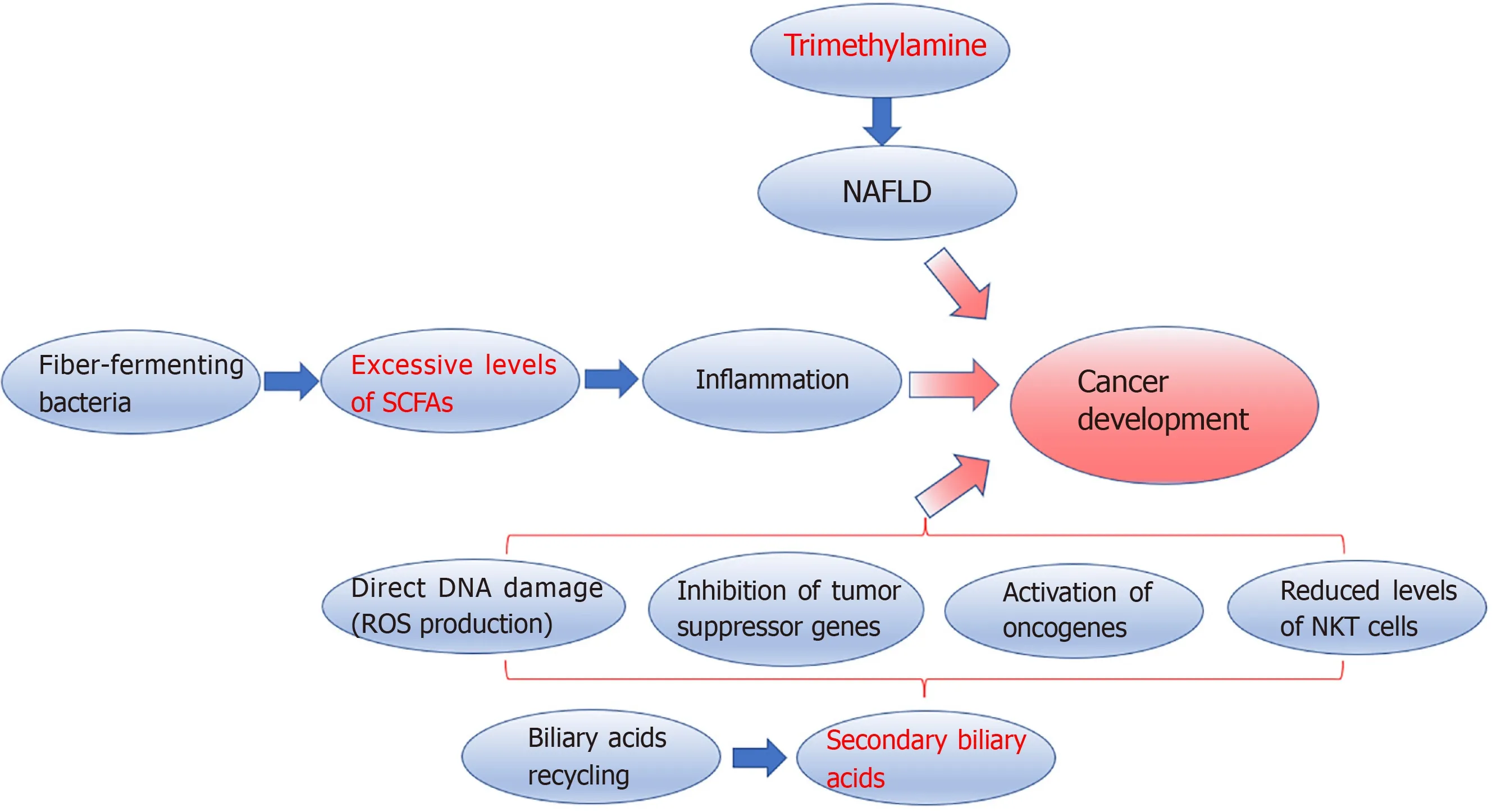
Microbial metabolites:Microbiota metabolites may also have a causal role in liver cancer development(Figure 2).Trimethylamine(TMA)is an example of microbial metabolites involved in the pathogenesis of NAFLD[8].Experimental studies on mice showed that excessive intake of soluble dietary fibre is associated with excessive proliferation of fibre-fermenting bacteria,includingClostridiumthat produces shortchain fatty acids(SCFAs),which showed immunomodulatory functions[68,69].Excessive levels of SCFAs,particularly butyrate,promote inflammation having a causal role in cholestasis,NAFLD,and HCC development,as reported in metabolomic studies[8,53,70].Faeces and serum levels of butyrate resulted higher in patients with NAFLD-related HCC than those with NAFLD-related cirrhosis.Furthermore,butyrate can impair cytotoxic CD8+T cell activity[53].Conversely,propionate seems able to inhibit cancer progression[71].Gut microbiota metabolises choline into several metabolites,including TMA,that the liver metabolises into TMA oxide,and TMA oxide is related to liver inflammation[72].

Figure 2 Microbiota metabolites causal role in liver cancer development.Gut microbiota metabolises choline into several metabolites,including trimethylamine(TMA)that the liver metabolises into TMA oxide,and TMA oxide is related to liver inflammation.Excessive intake of soluble dietary fibre is associated with excessive proliferation of fibre-fermenting bacteria,including Clostridiumthat produces short-chain fatty acids.Excessive levels of short-chain fatty acids,particularly butyrate,promote inflammation.The intestinal microbiota has a fundamental role in bile acid(BA)production and recycling.BAs are synthesised by the liver and metabolised by gut bacteria into secondary BAs,which are sensed by the farnesoid X-activated receptor of the epithelial cells.Farnesoid X-activated receptor provides feedback to the liver.Secondary BAs can cause direct DNA damage by producing reactive oxygen species,inhibiting tumour suppressor genes,activating oncogenes,and negatively affecting natural killer T cell infiltration.Natural killer T cells can control both primary and secondary cancer development.NAFLD:Non-alcoholic fatty liver disease;SCFAs:Short-chain fatty acids;NKT cell:natural killer T cell;ROS:Reactive oxygen species.
The intestinal microbiota also has a fundamental role in bile acids(BA)production and recycling.BAs are synthesised by the liver and metabolised by gut bacteria into secondary BAs,which are sensed by the epithelial cells’ farnesoid X-activated receptor(FXR).FXR provides feedback to the liver[73].BA excess is another recognised pathogenetic factor in carcinogenesis.Secondary BAs can cause direct DNA damage by producing reactive oxygen species,inhibiting tumour suppressor genes,and activating oncogenes[74].Furthermore,the deoxycholic acid,a secondary BA,binding to the TLR2 in hepatic stellate cells,can induce cyclooxygenase-2 expression,enhancing the inhibition of the antitumour activity prostaglandin E2-mediated[75].Obesity can increase BA conversions[30].On the contrary,the inhibition of 7αdehydroxylation responsible for secondary BA metabolization is associated with a lower incidence of HCC in mice[76].In both animal models and humans,conversion of primary BA to secondary BA is also negatively related to NKT cell infiltration.NKT cells can control both primary and secondary cancer development[77,78].
Complete knowledge of these pathways may allow the design of further studies on several appealing preventive options,including agents able to reestablish a correct balance between the different microbial species,selective agents against pathogenic bacteria,inhibitors of bacterial pathogenic metabolites production and gut barrier improvement[3].
Specific pathways in CCA:Infection ofOpisthorchis viverriniandClonorchis sinensisis a well-known risk factor for CCA development.Besides direct mechanical damage on the biliary tract epithelium and sustained inflammation,dysbiosis in local microbiota with bacterial translocation from the duodenum may contribute to CCA development[79].
CANCER TREATMENT
Surgical resection is the elective treatment for HCC,CCA or liver metastasis from several primary cancers,mostly colorectal adenocarcinoma,whenever possible.In the setting of advanced disease,chemotherapy and novel pharmacologic treatments,including immunotherapy and targeted therapies,should be preferred.
Immunotherapy
Immune checkpoint inhibitors,including the tremelimumab,a monoclonal antibody against cytotoxic T-lymphocyte-associated antigen 4,and nivolumab or pembrolizumab that are monoclonal antibodies against programmed cell death ligand 1[7],show a response rate in HCC patients that is reported to be up to 20%[80].Immunotherapy may also be combined with locoregional therapies,showing a synergistic effect[81].Tremelimumab showed greater efficacy in hepatitis C-related HCC since it can enhance CD8+T cell infiltration and,consequently,lower the viral load[81].
Since gut microbiota seems to impact these systemic treatments' efficacy,the microbiota’s modulation to enhance treatments’ response appears as a promising therapeutic target[7].It has been reported that while antibiotics may reduce the efficacy of the checkpoint inhibitors lowering the gut microbiota biodiversity,there are specific overrepresented taxa associated with more significant responses[82].
Zhenget al[83]showed higher levels ofAkkermansia muciniphilaand bacteria from the family of theRuminococcaceaein faecal samples of anti-programmed cell death ligand 1 immunotherapy responders.Conversely,in non-responders patients,higherProteobacterialevels were found from week 3 of therapy,and a predominance ofProteobacteriawas found at week 12[83].
The use of epigenetic drugs,including DNA methyltransferase enzymes-mediated hypermethylation and histone deacetylases-mediated histone modification,is under evaluation showing promising results in combination with conventional immunotherapy in murine models[84].
The microbiota evaluation may help in better selecting the candidate for a specific treatment hypothesising the response rate.Furthermore,the possibility to target both innate and adaptive immune systems could represent an appealing therapeutic option.In particular,actions on the innate arms may allow improvements in cytotoxic effect,stimulate the adaptive immune system and reduce the tumour-promoting effect[10].
Anticancer peptides
Antimicrobial peptides(AMPs)are constitutively or inducibly expressed in the tissues,which may be in contact with pathogens[85].AMPs are present in the great majority of vertebrates,invertebrates and vegetables.The antimicrobial effect of the AMPs can be exerted through cellular membrane damages,inhibition of cellular replication and through their immunomodulatory abilities[85-87].
Some AMPs showed anticancer properties,also causing cancer cell apoptosis.Furthermore,the healthy or cancer cell membrane composition differs,and tumour cells are more easily damaged by the anticancer peptides(ACPs).ACPs can interact with LPS or other bacterial products resulting in an anti-inflammatory effect[50].The use of ACPs,including TLR agonist and tumour-associated antigens-derived peptides,may represent a promising therapeutic option in HCC treatment[50,63,88].To be effective,some ACPs would have to be delivered.Delivery systems may include peptide-derived vaccines,nanoparticles and liposomes,each related to advantages and limitations[50].
More specific details about the design and the delivery of these molecules are reviewed elsewhere[50,89].
Microbiota,immune system and treatments response
Sorafenib is a tyrosine kinase inhibitor worldwide used in advanced HCC that can suppress abnormal cell proliferation and angiogenesis.Microbiota can influence sorafenib’s blood levels,affecting enterohepatic recirculation.Drug blood levels are related to the chance of suffering from the side effects[90].Two common side effects include diarrhoea and hand-foot syndrome and require reducing the administered drug[91].Butyric acid showed a protective action toward the inflamed intestinal mucosa by stimulating the Tregs and IL-10 secretion.IncreasedButyricimonas,a butyric acid producer,have been found in patients not experiencing diarrhoea[91,92].Dysbiosis and increased levels in the gut of bacteria typically found in the mouth(Veillonella,Bacillus,Enterobacter)have been found in patients not experiencing the hand-foot syndrome[91].
On the contrary,reduced Treg levels allow the achievement of better outcomes through the enhancement of the CD8+T cell antitumour activity.Furthermore,the baseline CD4+T effector/Tregs ratio has a prognostic value[93].
FUTURE PERSPECTIVES:OPTIONS FOR CANCER PREVENTION
Complete knowledge of the interaction between gut microbiota and liver cancer steps may help design new and tailored therapeutic options[3].
Microbiota modulation
The only actual method to prevent primary liver cancer development is to prevent and cure the underlying chronic liver disease whenever present.Although the gut microbiota role in these pathologies is still not wholly understood,microbiota modulation may be a promising target to reduce cancer.There are conditions in which microbiota modulation would have a marginal role,including perinatal viral hepatitis infections or cancers occurring on “healthy” livers[3].
The environment,diet,lifestyle,the use of antibiotics or pre/probiotics and several diseases may change the gut microbiota composition.It has been reported that a vegetable-enriched diet may lower the incidence of primary liver cancers,mainly in the male population.Conversely,a high-fat diet favours gram-negative bacteria overgrowth with increased LPS levels,and a high-fructose diet reduces the population ofBifidobacteriumandLactobacillus[94,95].
On a theoretical basis,using non-selective antibiotics may lower the entire gut microbiota population reducing the chance of bacterial translocation and the induction of a pro-inflammatory status.Treatments with selective antibiotics,if available,could reduce only those species producing cancer-promoting metabolites[3,76].
Experiments on rats demonstrated that non-absorbable antibiotic administration could positively affect steatosis and the inflammatory status.Studies on murine models showed that metronidazole administration might decrease the risk of cholestasis and HCC development by reducing the population of bacteria production of butyrate,which shows a health-promoting effect in other circumstances[70].Similarly,vancomycin may reduce gram-positive bacteria producing secondary BAs[96].The administration of the combination of ampicillin,neomycin,metronidazole and vancomycin showed a more powerful effect against late stages of HCC carcinogenesis compared to earlier stages[39].On the contrary,penicillin intake was reported to be related to a higher risk of HCC development in rats[97].
The non-absorbable oral norfloxacin and rifaximin showed a good safety profile and microbiota-related positive effects in cirrhotic patients and mice with HCC[3].An experimental study on the subcutaneous implantation model of thymoma on mice showed that an antibiotic combination of vancomycin,neomycin and primaxin reduced the chance of developing liver metastasis,though without affecting the primary tumour[77].
Despite the lack of much data on humans,it is reasonable that a long-term antibiotic assumption may be burdened with several side effects,including depletion of beneficial bacteria,kidney damages or antibiotic resistance[3].Consequently,further studies are needed.
The use of probiotics may help resolve dysbiosis,increase the number of bacteria with favourable properties,improve the intestinal barrier functions,absorb carcinogens and interact with the immune system,causing a reduction of Th17[98]cells.While ongoing human trials evaluate the effects of probiotic administrations in patients suffering from chronic liver diseases,evidence-based data about HCC comes only from murine models[3].
The assumption of the so-called VSL#3,a mixture ofStreptococcus thermophilus,Bifidobacterium breve,Bifidobacterium longum,Bifidobacterium infantis,Lactobacillus acidophilus,Lactobacillus plantarum,Lactobacillus paracasei,Lactobacillus delbrueckiisubspecies andBulgaricusseemed to have positive effects on the pathway inflammation-fibrosis-HCC development being associated with an enriched population ofPrevotellaandOscillibacterand with Th12 cell differentiation[97,99].
Similarly,prebiotics are substances able to stimulate the overgrowth of beneficial bacteria.Some examples include prebiotics of fructooligosaccharides reported to reestablish eubiosis,improve intestinal barrier function and reduce inflammation.Lactulose is related to an overgrowth ofBifidobacteriumthat shows a healthy behaviour by reducing LPS serum levels.Therapies with synbiotics are based on the combined use of probiotics and prebiotics[100].
Finally,faecal microbiota transplantation(FMT)is another treatment option that can reduce the risk of HCC development.It has been reported that FMT may reduce steatohepatitis in mice[57].However,several concerns have been raised,including the possibility of a long-lasting efficacy and the risk of infection transmission.The opportunity to transplant only beneficial bacteria could represent an appealing option[3].
TLR4 antagonists
The LPS-TLR4 axis has a crucial role in the inflammation-fibrosis-cirrhosis-cancer pathway.Consequently,several antagonists of the TLR4 have been proposed.Some examples include polymyxin B,able to bind and sequestrate LPS;E5531 or eritoran,molecules interacting with other steps of this signalling pathway;resatorvid,able to target the TLR4;thalidomide,a TLR inhibitor[3].Further details are not the object of this review and can be found elsewhere[3,88].
However,the primary concern is the consequent status of immunosuppression that could be detrimental in patients with chronic liver disease or HCC[3].Furthermore,the results of published studies are sometimes controversial due to the complexity of the known and unknown interactions[88].Consequently,further long-term studies are needed.
PPARs agonists
Several studies reported the beneficial effects of both natural and synthetic PPARs agonists in chronic liver disease development through microbiota modulation.Although specific studies on cancer progression are lacking,targeting the PPARs could represent,at least,a cancer prevention strategy.Further details can be found elsewhere[65].
Gut barrier function improvement
The integrity of the gut barrier is vital for healthy individuals.A high caloric diet seems to impair the intestinal barrier[101].Conversely,physical exercise improves short-term and long-term gut permeability through effects on the immune system and the microbiota,increasing theBacteroidetes/Firmicutesratio[6,102].
Cisapride is a prokinetic medication that resulted in reducing both bacterial overgrowth and translocation,fastening the intestinal transit time[103].Some nonselective β-adrenergic blockers showed similar properties.
BA influence the function of the gut barrier,and the FXRs are crucial in BA synthesis,other than in the regeneration of the liver and tumour growth suppression.The obeticholic acid is an FXR agonist and showed beneficial effects on damaged mucosa and reduced the gut barrier permeability,the inflammatory status,bacterial overgrowth and preventing the progression from non-alcoholic steatohepatitis to other complications,thus becoming an attractive potential treatment option[104].
Excessive TNF production is associated with increased gut barrier permeability reducing the tight junction proteins[105].Consequently,anti-TNF-based therapies could represent potential therapeutic options,but as previously stated,the related immunosuppression may be detrimental[3].However,n-3 polyunsaturated fatty acids(PUFA)showed anti-inflammatory properties in experimental models reducing the level of TNF and IL-1,thus resulting in an appealing option[106,107].Furthermore,in vitroexperiments demonstrated the ability of the n-3 PUFA to block β-catenin and cyclooxygenase-2[108].On the contrary,n-6 PUFA seems related to a pro-inflammatory status[109].
Early diagnosis
There is a continuous search for new,non-invasive biomarkers for diagnosis,and microbiota seems promising even in this field.Since there are different microbial signatures along with disease progression,microbial samples could represent appealing non-invasive biomarkers for an early diagnosis[16].
Furthermore,Ponzianiet al[110]demonstrated an inverse relation betweenAkkermansiaandBifidobacteriumand the well-known inflammatory marker calprotectin.Analysis on faecal samples of patients with primary liver cancers showed a significant link betweenVeillonellaand alpha-fetoprotein levels together with a negative connection betweenSubdoligranulumand alpha-fetoprotein levels[50].
Along with faecal samples,analysis of the tongue microbiota could represent another non-invasive biomarker.In particular,OribacteriumandFusobacteriumpresence could differentiate HCC patients from healthy subjects[17].
Jiaet al[1]reported that the plasma-stool ratio of two BAs,tauroursodeoxycholic and glycoursodeoxycholic acids,demonstrated the ability to identify patients with intrahepatic CCA from those with HCC or healthy people with an area under the curve of 0.801 and 0.906,respectively[1].Although some methodological and causeeffects concerns have been raised[111],this potential biomarker is appealing.Again,further studies are needed to obtain new markers that could be used independently or within algorithms[18,111].
CONCLUSION
In conclusion,a growing body of literature demonstrates a pathogenetic role of the gut microbiota-immunity axis in liver cancer development.Although there is an ongoing rapid development of metagenomic science,definitive and complete knowledge of this process is still far from being wholly acquired.However,targeting microbiota and the immune system may represent appealing therapeutic options alone or boost conventional treatments.Finally,the gut microbiota signature evaluation could represent a potential novel,non-invasive biomarker for early diagnosis.
 World Journal of Gastrointestinal Oncology2021年11期
- World Journal of Gastrointestinal Oncology的其它文章
- Hepatocellular carcinoma biomarkers,an imminent need
- Anatomical vs nonanatomical liver resection for solitary hepatocellular carcinoma:A systematic review and meta-analysis
- Atezolizumab plus bevacizumab versus sorafenib or atezolizumab alone for unresectable hepatocellular carcinoma:A systematic review
- Cell-free DNA liquid biopsy for early detection of gastrointestinal cancers:A systematic review
- Colorectal cancer in Arab world:A systematic review
- Induction chemotherapy with albumin-bound paclitaxel plus lobaplatin followed by concurrent radiochemotherapy for locally advanced esophageal cancer
