Self-Assembled Zn2+,Co2+ and Ni2+ Complexes Based on Coumarin Schiff Base Ligands:Synthesis,Crystal Structure and Spectral Properties
GUO Geng LI Juan WU Ya DING Wen-Min ZHANG Shu-Zhen SUN Yin-Xia
(School of Chemical and Biological Engineering,Lanzhou Jiaotong University,Lanzhou 730070,China)
Abstract:Mononuclear Zn2+,Co2+ complexes and binuclear Ni2+ complex based on coumarin Schiff base ligands,[Zn(L1)2](1)(HL1=6-((4-diethylamino-2-hydroxy-benzylidene)-amino)-benzopyran-2-one),[Co(L2)2](2)(HL2=6-((4-methoxy-2-hydroxy-benzylidene)-amino)-benzopyran-2-one)and[Ni2(L3)2(CH3OH)4](3)(H2L3=4-hydroxy-3-((4-methoxy-2-hydroxy-benzylidene)-amino)-benzopyran-2-one),were synthesized and characterized by elemental analysis,IR,UV-Vis,fluorescence spectra and X-ray single crystal diffraction analysis.The crystal structure analysis represents that the complexes 1 and 2 possess mononuclear structure composed of one metal ion(Zn2+ or Co2+)and two ligand units((L1)-or(L2)-).Whereas complex 3 had binuclear structure,containing two Ni2+ ions,two ligand units(L3)2- and four coordination methanol molecules.The crystals of complexes 1,2 and 3 are solved as monoclinic space group C2/c,triclinic space group P and triclinic space group P21/n,respectively.The spatial configurations of the central metal Zn2+ and Co2+ ions are four-coordinated tetrahedrons,and the Ni2+ ion is six-coordinated twisted octahedrons.In addition,UV-Vis studies demonstrate that the free ligands HL1 in DMF/H2O(4∶1,V/V)solution and HL2 in DMSO/H2O(4∶1,V/V)solution could selectively recognize Hg2+ and Zn2+,respectively.The detection limits were calculated to be 7.45 and 6.10 μmol·L-1,respectively.Fluorescence studies indicate that HL2 exhibits ability of selective recognition Zn2+ against other common cationic including Ag+,Ba2+,Ca2+,Cd2+,Cr3+,Cu2+,Fe3+,Mg2+, Mn2+ and Hg2+ in DMSO/H2O(4∶1,V/V),and the detection limit was calculated to be 2.91 μmol·L-1.CCDC:2034542,1;2034547,2;2034548,3.
Keywords:coumarin Schiff-base;transition metal complex;crystal structure;spectral property
0 Introduction
Coumarin is an excellent fluorescent compound with large Stokes shift,visible light excitation and emission,tunable light wavelength,two-photon effect,stable chemical properties,biocompatibility and high fluorescent rate[1-2].Different positions of coumarin ring can be replaced by different groups,which can obtain different absorption and fluorescent emission wavelengths,show different colors,and derive derivatives with strong fluorescence[3-5].In particular,coumarin modified with amino groups at positions 3 and 6 can react with aldehydes to prepare Schiff base compounds.Schiff base derivatives containing N and O donor site and fluorophore are appealing to tools for designing optical probes for metal ions[6-9].It is already established that Schiff base alone is weakly or nonfluorescent as the fluorescence is quenched due to the free rotation around C=N bond[10-15].Recently,numerous papers described the specific detection of various metal ions through the chelation of Schiff base probes[16-18].Coumarin,as an excellent chromophore,can be invoked as a fluorescent sensor to detect cations,anions and biologically related species[19-22].Meanwhile,coumarin Schiff base can form a variety of metal complexes because of their abundant coordinating atoms and the ability to participate in coordination is outstanding[23-26].Herein,three coumarin Schiff bases ligands and their corresponding complexes,[Zn(L1)2](1)(HL1=6-((4-diethylamino-2-hydroxy-benzylidene)-amino)-benzopyran-2-one),[Co(L2)2](2)(HL2=6-((4-methoxy-2-hydroxy-benzylidene)-amino)-benzopyran-2-one)and[Ni2(L3)2(CH3OH)4](3)(H2L3=4-hydroxy-3-((4-methoxy-2-hydroxy-benzylidene)-amino)-benzopyran-2-one),were synthesized and characterized.In addition,the photochemical properties of HL1,HL2,H2L3and their corresponding complexes 1,2 and 3 were discussed.Hg2+can be selectively identified in DMF/H2O(4∶1,V/V)solution by HL1based on UV-Vis spectrum,and HL2can detect Zn2+in DMSO/H2O(4∶1,V/V)solution by UV-Vis spectrum and the fluorescence spectrum.
1 Experimental
1.1 Material
4-hydroxyl coumarin and coumarin from Alfa Aesar were used without further purification.6-aminocoumarin and 3-amino-4-hydroxy-coumarin were synthesized according to an analogous method reported earlier[27-28].The other reagent and solvent were analytical grade from Tianjin Chemical Reagent Factory,and were used without further purification.
1.2 Instruments and methods
C,H and N analysis carried out with a GmbH VariuoEL V3.00 automatic elemental analyzer.FT-IR spectra were recorded on a VERTEX70 FT-IR spectrophotometer with samples prepared as KBr(400~4 000 cm-1)pellets.UV-Vis absorption spectra were recorded on a Hitachi UV-3900 spectrometer.Luminescence spectra in solution were recorded on a Hitachi F-7000 spectrometer.X-ray single crystal structures were determined on a Bruker Smart 1000 CCD area detectors.Melting points were measured by a X-4 microscopic melting point apparatus made by Beijing Taike Instrument Limited Company and were uncorrected.
1.3 Syntheses of ligands HL1,HL2and H2L3
The synthetic routes of ligands HL1,HL2and H2L3are shown in Scheme 1.

Scheme 1 Synthetic routes of HL1,HL2 and H2L3
1.3.1 Synthesis of HL1and HL2
6-amino coumarin was synthesized depending on an analogous method reported previously in literature[27].Firstly,coumarin(1.46 g,0.010 mol)and sodium nitrate(5.20 g)were placed in a flask,and 5.0 mL of H2O was added to the flask.The mixture was stirred thoroughly until the solid was completely dissolved.Concentrated sulfuric acid(12.0 mL)was slowly added dropwise into the solution under an ice-water bath,and the temperature was kept below 25℃.After stirred at room temperature for 4.0 h,the solution was poured into crushed ice,and a light-yellow solid was precipitated.After filtering,6-nitrocoumarin was obtained by recrystallization with glacial acetic acid.Secondly,3.5 mL water,0.86 g iron powder,0.3 mL glacial acetic acid and an ethanol solution of 6-nitrocoumarin(0.60 g,3.14 mmol)was added and refluxed at 95℃for 1.5 h.The solution was cooled slightly,then 20.0 mL warm saturated sodium carbonate solution was added.The mixture was filtered while it was hot.Yellow solid 6-aminocoumarin was obtained by vacuum filtration after the filtrate was cooled.Yield:67.8%,m.p.172~173℃.In the end,6-amino coumarin(1.00 g,9.50 mmol)and 4-diethylamino-salicylaldehyde(1.20 g,6.01 mmol)were placed in a flask,followed by 5.0 mL anhydrous ethanol and 2~3 drops of glacial acetic acid.The mixture was subjected to reflux at 75℃for 6.0 h and cooled to room temperature,and bright yellow solid precipitates were obtained.HL1was obtained by filtering.Yield:72.5%,m.p.155~156℃.Anal.Calcd.for C18H16N2O3(%):C,70.12;H,5.23;N,9.09.Found(%):C,70.21;H,5.19;N,9.02.
Ligand HL2was synthesized by a method analogous to that of HL1except substituting 4-diethylaminosalicylaldehyde with 4-methoxy-salicylaldehyde.Yield:77.4%.m.p.187~188 ℃.Anal.Calcd.for C17H13NO4(%):C,69.15;H,4.44;N,4.74.Found(%):C,69.19;H,4.45;N,4.72.
1.3.2 Synthesis of H2L3
3-amino-4-hydroxy-coumarin(1.23 g,6.90 mmol)and 4-methoxy-salicylaldehyde(1.00 g,6.60 mmol)were placed in a flask,then 15.0 mL absolute ethanol and 1~2 drops of acetic acid were added to the flask.The mixture was subjected to reflux at 70℃for 6.0 h.After returning to room temperature,the yellow precipitate was filtered and dried to obtain H2L3.Yield:78.6%,m.p.125~126 ℃.Anal.Calcd.for C17H13NO5(%):C,65.59;H,4.21;N,4.50;Found(%):C,65.60;H,4.22,N,4.54.
1.4 Synthesis of complexes 1,2 and 3
1.4.1 Synthesis of complex 1
A colorless methanol solution(3.0 mL)of zinc(Ⅱ)acetate dihydrate(3.0 mg,0.014 mmol)was added dropwise to a dichloromethane solution(3.0 mL)of HL1(4.6 mg,0.014 mmol)at room temperature.The mixing solution turned yellow immediately and was kept at room temperature for about one month.Clear light colorless single crystals of complex 1 suitable for X-ray structural determination were obtained.Anal.Calcd.for C40H38N4O6Zn(%):C,65.26;H,5.20;N,7.61.Found(%):C,66.58;H,5.36;N,7.85.
1.4.2 Synthesis of complexes 2 and 3
The complexes 2 and 3 were prepared by the same method as that of the complex 1 except changing the solvent ratios and metal ions.The solvent ratios of complexes 2 and 3 were adjusted from methanol/dichloromethane(3∶3,V/V)to methanol/dichloromethane(5∶5,V/V),and the metal salts were adjusted to the corresponding cobalt(Ⅱ) acetate tetrahydrate and nickel(Ⅱ)acetate tetrachloride.Complex 2:Anal.Calcd.for C34H24N2O8Co(%):C,63.07;H,3.74;N,4.33.Found(%):C,64.80;H,3.63;N,4.52.Complex 3:Anal.Calcd.for C38H38N2Ni2O14(%):C,52.82;H,4.43;N,3.24.Found(%):C,53.83;H,4.32;N,3.46.
1.5 Crystal structure determinations of complexes 1,2 and 3
The single crystals of dimensions 0.27 mm×0.23 mm×0.21 mm(1),0.29 mm×0.27 mm×0.25 mm(2)and 0.24 mm×0.22 mm×0.21 mm(3)were used to determine the crystal structures by X-ray diffraction technique on Bruker SMART 1000 CCD area-detector diffractometer.The reflections were collected using graphite-monochromatized MoKαradiation(λ=0.071 073 nm)at 291,295 and 293 K,respectively.The SMART and SAINT software packages[29]were used for data collection and reduction,respectively.Absorption corrections based on multi-scans using the SADABS software[30]were applied.The structures were solved by direct methods and refined by full-matrix least-squares againstF2using the SHELXL program[31].All nonhydrogen atoms were refined anisotropically.The positions of hydrogen atoms were calculated and isotropic fixed in the final refinement.Details of the crystal parameters,data collection,and refinements for complexes 1~3 are summarized in Table 1.

Table 1 Crystal data and structure refinement for complexes 1~3

Continued Table 1
CCDC:2034542,1;2034547,2;2034548,3.
2 Results and discussion
2.1 Crystal structures of complexes 1,2 and 3
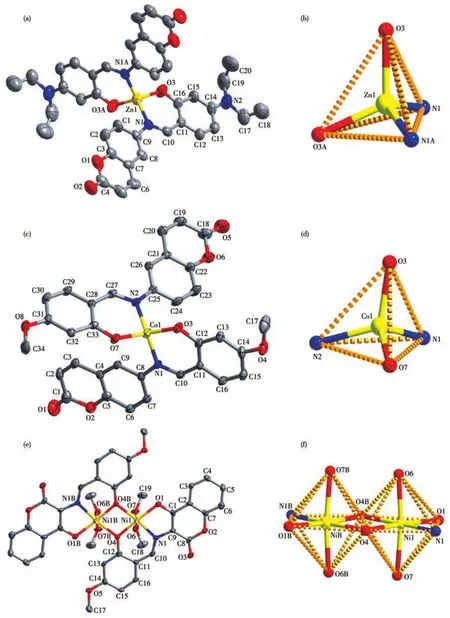
The molecular structure of complexes 1,2 and 3 are depicted in Fig.1.Selected bond lengths and angles for complexes 1~3 are listed in Table S1(Supporting information).X-ray crystallographic analysis shows that complexes 1 and 2 have similar mononuclear crystal structures.The crystals of complexes 1 and 2 are solved as monoclinic space groupC2/cand triclinic space groupP,respectively.Complexes 1 and 2 all consist of one central ion(Zn2+for 1 and Co2+for 2)and two units((L1)-for 1 and(L2)-for 2).The central metal(Zn2+and Co2+)of complexes 1 and 2 are all fourcoordinated by two deprotonated hydroxyl oxygen atoms(O3,O3A in 1 and O3,O7 in 2)and two nitrogen atoms(N1,N1A in 1 and N1,N2 in 2)from the ligand units((L1)-and(L2)-,respectively),which constitute the[Zn(L1)2](1)and[Co(L2)2](2)moieties,respectively(Fig.1a~1d).Therefore,the coordination geometry around the central Zn2+and Co2+of complexes 1 and 2 all can be described as a tetrahedron space configurations.
The Schiff base ligand H2L3moiety of complex 3 contains phenolate-O atoms that connect two neighboring nickel units by way of twoμ-O bridges to form binuclear structures,which crystallizes in monoclinic systemP21/nspace group,and the asymmetry unit consists one Ni2+ion,one(L3)2-ligand and two coordinated methanol molecules(Fig.1e).In complex 3,Ni2+center is characterized by a twisted octahedron coordination geometry,with the basal donor atoms coming from the phenolate-O(O4),coumarin′s hydroxyl-O(O1),and imine-N(N1)atoms of the Schiff base ligand,the symmetry-related phenolate-O(O4B)and the two coordinated methanol-O(O6,O7)(Fig.1f).The length of Ni—O and Ni—N falls in a range of 0.200 2~0.216 9 nm and 0.200 4 nm(Table S1),which are within the ranges reported for other similar nickel complexes.

Fig.1 Molecular structure of complexes 1(a),2(c)and 3(e)showing 30% probability displacement ellipsoids and their coordination pattern diagram for Zn2+(b),Co2+(d),Ni2+(f)
2.2 IR spectra of HL1,HL2,H2L3 and their corresponding complexes 1,2,3
The FT-IR spectra of the ligands HL1,HL2and H2L3and their corresponding complexes 1,2 and 3 exhibited various bands in the 400~4 000 cm-1region,and the important FT-IR bands are given in Table 2.The broad O—H group stretching bands at 3 419,3 442 and 3 439 cm-1for the free ligands HL1,HL2and H2L3,respectively,disappeared in complexes 1,2 and 3,which indicates that the oxygen atoms in the phenolic hydroxyl groups are completely deprotonated and coordinated to the metal ion[32-35].Whereas,the stretching bands at 3 483 cm-1in complex 3 are attributed to the stretching vibrations of the O—H group of coordinated methanol[36-37].The Ar—O stretching bands at 1 240,1 207 and 1 259 cm-1of complexes 1,2 and 3 shifted toward lower frequencies byca.13,15 and 24 cm-1,respectively,compared with that of free ligands HL1,HL2and H2L3at 1 253,1 222 and 1 293 cm-1,respectively.The lower frequency of the Ar—O stretching shift indicates that M—O bond is formed between the metal ions and the oxygen atoms of the phenolic groups[38-40].In addition,the free ligand HL1,HL2and H2L3exhibited characteristic band of stretching vibration of C=N group at 1 624,1 644 and 1 674 cm-1,respectively,and their corresponding complexes 1,2 and 3 were observed at 1 611,1 644 and 1 692 cm-1,respectively[41-42].Compared with ligand HL1and HL2,the C=N stretching frequency of complexes 1 and 2 shifted to a lower frequency byca.13 and 8 cm-1,respectively.While complex 3 shifted to a higher frequency byca.18 cm-1.It is indicated that the C=N bond frequencies decreased or increased due to the coordination bond between the metal atom and the ami-no nitrogen lone pair[43-44].The FT-IR spectrum of complex 1 showedν(M—N)andν(M—O)vibration frequencies at 615 and 483 cm-1(or 505 and 467 cm-1for complex 2,582 and 459 cm-1for complex 3).These assignments are consistent with the frequency values in literature[44-45].

Table 2 Important IR bands of free ligands HL1,HL2,H2L3 and their corresponding complexes 1,2,3 cm-1
2.3 UV-Vis absorption spectra analyses
The absorption spectra of ligands HL1,HL2,H2L3and their corresponding complexes 1,2,3 in 50 μmol·L-1DMSO solution are shown in Fig.2.As shown in Fig.2,the typical absorption peaks of HL1(HL2or H2L3)atca.276 nm(ca.285 nm orca.282 and 314 nm)was clearly observed.The peaks can be attributed respectively to theπ-π*transitions of the benzene ring[46-47].Whereas the peak at 382 nm(342 or 411 nm)may be attributed toπ-π*transition of the C=N group in the ligand[48-49].Upon coordination of Zn2+ion with HL1(Co2+ion with HL2or Ni2+ion with H2L3),the absorption peaks had evidently changed to 277 nm in complex 1(287 nm in complex 2 or 277 and 312 nm in complex 3).Compared with free ligands HL1at 276 nm(285 nm in HL2or 282 and 314 nm in H2L3),the absorption peak was red-shifted by about 1 nm in complex 1(2 nm in complex 2),and 282 and 314 nm were slightly shifted hypochromic to 277 and 312 nm in complex 3.Additionally,another absorption peak at 382 nm of ligand HL1(342 nm of HL2or 411 nm of H2L3)disappeared in the UV-Vis absorption spectrum of complex 1(complex 2 or complex 3),which indicates that the imine nitrogen atoms of the ligands HL1(HL2or H2L3)have coordinated to the metal Zn2+(Co2+or Ni2+)ions.The new peak at 467 nm of complex 3 is assigned to transition of ligand to metal charge-transfer[50].

Fig.2 UV-Vis spectra of(a)HL1 and its Zn2+ complex 1,(b)HL2 and its Co2+ complex 2,(c)H2L3 and its Ni2+ complex 3
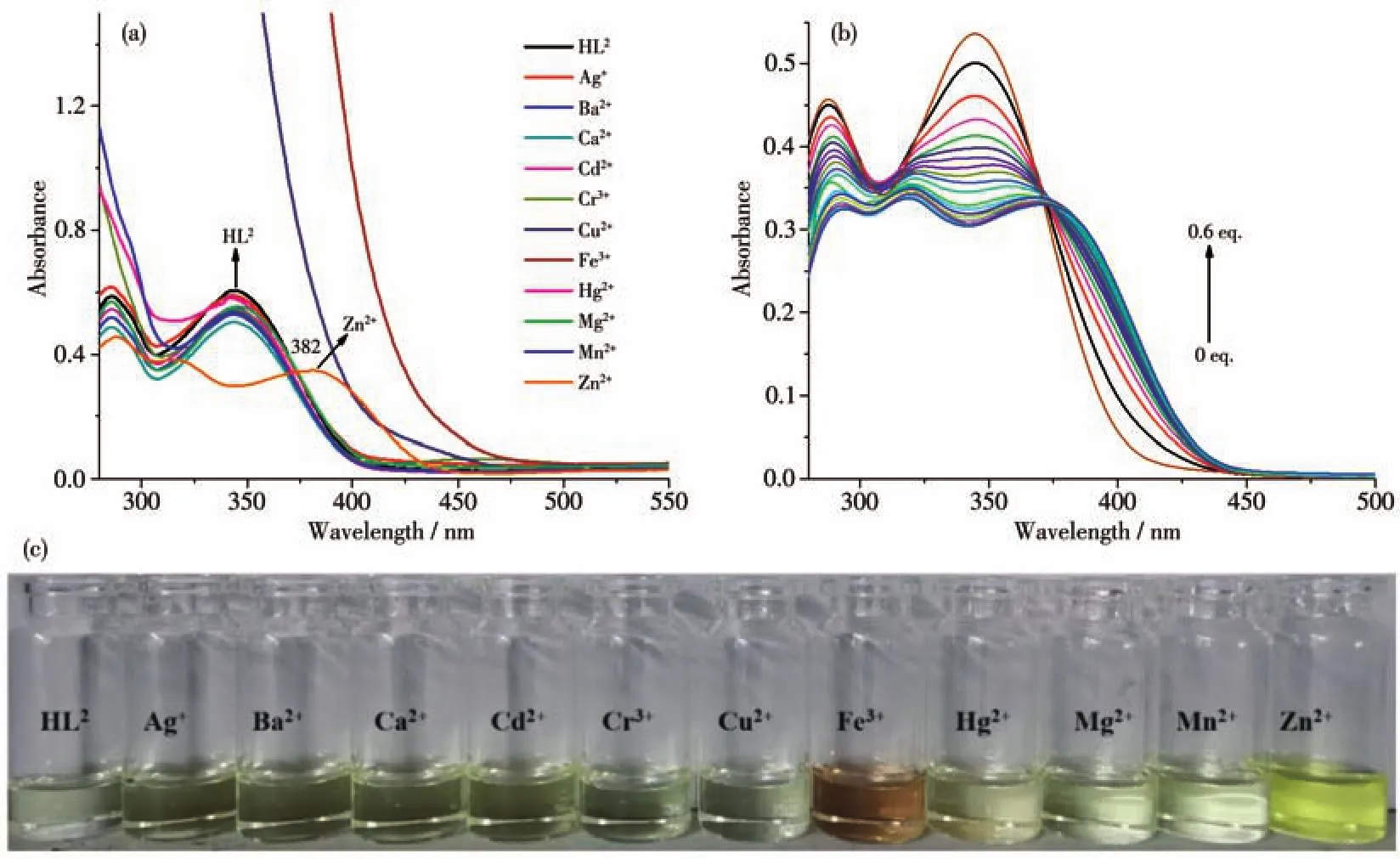
The UV-Vis specific responses of HL1or HL2to partial common cations(Ag+,Ba2+,Ca2+,Cd2+,Cr3+,Cu2+,Fe3+,Mg2+,Mn2+,Hg2+and Zn2+)were carried out in DMF/H2O(4∶1,V/V)or DMSO/H2O(4∶1,V/V)solutions(c=50 μmol·L-1).The UV-Vis spectra of HL1solution had a significant blue-shift from 383 to 348 nm when Hg2+ion was added,and the other measured ions could not lead to apparent changes of the spectrum,except for Hg2+ion and the self-absorption of Cu2+and Fe3+ions(Fig.3a).In order to study the variation of absorption peak intensity with time in UV-Vis spectra of HL1,the UV-Vis absorption intensity was measured at 1 min interval after the addition of Hg2+.It can be seen from Fig.3b that the UV-Vis peak position of HL1solution was significantly red-shifted from 383 to 424 nm after adding Hg2+.With the passage of time,the peak at 424 nm gradually disappeared and the peak intensity at 348 nm gradually increased,and remained stable after 18 min.As for HL2,the UV-Vis spectra of HL2solution had a significant red-shift from 342 to 382 nm when the Zn2+ion was added,and the other tested ions could not lead to obvious changes of spectra except for Zn2+ion and the self-absorption of Cu2+and Fe3+ions(Fig.4a).In the natural light,the color changes from yellow(or colorless)to colorless(or yellow)after adding Hg2+(or Zn2+)to the DMF/H2O(4∶1,V/V)(or DMSO/H2O(4∶1,V/V))solution of HL1(or HL2),and the color changes of other ions except Fe3+are not obvious,as shown in Fig.3d and Fig.4c.The results showed that the HL1and HL2represented excellent selectivity for Hg2+and Zn2+,respectively.

Fig.3 (a)UV-Vis spectra of HL1 upon addition of different cations(50 eq.)in DMF/H2O(4∶1,V/V)solution(c=50 μmol·L-1)at room temperature;(b)UV-Vis absorption intensity of HL1 varying with time;(c)UV-Vis spectra of HL1 in DMF/H2O(4∶1,V/V)solution(c=50 μmol·L-1)by adding 0~1.15 eq.of Hg2+;(d)Discoloration photos of HL1 in DMF/H2O(4∶1,V/V)solution after adding metal cations under natural light

Fig.4 UV-Vis spectra of HL2 upon addition of different cations(50 eq.)in DMSO/H2O(4∶1,V/V)solution(c=50 μmol·L-1)at room temperature;(b)UV-Vis spectra of HL2 in DMSO/H2O(4∶1,V/V)solution(c=50 μmol·L-1)by adding 0~0.6 eq.of Zn2+,(c)Discoloration photos of HL2 in DMSO/H2O(4∶1,V/V)solution after adding metal cations under natural light
The identification characteristics of HL1(or HL2)as Hg2+(or Zn2+)probe were confirmed by UV-Vis titration in DMF/H2O(4∶1,V/V)(or DMSO/H2O(4∶1,V/V))solution(Fig.3c and 4b).The absorption band of HL1(or HL2)decreased at 383 nm(or 342 nm),and a new absorption band appeared gradually at 348 nm(or 382 nm)when the concentration of Hg2+(or Zn2+)increased from 0 to 1.15(or 0.6)eq.,which signs the deprotonation of hydroxyl moiety and coordination with the Hg2+(or Zn2+)ions.The change in absorbance of HL1(or HL2)at 348 nm(382 nm)had a linear relationship with Hg2+(or Zn2+)ion.In the low concentration range,the detection limit of 7.45 μmol·L-1(or 6.10 μmol·L-1)for Hg2+(or Zn2+)was calculated(Fig.S7 and Fig.S8)[51-54].The modified Benesi-Hildebrand formula was used to calculate LOD(limit of detection)and LOQ(limit of quantitation)[55]:LOD=3σ/S,LOQ=10σ/S(N=20).As shown in Table S4,we compared probe HL1and HL2of our work with the previously reported Hg2+and Zn2+probes.The probes in the table all have lower detection limits and are applied in relatively less toxic solvents.The detection limit of HL1(or HL2)is similar with previously reported detection limit,and the obtained LOD value is below the value(76 μmol·L-1for Hg2+and 46 μmol·L-1for Zn2+)approved by World Health Organization(WHO)[56-57].
2.4 Emission spectra analyses
The emission spectra of free ligands HL1,HL2,H2L3and its corresponding complexes 1,2,3 in 50 μmol·L-1DMSO solution at room temperature are shown in Fig.5.As shown in Fig.5a and 5b,the free ligand HL1(or HL2)exhibited a relatively strong emission peak atca.546 nm(or 498 nm)and a weaker emission peak at 458 nm(or 384 nm)upon excitation at 380 nm(or 340 nm),which could be assigned to the intra-ligandπ*-πtransition[58-59].Compared with the free ligand HL1(or HL2),the emission intensity of complex 1(or 2)decreased significantly at 546 nm(or 498 nm),but increased at 458 nm(or 384 nm),indicating that the introduction of M2+ions(Zn2+or Co2+)affected the fluorescence properties.These transitions may be related to the coordination of ligand HL1or HL2and Zn2+or Co2+ions,which allows the ligand to develop towards a more stable complex[60].Both H2L3and its complex 3 showed strong fluorescence emission at 482 and 396 nm with excitation at 300 nm,respectively(Fig.5c).Compared with H2L3,the fluorescence intensity of complex 3 reduced slightly and blue-shiftedca.86 nm,which might be related to the coordination of the N and O atom to the metal ions,and allows H2L3to develop towards a more stable complex.

Fig.5 Emission spectra of(a)HL1 and complex 1(λex=380 nm),(b)HL2 and complex 2(λex=340 nm),(c)H2L3 and complex 3(λex=300 nm)in DMSO solutions at room temperature
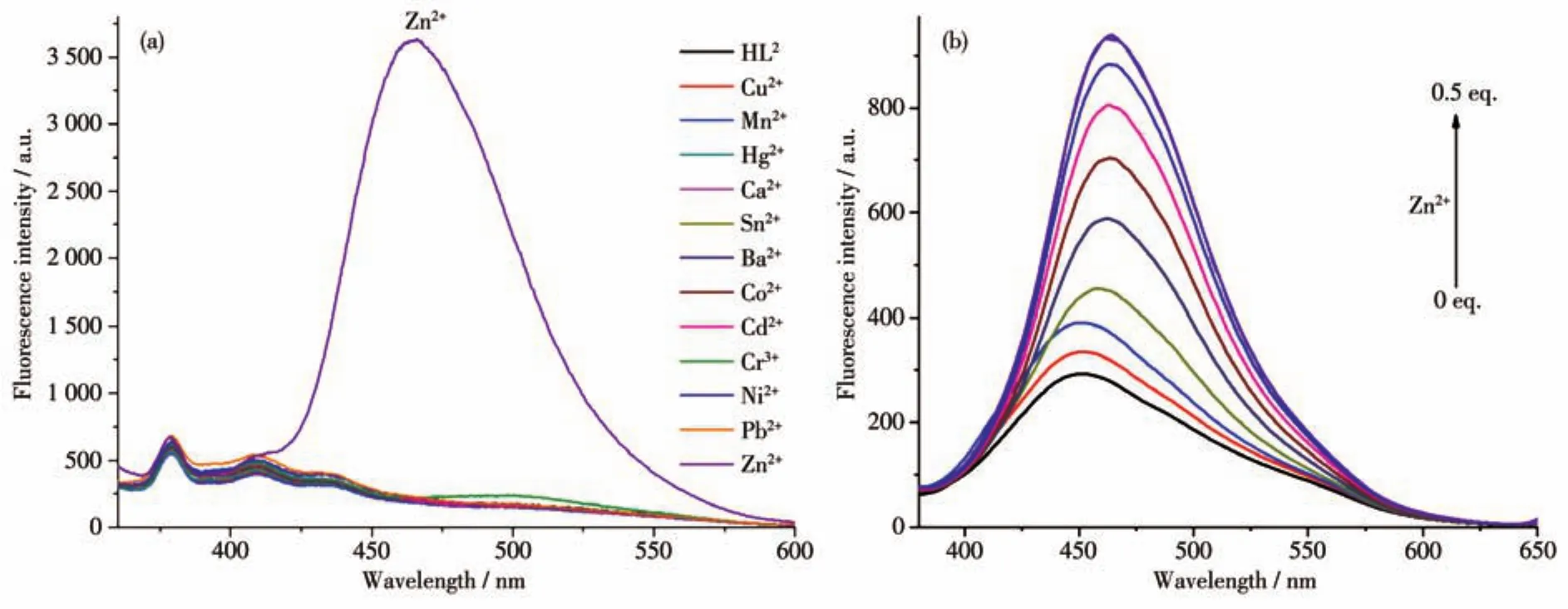
Under the excitation wavelength of 380 nm,the fluorescence response of HL2to various metal ions(Ba2+,Ni2+,Pb2+,Cu2+,Cd2+,Co2+,Cr3+,Mn2+,Hg2+,Ca2+,Sn2+and Zn2+)was studied.The DMSO/H2O(4∶1,V/V)solution of HL2(50 μmol·L-1)was added to 50 eq.of the solution with above metal ions(10 mmol·L-1).As shown in Fig.6a,the experimental results showed that when Zn2+ion was added,the fluorescence intensity of HL2solution changed significantly,and a new strong fluorescence emission peak appeared at 464 nm,which may be caused by theπ*-πtransition in the molecules.After the addition of other metal cations,the fluorescence intensity of HL2solution did not change,which indicates that HL2has a single and efficient recognition of Zn2+ion.

Fig.6 (a)Fluorescence emission spectra of HL2 upon addition of different cations(50 eq.)in DMSO/H2O(4∶1,V/V)solution(c=50 μmol·L-1)at room temperature;(b)Fluorescence titration spectra of HL2 with increasing concentration of Zn2+(0~0.5 eq.)in DMSO/H2O(4∶1,V/V)solution
Fluorescence titration experiments were used to detect the binding affinity of HL2to Zn2+ion.The Zn2+ion solution was gradually added to the DMSO/H2O(4∶1,V/V)solution of HL2.With the increase of the amount of Zn2+ion,the fluorescence emission intensity of HL2gradually increased at 464 nm until the amount of Zn2+ion reached 0.5 eq.After adding Zn2+,the fluorescence intensity did not change any more,as shown in Fig.6b.It is worth noting that the change in fluorescence intensity of HL2at 464 nm had a linear relationship with Zn2+ion in a range of 0~0.5 eq.The experimental results show that the coordination ratio of HL2to Zn2+is 2∶1.
In addition,the binding constant of Zn2+ion to HL2was determined by Hill′s equation to beKsv=8.91×10⁴L·mol-1(Fig.S9a).The calculated LOD(2.91 μmol·L-1)and LOQ(0.808 μmol·L-1)of HL2for Zn2+ions were calculated from the titration spectra of HL2containing Zn2+,as shown in Fig.S9b.The detection limit was also lower than the WHO approved LOD value.
3 Conclusions
Two mononuclear complexes 1,2 and one binuclear complex 3 based on coumarin Schiff base ligands(HL1,HL2and H2L3)were synthesized and structural characterized.The crystal structure analyses of complexes 1 and 2 illustrated that the Zn2+and Co2+are all four-coordinated distorted tetrahedrons,and the Ni2+in complex 3 are six-coordinated distorted octahedrons.Meanwhile,the optical properties of complexes 1,2 and 3 indicate that the fluorescence and UV-Vis variations of HL1,HL2and H2L3are due to the coordination of Zn2+,Co2+and Ni2+,respectively.UV-Vis studies demonstrate that the free ligands HL1(in DMF/H2O(4∶1,V/V)solution)and HL2(in DMSO/H2O(4∶1,V/V)solution)could selectively recognize Hg2+and Zn2+,respectively.The detection limits were calculated to be 7.45 and 6.10 μmol·L-1,respectively.Fluorescence studies indicate that HL2exhibits selective recognition ability of Zn2+against other common cationic including Ag+,Ba2+,Ca2+,Cd2+,Cr3+,Cu2+,Fe3+,Mg2+,Mn2+and Hg2+in DMSO/H2O(4∶1,V/V).The sensing capability of HL2for Zn2+against other common cationic with a low limit of 2.91 μmol·L-1renders it a candidate probe for Zn2+detection.
Supporting information is available at http://www.wjhxxb.cn
- 无机化学学报的其它文章
- Synthesis and Characterization of Palladium Nanoparticles with High Proportion of Exposed(111)Facet for Hydrogenation Performance
- Dielectric Phase Transition Induced by Halogen Substitution Based on 1,4-Diazabicyclo[2.2.2]octane-Derivatives
- 不同晶体生长活化能对SrZrO3∶Ce发光性能及微观组织影响
- 炭球修饰g-C3N4材料的制备及其可见光光催化性能
- Er3+掺杂 Li2O-SrO-ZnO-Bi2O3玻璃中 Er3+离子在1.53 μm处的荧光发射特性
- 3-((5-(3-吡啶基)-2-(1,3,4-噁二唑基))硫代)-2,4-戊二酮Cu(Ⅱ)/Zn(Ⅱ)/Mn(Ⅱ)配合物的合成及其晶体结构

