Differential induction of TLR2/c-Jun and NF-κB signaling pathway in human keratinocytes infected by Trichophyton rubrum and Microsporum gypseum
, , , , , ,
The Third Affiliated Hospital of Sun Yat-sen University, Guangzhou 510630, China
[Abstract] Objective:To explore the activation of TLR2/c-Jun and NF-κB signaling pathway in human keratinocytes infected by different ecological classifications of dermatophytes. Methods: Total and phosphorylated p65, relb, IκB, and c-Jun in HaCaT cells were detected by immunoblotting following co-culture with either Trichophyton rubrum(T. rubrum) or Microsporum gypseum(M. gypseum) for 24 h. Immunoblotting was used to assess expression levels of TLR2, c-Jun, and phosphorylated c-Jun after co-culture of TLR2 deficient HaCaT cells (HaCaT TLR2-/- cells) with T. rubrum and M. gypseum for 24 h. Results: Co-culture of HaCaT cells with M. gypseum, but not T. rubrum, significantly increased the levels of phosphorylated c-Jun. However, neither T. rubrum nor M. gypseum induced significant changes in the total and phosphorylated p56, relb, and IκB in HaCaT cells. Following TLR2 knockout in HaCaT cells, the level of phosphorylated c-Jun decreased significantly in HaCaT TLR2-/- cells co-cultured with T. rubrum, but not M. gypseum.Conclusions: T. rubrum and M. gypseum can activate c-Jun signaling pathway through TLR2 in keratinocytes, without activation of NF-κB signaling pathway. TLR2, together with other receptors, may dominate the differential induction of c-Jun pathway in keratinocytes infected by T. rubrum and M. gypseum.
[Keywords] keratinocytes; Trichophyton rubrum; Microsporum gypseum; TLR2; c-Jun signaling pathway
Dermatophytosis, affecting millions of people, remains an unsolved worldwide public health problem, and costs more than 500 million dollars annually in treatment. It is estimated that more than eight million dollars are spent on the treatment of dermatomycosis in Unite States[1].T.rubrum, accounting for about 80% of global dermatophytes, is the main pathogen of dermatophytosis[2-3], and can cause mild inflammation[4].M.gypseum, a geophilic strain, is a rare cause of human skin infection and tends to induce acute inflammatory response[4]. The differences in inflammatory responses caused by different ecological dermatophytes have long been recognized as the common clinical sense, but the mechanisms are still unclear. However, our previous study indicated that c-Jun gene might play an important role in keratinocytes in response toM.gypseuminfection. In contrast,T.rubruminfection does not activate c-Jun signaling pathway[5], suggesting the role of c-Jun contributes to the differences in inflammatory responses betweenT.rubrumandM.gypseum.
Dermatophytes mainly invade the epidermis and its appendages in humans. The pattern recognition receptors (PPRs) on the surface of epidermal keratinocytes recognize certain components of the fungal cytoderm and induce innate immune response. Toll-like receptor (TLR2) is one of PPRs on the surface of keratinocytes, which plays an important role in antifungal immunity. After pathogen-associated molecular pattern (PAMP) recognition, MAPK (ERK, P38, JNK) and NF-κB signaling pathways are activated through Myd88-dependent pathway, followed by regulating the expression of cytokines and chemokines through the transcription factors such as AP-1 (c-Jun/c-Fos) and NF-κB[6], which play an important role in inflammation reaction[7]. In this study, we first examined the activation of c-Jun and NF-κB signaling pathways in keratinocytes infected byT.rubrumandM.gypseum. Then TLR2 deficient HaCaT cells were used to investigate whether the upstream TLR2 causes the different extent of activation of c-Jun in keratinocytes infected byT.rubrumandM.gypseumin co-culture models.
1 MATERIALS and METHODS
1.1 Dermatophyte culture
T,rubrumstrains T1a were obtained from China General Microbiological Culture Collection Center, andM.gypseumstrains were isolated from patients. These two strains were identified as standard strains by morphological identification, ITS region sequencing and ribosomal RNA large subunit D1-D2 region sequencing. Both strains were cultured on Sabouraud dextrose agar (SDA) in fungal incubator at 30 ℃ for 14 days.
1.2 Cell culture
The immortalized human keratinocyte cell lines (HaCaT cells) were purchased from iCell Bioscience Inc, Shanghai. HaCaT cells were cultured in DMEM culture medium (Thermo Fish Scientific) containing 10% fatal bovine serum (FBS) and 1% antibiotics (100 μg/mL of streptomycin and 100 Ul/mL of penicillin) and incubated in cell incubator with 0.5% CO2at 37 ℃. The cells were digested with trypsin after incubation for about two or three days and then counted using cell counting board.
1.3 Construction of lentiCRISPR v2-TLR2-sgRNA
The sgRNA of TLR2 gene was designed at http://chopchop.cbu.uib.no/ website based on the design rules and the sequence of sgRNA is 5′-TGGAAACGTTAACAATCCGG-3′. Subsequently, lentiCRISPR v2 vector was linearized by BsmBⅠ restriction endonuclease (New England Biolabs) and then recovered by agarose gel electrophoresis. In the meantime, single-stranded oligonucleotide of sgRNA was annealed to form double strands. Finally, the lentiCRISPR v2-TLR2-sgRNA vector was constructed by using T4 ligase (New England Biolabs) at 16 ℃ for 30 min following the instruction. The lentiCRISPR v2 vector was preserved in our laboratory.
1.4 Sequencing identification and plasmid amplification
The constructed vector was introduced intoEscherichiacoliTrans 5α (TransGene Biotech) and incubated in a shaker at 37 ℃. After amplification, the monoclone was picked out for sequencing. The plasmids which sequence was verified were cultured and amplificated in LB medium containing ampicillin, followed by the plasmid extraction according to the instruction of NucleoBond Xtra Midi EF kit (MACHEREY-NAGEL).
1.5 Construction of TLR2-/- HaCaT cells
HaCaT cells were incubated in six-well plate and transfected with the knockout plasmid lentiCRISPR v2-TLR2-sgRNA and the control plasmid respectively using lipo2000 liposomes (Invitrogen). After transfection for 24 h, puromycin (the final concentration 1 μg/mL) was added to screen TLR2-/-HaCaT cells and the screening was completed after all cells transfected with empty vector died. Afterwards, the screened cells were amplified and seeded in a 96-well plate at a density of one cell per well to screen monoclonal cells, then the DNA was extracted from monoclonal cells and performed TA clone to verify the knocked-out gene sequence. Finally, immunoblotting essay was performed by total proteins extraction to detect the expression of proteins.
1.6 Dermatophyte conidia collection
The conidia were scraped gently with PBS after dermatophytes were cultured for 14 d, followed by washing twice and centrifugation at 3 500 rpm/min for 15 min. Specifically, the conidia ofT.rubrumwere filtered with Whatman qualitative Grade 1 filter paper (Whatman) to remove the hyphae while the conidia ofM.gypseumwere filtered with Miracloth (Millipore). The collected conidia were cultured in YEPD medium (10% yeast extract, 20% peptone, 2% glucose) in water-bath at 30 ℃ overnight[8].
1.7 Co-culture
Both HaCaT cells and TLR2-/-HaCaT cells were incubated in six-well plate at the density of 5×10⁵per well. Then the medium was changed after 24 h and the conidia ofT.rubrumandM.gypseumwere co-cultured with cells at the same density of MOI=1 for 24 h.
1.8 Immunoblotting
The total protein of co-cultured cells was extracted with RIPA lysis buffer, followed by protein quantification using BCA method. Total of 15 μg of protein was electrophoresed on 10% SDS-PAGE, then blotted onto polyvinylidene fluoride (PVDF) membrane. Afterwards, the membrane was incubated with the primary antibodies (Cell Signaling Technology), including p65, relb, IκB, c-Jun protein and their phosphorylated protein, TLR2 protein and β-actin protein (internal control) overnight at 4 ℃. The membrane was developed by chemiluminescence after incubation with the secondary antibodies at the room temperature for 1 h.
2 RESULTS
2.1 DNA Sequencing validation of TLR2-/- HaCaT cells
The sequencing results were shown in the Figure 1. A T base was inserted into the target site of TLR2 gene to induce frameshift mutation, resulting in homozygous monoclone which did not express TLR2.

Fig.1 The gene sequencing verification of TLR2-/- HaCaT cells.
2.2 Immunoblotting validation of TLR2-/- HaCaT cells
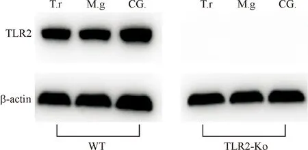
As shown in Figure 2, there were no significant changes in the expression of TLR2 protein in HaCaT cells co-culture with eitherT.rubrumorM.gypseumfor 24 h while expression of TLR2 protein was absent in TLR2-/-HaCaT cells stimulated with or without dermatophytes, indicating complete knockout of TLR2 gene.

Fig.2 The immunoblotting results of TLR2 protein in HaCaT cells co-cultured with T. rubrum and M. gypseum for 24 h and the verification of TLR2-/- HaCaT cells. Date are presented from one of three independent experiments.(T. r represents as T. rubrum; M. g represents as M. gypseum; CG. represents as control group)
2.3 Immunoblotting of proteins of c-Jun, and NF-κB signaling pathways in HaCaT cells infected by T. rubrum and M. gypseum, respectively
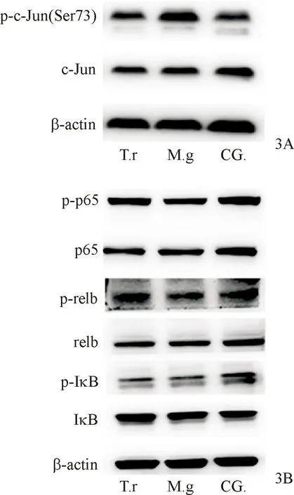
As shown in Figure 3A, the level of phosphorylated c-Jun protein did not change markedly in HaCaT cells co-cultured withT.rubrumfor 24 h, while co-culture of HaCaT withM.gypseumsignificantly increased phosphorylated c-Jun expression. However, neitherT.rubrumnorM.gypseumsignificantly changed the levels of total proteins and the phosphorylated p65, relb, and IκB in HaCaT cells after 24-hour co-culture (Figure 3B).
2.4 Immunoblotting of phosphorylated c-Jun protein in TLR2-/- HaCaT cells infected by T. rubrum and M. gypseum, respectively
The level of phosphorylated c-Jun decreased dramatically in TLR2-/-HaCaT cells co-cultured withT.rubrumfor 24 h while co-culture of TLR2-/-HaCaT cells withM.gypseumdid not change the level of phosphorylated c-Jun significantly (Figure 4). Data were from one of two independent experiments.

Fig.3 Immunoblotting results of relevant proteins of c-Jun signaling pathway (3A) and NF-κB signaling pathway (3B) in HaCaT cells co-cultured with T. rubrum and M. gypseum for 24 h. Date are from one of three independent experiments.

Fig.4 Immunoblotting results of proteins from c-Jun signaling pathway in TLR2-/- HaCaT cells co-cultured with T. rubrum and M. gypseum for 24 h. Data are from one of two independent experiments.
3 DISCUSSION
Dermatophytes are keratinophilic fungi that infect the epidermis and its appendages where they degrade keratin. Keratinocytes are the predominant cells of the epidermis. HaCaT cell line, a cell line derived from natural immortalized human keratinocytes[9], is the well-established research model of human keratinocytes to simulate dermatophytes infection[10].
Since NF-κB and MAPK signaling pathways are two classical pathways related to antifungal immunity, we studied the role of these two pathways in keratinocytes infected byT.rubrumandM.gypseum. The NF-κB transcription factor family is mainly composed of p65, relb and other proteins. Upon stimulation and activation, IκB is phosphorylated, and NF-κB is released to regulate gene expression[11]. The present study demonstrated no noticeable changes in the levels of total proteins and phosphorylated p65, relb and IκB in HaCaT cells co-cultured withT.rubrumandM.gypseumfor 24 h, suggesting thatT.rubrumandM.gypseuminfection do not activate NF-κB signaling pathway in keratinocytes. Therefore, the difference betweenT.rubrumandM.gypseuminfection of keratinocytes might be primarily associated with MAPK signaling pathway. Moreover, our previous studies of gene expression profile sequencing showed that c-Jun signaling pathway was activated in keratinocytes infected byM.gypseum, but not byT.rubruminfection[5]. Hence, c-Jun signaling pathway may be the main signaling pathway which contributes to the difference in inflammation between these two dermatophytes.
Achterma et al.[8]showed that MAPK signaling pathway was the crucial pathway activated by dermatophytes in primary keratinocytes and organotypic epidermal models. The activation of MAPK signaling cascade can activate AP-1[12], regulating the expression of cytokines, which play a key role in inflammatory responses. The c-Jun protein, encoded by proto-oncogene Jun gene, is the major component of transcription factor AP-1[13]. It is generally recognized that the phosphorylation of c-Jun protein can activate c-Jun/AP-1[14-16].
Our studies further validated that the levels of phosphorylated c-Jun were not significantly altered in HaCaT cells co-cultured withT.rubrum. In contrast,M.gypseumsignificantly increased phosphorylated c-Jun, indicating that the c-Jun signaling pathway is activated in HaCaT cells infected byM.gypseum, but not byT.rubrum. These results indicate thatT.rubrumandM.gypseuminfection differentially regulate expression of transcription factor c-Jun in MPAK pathway[5].
Prior studies have revealed the importance of TLR2 in antifungal immunity. For example, TLR2 on the surface of phagocytes mediates the phagocytosis of conidia ofT.rubrum[17], and keratinocytes can recognizeT.rubrumby TLR2 on the surface[18]. Here, we generated TLR2-deficient HaCaT cell using CRISPR-Cas9 technology to further investigate the role of TLR2 in the different clinical manifestations betweenT.rubrumandM.gypseuminfection which is mediated by c-Jun signaling pathway. Compared with RNAi technology[19]and antibody blocking method, CRISPR-Cas9 technology can completely block the expression of TLR2 at the gene level. We demonstrated that stimulation of TLR2-/-HaCaT cells withT.rubrummarkedly decreased the levels of phosphorylated c-Jun, indicating the requirement of TLR2 in activation of c-Jun signaling pathway in keratinocytes uponT.rubruminfection. However, it is puzzled that infection of normal keratinocyte withT.rubrumdoes not activate c-Jun signaling pathway. One possibility is that the ability of activating downstream c-Jun signaling by TLR2 is attenuated to some extent byT.rubrumbecauseT.rubrumcan suppress the expression of TLR2 in keratinocytes[18]. In addition, many other surface receptors in keratinocytes such as TLR4[20]and C-type lectin receptors (CLRs)[21]could also be involved in the recognition ofT.rubrum. An equilibrium state of inhibition of some receptors and the agonistic effect of TLR2 can result in no activation of c-Jun signaling pathway in normal keratinocytes infected byT.rubrum. But no significant change was observed in the levels of phosphorylated c-Jun in TLR2-/-HaCaT cells after stimulated byM.gypseum, indicating that c-Jun signaling pathway is not activated inM.gypseuminfection after TLR2 was knocked out. Collectively, we can infer that keratinocytes can recognizeM.gypseummainly through TLR2, and then activate c-Jun signaling pathway.
4 CONCLUSION
EitherT.rubrumorM.gypseuminfection does not activate NF-κB signaling pathway. BothT.rubrumandM.gypseumcan be recognized by TLR2 in keratinocytes and activate c-Jun signaling pathway.T.rubrum, but notM.gypseum, can inhibit c-Jun signaling pathway by some other receptors. On the whole,T.rubrumcannot activate c-Jun signaling pathway resulting mild inflammation, whileM.gypseumtrigger a series of strong inflammatory response by activating c-Jun signaling pathway. Further studies are needed to screen and identify which receptor mediates the inhibitory effect ofT.rubrumon c-Jun signaling pathway in keratinocytes and its mechanisms. This may help us understand the molecular mechanism of distinct immune responses of different ecological dermatophytes.

