Thoracic pyogenic infectious spondylitis presented as pneumothorax: A case report
Mi-Kyung Cho, Byeong-Ju Lee, Jae-Hyeok Chang, Young-Mo Kim
Mi-Kyung Cho, Byeong-Ju Lee, Jae-Hyeok Chang, Young-Mo Kim, Department of Rehabilitation Medicine, Pusan National University Hospital, Busan 49241, South Korea
Abstract BACKGROUND Pyogenic infectious spondylitis (PIS) is a rare condition, with an incidence between 0.2 and 2 cases per 100000 per annum. It’s most common symptom—back or neck pain—occurs in more than 90% of cases. Herein, we reported a case of thoracic PIS accompanied by pneumothorax in a 65-year-old male patient.CASE SUMMARY A 65-year-old man presented with right chest pain and dyspnea. The initial erect posteroanterior chest radiography revealed pneumothorax, which was further evaluated by chest computed tomography, revealing pleural effusion in the right lung and a paravertebral abscess with bony destruction of vertebral body. Based on magnetic resonance imaging, the patient was diagnosed with thoracic infectious spondylitis with an anterior paravertebral abscess. He was prescribed antibiotics and underwent neurosurgery due to aggravated symptoms and neurologic deficit. Tissue examination revealed that the cause of pleural effusion and pneumothorax was Staphylococcus aureus infection contiguously spread to lung pleura. After several surgical treatments with intravenous antibiotic therapy for two months and transition to oral antibiotics (rifampin 600 mg qd and ciprofloxacin 500 mg bid), the patient received physical therapy to recover balance. One month after discharge, the patient had no chest pain or dyspnea, and exhibited no elevation in inflammatory markers or new thoracic lesions.CONCLUSION To our knowledge, this is the very first report of a case of thoracic PIS with pneumothorax.
Key Words: Chest pain; Pneumothorax; Pleural effusion; Neurologic deficits; Spondylitis;Case report
INTRODUCTION
Pyogenic infectious spondylitis (PIS) is a rare infectious disorder, with an annual number of incident cases between 0.2 and 2 cases per 100000 people[1-3]. This disorder is defined as an inflammatory process of the spine and disc that may extend into the epidural space and paraspinal soft tissues. Spontaneous PIS manifests as a wide range of symptoms and a usually insidious progression. Back or neck pain is the most common symptom in more than 90% of PIS cases. Neurologic deficits and fever — two other major symptoms — are observed in 30% and less than 20% of cases, respectively.Other symptoms of PIS include nausea, vomiting, anorexia, weight loss, lethargy, and confusion[4-8]. Herein, we report a rare case of PIS that accompanied pneumothorax in a 65-year-old patient who complained of chest pain and dyspnea.
CASE PRESENTATION
Chief complaints
A 65-year-old man presented to a primary health care institution complaining of right chest pain and dyspnea.
History of present illness
The patient’s symptoms manifested approximately two months ago and had worsened within the last 24 h.
History of past illness
The patient had a medical history of type 2 diabetes, the most predisposing risk factor for PIS, and his glycated hemoglobin level was 9.4%.
Personal and family history
He had no remarkable family medical history. He smoked 10 cigarettes a day for 20 years. He drank 180 mL of 17 % alcohol three times a week. The amount of alcohol he drank per day was 13.1 g.
Physical examination
The patient complained of right chest pain with a rate of 7 in the visual analog scale and mild dyspnea. The patient’s temperature was 36.5 °C, heart rate was 84 bpm,respiratory rate was 16 breaths per minute, blood pressure was 100/60 mmHg, and oxygen saturation in room air was 97%. Neurological examination revealed no abnormalities.
Laboratory examinations
Blood analysis revealed no leukocytosis (white blood cell: 5080/μL) with mildly elevated serum C-reactive protein (1.6 mg/dL, normal range: < 0.5 mg/dL). Blood biochemistry parameters and urinalysis were normal.
Imaging examinations
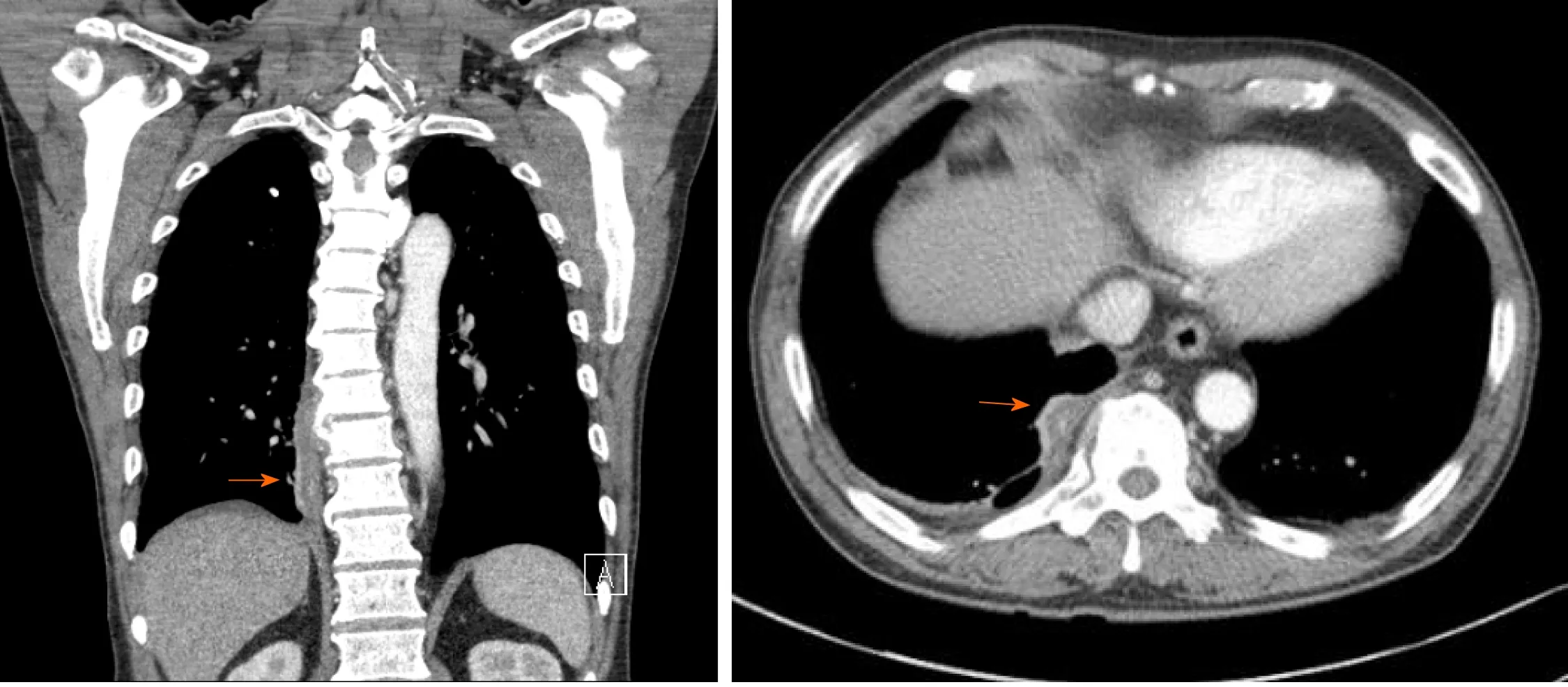
An initial erect posteroanterior chest radiography (CXR) performed at a previous primary health care institution revealed pneumothorax (Figure 1). When the CXR was repeated at our hospital, however, it revealed no abnormalities. On chest computed tomography (CT) scan, we observed pleural effusion in the right lung and a paravertebral abscess with bony destruction of the T9 vertebral body (Figure 2).
FINAL DIAGNOSIS
The final diagnosis is thoracic PIS due toStaphylococcus aureus.
TREATMENT
After several surgical procedures and intravenous antibiotic therapy for about two months, the patient’s treatment was transitioned to oral antibiotic therapy (rifampin 600 mg once and ciprofloxacin 500 mg twice daily). One month after the last surgery,he was transferred to the Department of Rehabilitation Medicine. A manual muscle test was conducted (scale: 0-5), and his upper extremities received a grade of 5 and lower extremities received a grade of 4. His feet exhibited severely impaired proprioception (2 successes in 10 trials), and he received a score of 8/56 on the Berg balance scale (BBS). He was provided physical therapy for balance and gait training for 2 wk.
OUTCOME AND FOLLOW-UP
At follow-up visit one month after discharge, the patient had no chest pain or dyspnea.Laboratory examination revealed no elevation in inflammatory markers and chest radiograph showed no new lesions. Additionally, the patient was able to walk independently with an assistive monocane. Over a 4-mo period, his proprioception improved from 2 to 4 successes, and his BBS score increased to 20.
DISCUSSION
PIS is a relatively rare condition. The predisposing factors of PIS included diabetes mellitus, long-term steroid therapy, malignancy, liver cirrhosis, chronic renal failure,malnutrition, substance abuse, human immunodeficiency virus infection, and septicemia. Excessive alcohol ingestion may also be counted among the major predisposing factor[4]. In this patient, regular alcohol intake and diabetes mellitus might had been predisposing factors. This disease predominantly affects people aged 50 years or older, and the incidence increases with every 10 years thereafter. Men are more susceptible than women to PIS, with a case ratio of 1.5-3:1. The most common symptoms include pain, fever, limited range of motion, and localized tenderness near the affected area, whereas neurological complications are rarely found. A diagnosis of suspected PIS is based on clinical symptoms.
PIS could result in potentially systemic (e.g.,endocarditis, liver abscess, and peritonitis) or local infectious complications (e.g.,psoas muscle abscess, and esophageal involvement) in rare cases[9-12]. Rushworthet al[13]reported an infant that was first diagnosed with pneumonia caused by a spinal epidural abscess withStaphylococcus aureusinfection. Except for this pediatric case, no other cases of pulmonary complications accompanied by PIS have been reported to the best of our knowledge.
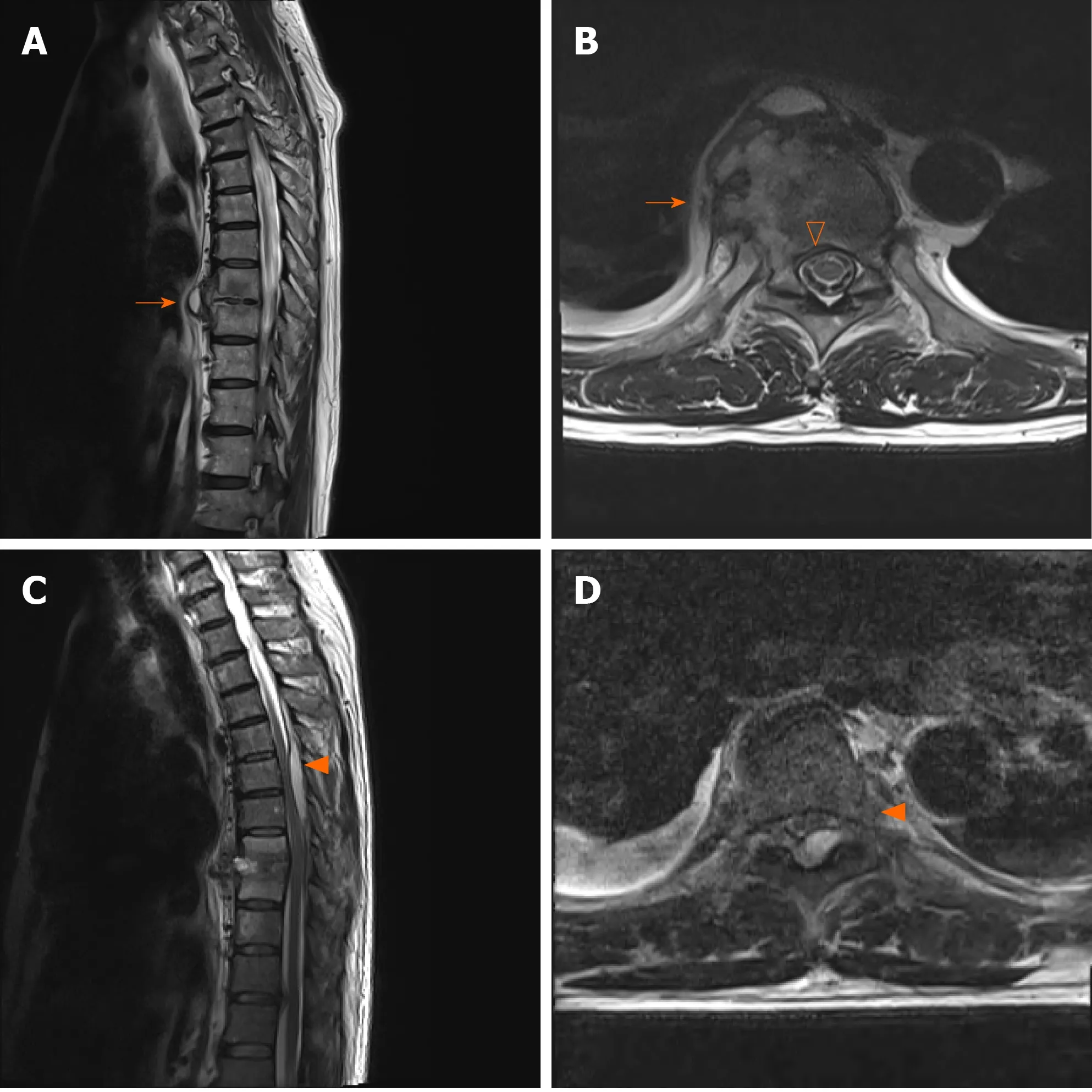
Anatomically, abscesses usually occur in the posterior region of the spine because lesions are more likely to develop in larger epidural spaces that contain infectionprone fat[14,15]. However, in this case anterior paravertebral abscess was connected with the pleural effusion with extension to T8/9 disc, right rib and anterior epidural space(Figure 3). It might have spread contiguously to the lung pleura to cause pleural effusion and pneumothorax. This would explain why the patient complained of the nonspecific symptoms of PIS, chest pain and dyspnea.

Figure 1 Visible pneumothorax (arrow) on an initial erect posteroanterior chest radiograph.

Figure 2 Initial chest computed tomography showing a paravertebral abscess (arrow) connected with the pleural effusion.
Pneumothorax can be divided into three categories: primary spontaneous pneumothorax (PSP), secondary spontaneous pneumothorax (SSP), and traumatic pneumothorax. PSP tends to occur in young adult patients without underlying lung problems and usually causes limited symptoms of chest pain and mild breathlessness.In contrast, SSP can be assumed in a patient with a history of lung disease or an age of 45 years or older with a habit of smoking (Table 1).
The clinical categorization of pneumothorax into PSP and SSP is of great importance, as the treatment of SSP differs from that of PSP. Schnellet al[16]suggested that chest CT scans should be performed in cases where findings are unclear or if the patients are suspected to suffer from SSP. According to the algorithm[16], the patient underwent a CT scanning, was given a final diagnosis of PIS, and received proper surgical treatment. To our knowledge, this is the very first report of a case of thoracic PIS with pneumothorax.
CONCLUSION
PIS is a rare infectious disorder. Back or neck pain is the most common symptom in over 90% of cases along with two other major symptoms, neurologic deficits and fever.We reported a rare case of thoracic PIS accompanied by pneumothorax with a chief complaint of chest pain and dyspnea. To our knowledge, this is the very first report of a case of PIS with pneumothorax.

Table 1 Characteristics of primary and secondary pneumothorax (according to Bobbio et al[17])

Figure 3 Magnetic resonance imaging of the thoracic spine. A and B: Initial magnetic resonance imaging (MRI) scan showing a paravertebral abscess with extension to T8/9 disc, right rib (arrow), and anterior epidural space (empty arrowhead); C and D: Follow-up MRI scan showing an epidural abscess (arrowhead)at the T4-7 Level with spinal cord compression at T6-7.
 World Journal of Clinical Cases2021年6期
- World Journal of Clinical Cases的其它文章
- Interactive platform for peer review: A proposal to improve the current peer review system
- Animal models of cathartic colon
- New indicators in evaluation of hemolysis, elevated liver enzymes,and low platelet syndrome: A case-control study
- Analysis of hospitalization costs related to fall injuries in elderly patients
- Effect of alprostadil in the treatment of intensive care unit patients with acute renal injury
- Etomidate vs propofol in coronary heart disease patients undergoing major noncardiac surgery: A randomized clinical trial
