Healthy individuals vs patients with bipolar or unipolar depression in gray matter volume
Yin-Nan Zhang, Hui Li, Zhi-Wei Shen, Chang Xu, Yue-Jun Huang, Ren-Hua Wu
Yin-Nan Zhang, Department of Rehabilitation Medicine, Mental Health Center of Shantou University, Shantou 515000, Guangdong Province, China
Hui Li, Mental Health Center of Shantou University, Shantou 515000, Guangdong Province,China
Zhi-Wei Shen, Philips Healthcare China, Beijing 100000, China
Chang Xu, Mental Health Center of Shantou University, Shantou 515000, Guangdong Province,China
Yue-Jun Huang, Department of Pediatrics, The Second Affiliated Hospital of Shantou University Medical College, Shantou 515000, Guangdong Province, China
Ren-Hua Wu, Department of Medical Imaging, The Second Affiliated Hospital of Shantou University Medical College, Shantou 515041, Guangdong Province, China
Abstract BACKGROUND Previous studies using voxel-based morphometry (VBM) revealed changes in gray matter volume (GMV) of patients with depression, but the differences between patients with bipolar disorder (BD) and unipolar depression (UD) are less known.AIM To analyze the whole-brain GMV data of patients with untreated UD and BD compared with healthy controls.METHODS Fourteen patients with BD and 20 with UD were recruited from the Mental Health Center of Shantou University between August 2014 and July 2015, and 20 nondepressive controls were recruited. After routine three-plane positioning, axial T2WI scanning was performed. The connecting line between the anterior and posterior commissures was used as the scanning baseline. The scanning range extended from the cranial apex to the foramen magnum. Categorical data are presented as frequencies and were analyzed using the Fisher exact test.RESULTS There were no significant intergroup differences in gender, age, or years of education. Disease course, age at the first episode, and Hamilton depression rating scale scores were similar between patients with UD and those with BD.Compared with the non-depressive controls, patients with BD showed smaller GMVs in the right inferior temporal gyrus, left middle temporal gyrus, right middle occipital gyrus, and right superior parietal gyrus and larger GMVs in the midbrain, left superior frontal gyrus, and right cerebellum. In contrast, UD patients showed smaller GMVs than the controls in the right fusiform gyrus, left inferior occipital gyrus, left paracentral lobule, right superior and inferior temporal gyri, and the right posterior lobe of the cerebellum, and larger GMVs than the controls in the left posterior central gyrus and left middle frontal gyrus.There was no difference in GMV between patients with BD and UD.CONCLUSION Using VBM, the present study revealed that patients with UD and BD have different patterns of changes in GMV when compared with healthy controls.
Key Words: Bipolar disorder; Unipolar depression; Gray matter; Functional magnetic resonance imaging; Classification techniques; Voxel-based morphometry
INTRODUCTION
Bipolar disorder (BD) is a severe mental illness featured by episodes of mania and depression in an alternating pattern, accompanied by changes in activity or energy and cognitive, physical, and behavioral symptoms[1,2]. The overall incidence of BD worldwide is approximately 2%, with the disease's subthreshold forms affecting another 2% of the global population[3]. The estimated lifetime prevalence of BD based on data from 11 countries is 0.4%-1.4%[4]. According to data from the World Health Organization, the disability rate associated with BD ranks sixth in the youth population, causing a heavy financial burden on families and society[5].
In clinical practice, BD type I is characterized by at least one episode of mania. In comparison, BD type II is characterized by at least one episode of depression and at least one hypomanic episode, without mania episodes[1,2]. However, since the disease episodes must be clinically monitored to determine the diagnosis, the two types are challenging to identify in the disease's early stages. As a result, diagnosis may be delayed by up to 10 years or even longer in one-third of patients with BD[6], and 60% of patients with BD who sought treatment for depressive symptoms were initially misdiagnosed as having unipolar depression (UD)[7]. Thus, the similarities in the clinical symptoms of BD and UD cause difficulties in a timely and accurate diagnosis of the two conditions[8].
Several clinical studies have attempted to identify biological indicators that can distinguish UD from BD[9-11]. Elevated plasma uric acid levels may be associated with emotional instability in patients with BD, while low uric acid levels may be related to UD patients’ depressive mood[10]. Similarly, patients with BD show high plasma levels of nerve growth factor (NT)-3 and NT-4/5[11]. Likewise, neuroelectrophysiological studies have suggested that the auditory steady-state response is a potential electrophysiological marker for distinguishing between BD and UD[9]. While patients with BD and UD show some differences in electrophysiological and biochemical indicators, the evidence for using these indicators in differential diagnosis remains inadequate.
Voxel-based morphometry (VBM) is an automatic, comprehensive, and objective technique for the analysis of brain structural magnetic resonance (MR) images[12]. In VBM, gray and white matter lesions in the brain are evaluatedviaquantitative analysis of the changes in brain gray and white matter volumes of each voxel of MR images.VBM is widely used in research to study a variety of neuropsychiatric diseases such as Alzheimer’s disease[13], depression[14,15], bipolar disorder[15,16], and schizophrenia[17],providing reliable imaging data regarding the changes in brain morphology in those diseases.
Previous VBM studies have reported gray matter structural abnormalities in patients with BD and depression. In patients with UD, smaller gray matter volume(GMV) are observed in the anterior cingulate gyrus[18], hippocampus[19], amygdala[18,19],caudate nucleus[14], frontal lobe[20], parietal lobe[20], temporal lobe[21], and cerebellum[21].In contrast, the brain regions reported to show smaller GMVs in patients with BD include the frontal lobe[22]and the temporal lobe[23]. Since very few studies have directly compared the VBM findings in the two diseases, the exact differences in GMV changes between patients with BD and UD remain unclear.
The results of the available VBM studies also lacked consistency because of methodological differences[24,25]. For example, De Azevedo-Marques Pericoet al[24]showed that the GMV of the right lateral anterior cingulate cortex in BD patients was larger than that in normal controls. In comparison, the GMV of the bilateral dorsolateral prefrontal cortex in patients with UD was smaller than that in controls.Patients with UD and BD showed significant GMV differences at the level of the right dorsolateral prefrontal lobe. In contrast, Wiseet al[25]performed a meta-analysis of the VBM results in patients with BD and UD and showed that the GMVs of the bilateral insular lobe and the dorsomedial and ventromedial frontal cortices are smaller in both groups in comparison with normal controls, while the GMVs of the right frontal gyrus,left hippocampus and parahippocampal gyrus, right inferior temporal gyrus and fusiform gyrus, left inferior parietal lobule, and right cerebellar vermis are smaller in patients with UD than in those with BD[25]. Redlichet al[26]showed reduced GMV in the anterior cingulate gyrus in American and German patients with UD compared with BD and suggested a pattern classification (based on a multivariable analysis of GMV,white matter volume, and structural abnormalities) that had 79% accuracy. From Singapore, Caiet al[27]showed that patients with BP1 and UD have lower GMV in the right frontal gyrus, but that lower GMV in the right middle cingulate gyrus might be specific to BP1.
Nevertheless, although differences in the GMV distributions in patients with UD and BD have been reported across multiple studies, only rare studies[26,27]have attempted to determine whether these differences can be used to distinguish between BD and UD. Because of the possible cultural, language, ethnic, and technical [magnetic resonance imaging (MRI) sequences] differences in VBM results, this study aimed to use VBM to analyze the whole-brain GMV data of patients with untreated UD and BD compared with healthy controls.
MATERIALS AND METHODS
Study design and subjects
This study included 14 patients with BD and 20 patients with UD admitted to the outpatient department of the Mental Health Center of Shantou University between August 2014 and July 2015. The study protocol was approved by the ethics committee of Shantou University Medical College ([2017]0301). All patients voluntarily participated in the study and signed an informed consent form.
All patients were diagnosed according to the diagnostic criteria of the Diagnostic and Statistical Manual of Mental Disorders (DSM)-IV[28]. Intelligence quotient (IQ) was tested using the Wechsler Adult Intelligence Scale-Revised in China[29,30]. Depression was evaluated using the Hamilton depression rating scale (HDRS)[31], validated in Chinese[32].
For the UD group, the inclusion criteria were: (1) Meeting the diagnostic criteria for recurrent depression in DSM-IV (code 32.4); (2) 18-60 years of age; (3) HDRS score ≥17; (4) Right-handedness; (5) IQ > 70; and (6) No history of drug consumption for at least 2 wk. For the BD group, the inclusion criteria were as follows: (1) Meeting the diagnostic criteria for bipolar disorder depressive episodes in DSM-IV (codes 31.5 and 31.6); (2) 18-60 years of age; (3) HDRS score ≥ 17; (4) Right-handedness; (5) IQ > 70; and(6) No history of drug consumption for at least 2 wk. The exclusion criteria were: (1)Schizophrenia or schizoaffective psychosis; (2) Substance dependence; (3) Severe physical and Neurological diseases; (4) Pregnancy or lactation; (5) Intrauterine contraceptive ring usage; (6) Presence of a cardiac pacemaker or other implantable metal devices; and (7) Left-handedness.
Twenty non-depressive individuals were recruited as controls, based on the following criteria: (1) No history of significant physical or mental illness; (2) 18-60 years of age; (3) Right-handedness; and (4) IQ > 70.
Head MRI
After routine three-plane positioning, axial T2WI scanning was performed. The connecting line between the anterior and posterior commissures was used as the scanning baseline. The scanning range extended from the cranial apex to the foramen magnum. The scanning parameters were as follows: Repetition time (TR), 7000 ms;Echo time (TE), 107.3 ms; Layer thickness, 5 mm; Layer spacing, 1 mm; Matrix, 384 ×384; Field of view (FOV), 240 × 240 mm2; Bandwidth, 62.5; And number of excitations(NEX), 2. T1 fluid-attenuated inversion recovery scanning was performed with the following parameters: TR, 1750 ms; TE, 24 ms; Matrix, 320 × 224; and FOV, 240 × 240 mm2. Axial scanning was performed using the 3D-BRAVO sequence (T1WI) with the following parameters: TR, 6 ms; TE, 1.8 ms; Flip angle, 15°; Layer thickness, l mm;Continuous scanning with no intervals; FOV, 256 mm × 256 mm; Matrix, 256 × 256;NEX, l; Bandwidth, 41.67 Hz; number of scanning layers, 168; Voxel size, 1 mm × 1 mm × 1 mm; And scanning time, 2 m 59 s.
Data collection
Information about demographics (gender, age, income, employment status, and years of education) and clinical characteristics (age at the first episode, disease course, and HDRS scores) of the subjects were collected.
Image processing and data analysis
The VBM8 and DARTEL toolkits in the SPM8 software package (Statistical Parametric Mapping, http://www.fil.ion.ucl.ac.uk/spm) were used to process structural image data, as described previously[33]. Post-processing was performed using the DARTEL algorithm. The specific processing steps were: (1) Format conversion: The original data in DICOM format was converted to the NIfTI format; (2) Segmentation: Images of the individual brain structures were segmented to obtain white matter, gray matter, and cerebrospinal fluid images; (3) Template establishment: Three groups of subjects (BD,UD, and controls) were used to generate the template, and the DARTEL algorithm was used to estimate the optimal registration template for each participant; (4) Volume modulation map generation: Space was normalized to the Montreal Neurological Institute (MNI) space[34]to generate the volume modulation map; (5) Data smoothing:A Gaussian kernel with a full-width at half maximum of 8 mm was used for smoothing the data; and (6) Statistical analysis: Statistical analysis was performed on the data by using a two-samplettest in SPM8. The Alpha Sim application was used for correction for multiple comparisons. Age, sex, and total intracranial volume were used as covariates. After false discovery rate correction,P< 0.05 and a voxel threshold of >100 were considered significant, at least statistically. The voxels with statistical significance were superimposed on 3D MNI standard templates to generate pseudocolor maps. The characteristics of GMV changes in the whole brains of the three groups were analyzed.
Categorical data are presented as frequencies and were analyzed using the Fisher exact test. Continuous data are presented as the mean ± SD and were analyzed using one-way analysis of variance (ANOVA) with the SNK post hoc test. SPSS 21.0 (IBM,Armonk, NY, United States) was used for statistical analysis.Pvalues < 0.05 were considered statistically significant.
RESULTS
Characteristics of the subjects
MRI was performed in 14 patients with BD, 20 patients with UD, and 20 nondepressive volunteers. One patient with BD who showed a subarachnoid cyst was excluded. There were no significant intergroup differences in gender, age, or years of education (P> 0.05 for all three characteristics, ANOVA). Disease course, age at the first episode, and HDRS scores were similar between patients with UD and those with BD (Table 1).
GMV analyses in BD patients
The BD group showed significantly smaller GMVs in the right inferior temporal gyrus,left middle temporal gyrus, right middle occipital gyrus, and right superior parietal gyrus compared with healthy controls. The GMVs in the midbrain, left superior frontal gyrus, and right cerebellum in the BD group were larger than those in the controls(Table 2 and Figures 1 and 2).
GMV analyses in UD patients
The UD group showed smaller GMVs in the right fusiform gyrus, left inferior occipital gyrus, left paracentral lobule, right superior temporal gyrus, right inferior temporal gyrus, and right posterior cerebellar lobe compared with healthy controls. The GMVs in the left post central gyrus and left middle frontal gyrus in the UD group were larger than those in the controls (Table 3 and Figures 3 and 4).
GMV in BD vs UD
Between patients with UD and those with BD, the GMVs in the superior frontal gyrus(orbital part; 963 mm3vs958 mm3), middle frontal gyrus (3294 mm3vs3301 mm3),insula (1770 mm3vs1761 mm3), cuneus (1488 mm3vs1495 mm3), amygdala (114 mm3vs109 mm3), thalamus (1667 mm3vs1678 mm3), caudate (994 mm3vs989 mm3), and supramarginal gyrus (1973 mm3vs1981 mm3) were not significantly different (Table 4 and Figure 5;P> 0.05 for all).
DISCUSSION
BD is often misdiagnosed as UD[8], and none of the existing biochemical or neuroelectrophysiological markers have been validated for the differential diagnosis of these two diseases[9-11], and most functional imaging studies did not directly compare the two conditions or yielded inconsistent findings[24-27,35]. Therefore, this study examined whether the GMV changes identified in VBM could distinguish UD from BD. The results show that both UD and BD patients showed significant changes in GMV when compared with healthy controls and that the patterns of changes were different. Still, the differences did not reach statistical significance between the UD and BD groups, probably because of study limitations. Although different patterns of change were observed, the present study does not suggest VBM as a supplementary technique for the differential diagnosis of UD and BD. Additional studies with a larger sample size is necessary to confirm this result.
The study by Redlichet al[26]was conducted in parallel at two centers (one in the United States and one in Germany). They showed that UD and BD could be distinguished using a multivariable model that includes GMVs (anterior cingulate gyrus), and consistent results were obtained between the two centers. Of note, the two centers shared similar patients,i.e, Caucasians of European ancestry and languages sharing the same root. Caiet al[27]showed significant differences in GMVs between BP1 and UD patients in the right middle cingulate gyrus, and that this region could be used to differentiate the two diseases. Chenet al[35], after adjusting for age, sex, body mass index, duration of illness, trivalent inactivated virus vaccine, lithium, and symptoms, showed that GMVs in the right orbitofrontal cortex and left lingual gyrus were significantly different between UD and BD patients. Besides, Liet al[36]and Tanet al[37]revealed differences in neuromatabolite levels between UD and BD, based on multi-voxel proton MR spectroscopy. Here, the results showed no significant differences between UD and BD for any part of the brain, which contradicts the studies above. Some factors could be responsible for those discrepancies. Chinese patients without drugs for at least 2 wk were recruited in the present study. Cultural and ethnic differences can be observed in the functional MRI data[38,39]and could accountfor the discrepancies. Of course, drugs used in psychiatry affect brain functions[40]. In addition, the MRI sequences are known to affect the brain function results[41,42]. The present study used the 3D-BRAVO protocol, while Caiet al[27]and Redlichet al[26]used T1W images, and Chenet al[35]used the BRAVO sequence. The discrepancies can also be attributed to the small number of participants. Another limitation was the lack of follow-up and re-imaging examinations after treatment, precluding comparisons of patients before and after treatment. Additional studies are still necessary to determine whether VBM can distinguish between UD and BD, especially in the context of interpopulation variability. As suggested by Redlichet al[26], the answer may lie in multivariable models or, as suggested by Chenet al[35], in a combination of VBM and biochemical parameters.

Table 1 Demographic and clinical data of the subjects
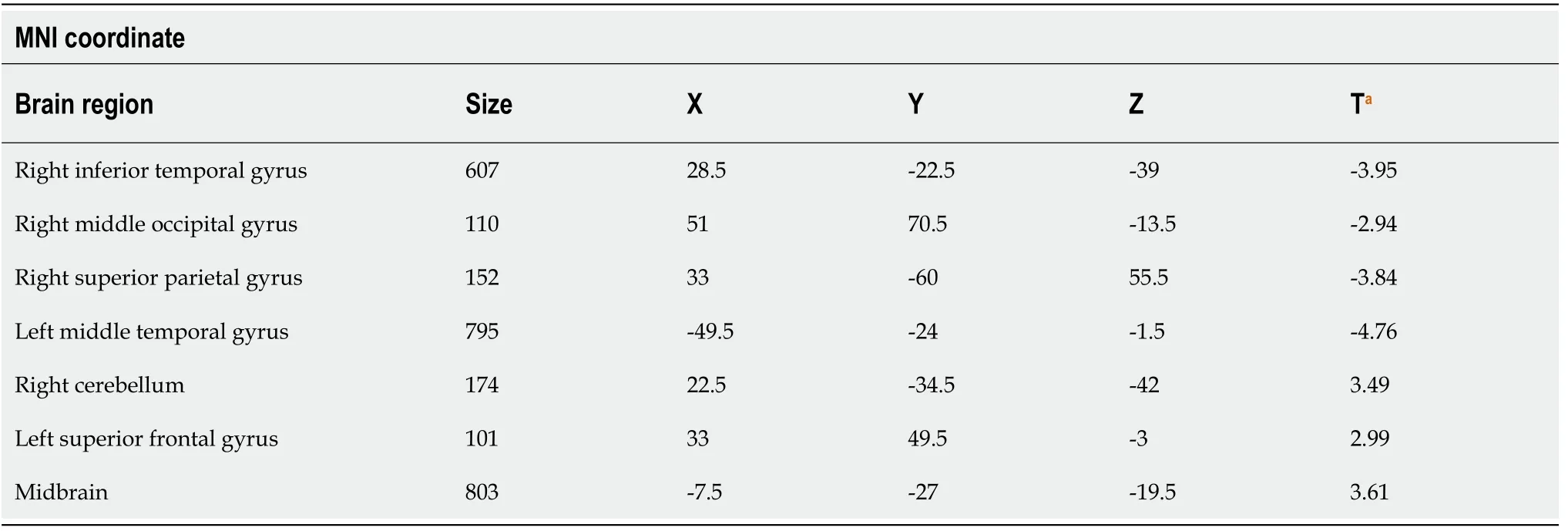
Table 2 Distribution of brain regions with larger/smaller gray matter volume in the bipolar depression group in comparison with the control group
The present study revealed two different patterns of changes in GMV between healthy controls and UD and BD. Still, no statistically significant difference was observed in GMV between UD and BD patients. The two groups showed the involvement of different brain regions compared to the normal control group, which possibly supports the differences in the pathophysiological mechanisms between the two groups. In addition, even though the patients did not receive treatment for at least 2 wk before undergoing functional imaging, they were not newly diagnosed patients.Some previously administered drugs may have had mid- and long-term effects on the brain's functional areas. Additional studies are essential to determine the exact mechanisms involved in the pathogenesis of BD and UD. Most studies reported that BD and UD patients had smaller cortical volumes in some brain regions; Some studies have also reported larger cortical volumes of local brain regions in depressive patients.For example, Qiuet al[43]studied patients with first-episode depression without medication and found that the GMVs of the right orbitofrontal cortex, right middle frontal gyrus, and right superior margin gyrus were larger than those in the controls.Similarly, Papmeyeret al[44]reported that patients with depression had larger leftinferior frontal gyrus volume compared with the controls. Since compensatory changes in the GMV in different regions have been previously reported in patients with vestibular neuritis[45], the larger GMV in the left superior frontal gyrus in patients with BD and the left middle frontal gyrus in patients with UD might be compensatory responses to the reduction in other regions’ GMV. Still, this hypothesis has to be confirmed in future studies.
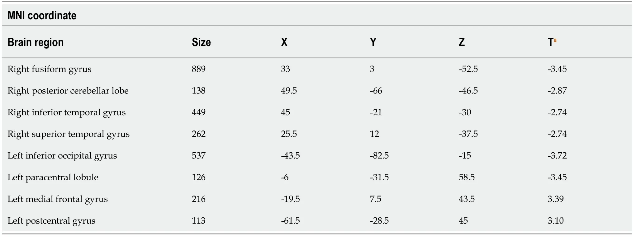
Table 3 Distribution of brain regions with larger/smaller gray matter volume in the unipolar depression group in comparison with the control group

Table 4 Gray matter volumes in brain regions of the unipolar depression group in comparison with the bipolar depression group
Compared with controls, the present study showed that patients with UD had a smaller GMV in the right cerebellum posterior lobe, while patients with BD had a larger GMV in the right cerebellum. The cerebellum is located behind the cerebral hemisphere and consists of the bilateral cerebellar hemispheres and the cerebellar vermis in the middle. Although the cerebellum was previously thought to mainly regulate the movement and balance functions of the human body, the cerebellum is also involved in higher brain function regulation, including cognition and emotions through multiple afferent and efferent neural pathways[46]. The cerebellum is involved in the pathogenesis of BD and UD[47-49]. Here, the results suggest that the pathogenesis of BD and UD involves the cerebellum, but the specific mechanisms need to be elucidated in future studies.
The temporal lobe is located below the frontal and parietal lobes, in front of the occipital lobe, and below the lateral fissure. It is divided into the superior temporal gyrus, middle temporal gyrus, inferior temporal gyrus, and superior temporal sulcus and inferior temporal sulcus[50,51]. The temporal lobe is responsible for processing auditory information and is also related to memory and emotions. The temporal and parietal lobes are crucially involved in regulating emotion by the neural circuits connected with the nerve fiber bundle and frontal lobe[52,53]. Here, patients with BD had smaller GMV of the right temporal lobe, as supported by Chenet al[54], who suggested that the temporal lobe was damaged and that the extensive damage to the temporal lobe structure might be involved in the pathogenesis of BD.
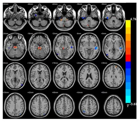
Figure 1 Distribution of brain regions with larger/smaller gray matter volume in the bipolar depression group compared with the control group. Red: Larger gray matter volume (GMV); Blue: Smaller GMV.
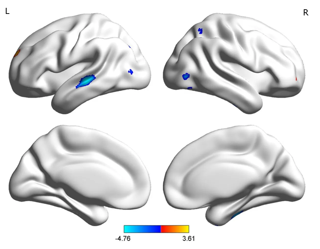
Figure 2 Comparison of voxel-based morphometry results between the bipolar depression group and the normal control group (Brainnet viewer). Red: Larger gray matter volume; Blue: Smaller gray matter volume.
Based on the combination of anatomical nerve connections and functional regions,Koenigset al[55]proposed dividing the prefrontal cortex (PFC) into the dorsolateral PFC(dlPFC) and the ventromedial PFC. The dlPFC mainly includes the middle frontal gyrus and the superior frontal gyrus and mainly receives specific signals from the sensory cortex. It is closely associated with the premotor and oculomotor areas of the frontal lobe and the lateral parietal cortex. Therefore, it is mainly responsible for cognitive and executive functions, including operational working memory, purposeful behavior, abstract thinking, and attention control[56]. Thus, the frontal lobe plays an important role in the pathogenesis of BD and UD[57].
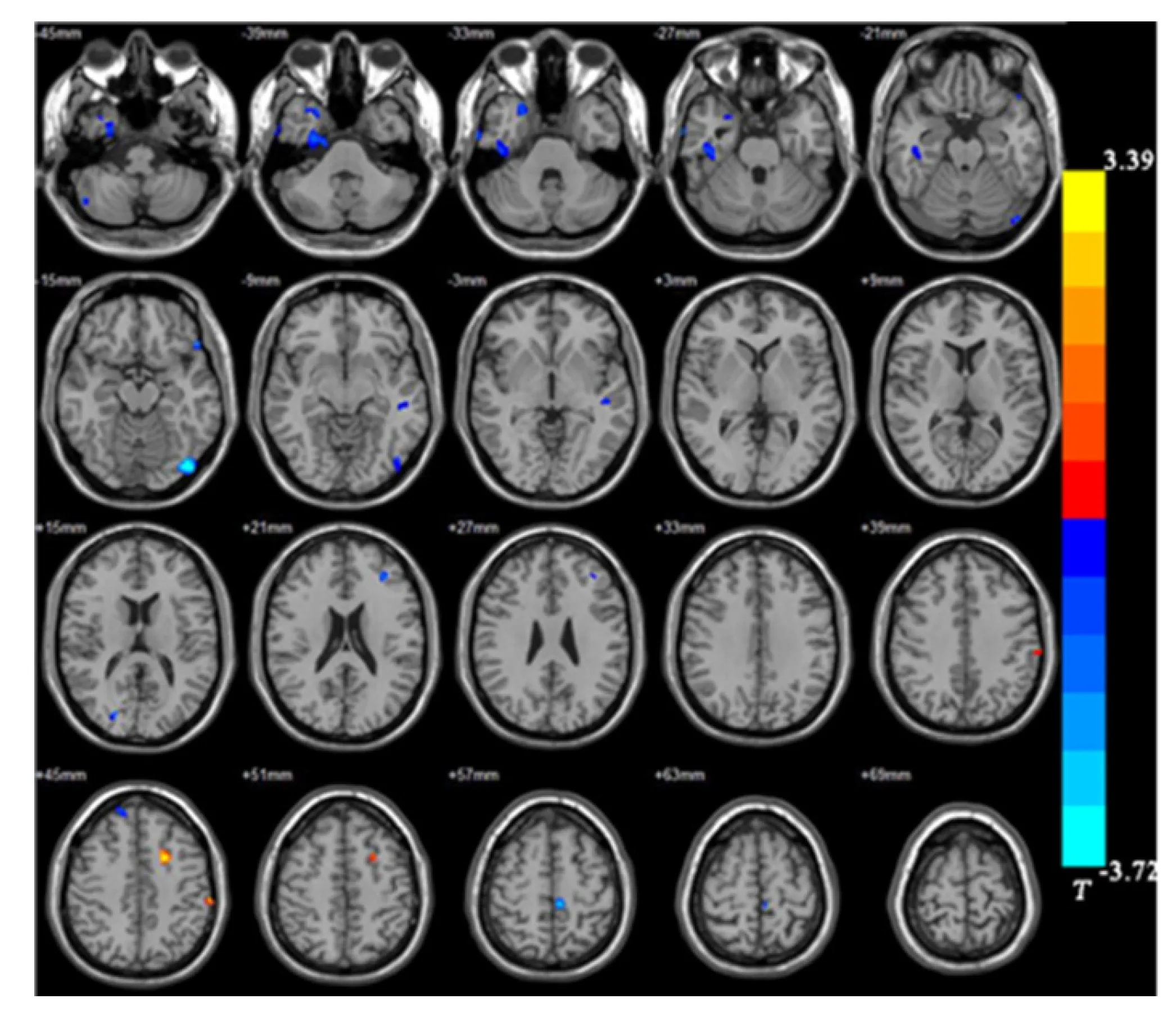
Figure 3 Distribution of brain regions with larger/smaller gray matter volume in the unipolar depression group compared with the control group. Red: Larger gray matter volume (GMV); Blue: Smaller GMV.

Figure 4 Comparison of voxel-based morphometry results between the unipolar depression group and the normal control group(Brainnet viewer). Red: Larger gray matter volume (GMV); Blue: Smaller GMV.
The midbrain is located above the pons and is the midpoint of the whole brain. It is the reflection center for vision and hearing[58]. Lauterbachet al[59]reported that midbrain lesions could cause BD, while Wanget al[60]found that reduced functional connections in the midbrain were associated with a polygenic genetic risk in patients with BD[60]. Another study[61]also reported abnormal connections between the striatum and midbrain in patients with BD and bipolar mania. These results support the involvement of the midbrain in the pathogenesis of BD. The present study showed a larger GMV in the midbrain of patients with BD compared to the controls, which might also be a compensatory midbrain response to the smaller GMV in the primary center. However, this hypothesis has to be validated in future studies as well.
This study has limitations. The small sample size is, of course, a significant limitation, especially when using a whole-brain approach (instead of a priori regions of interest to reduce risk of type II error) and performing a comparison (BDvsUD)where large effect sizes are not expected[62]. This could have resulted in a lack of statistical power, especially when comparing UDvsBD. In addition, sex representation was unbalanced in the UD group.
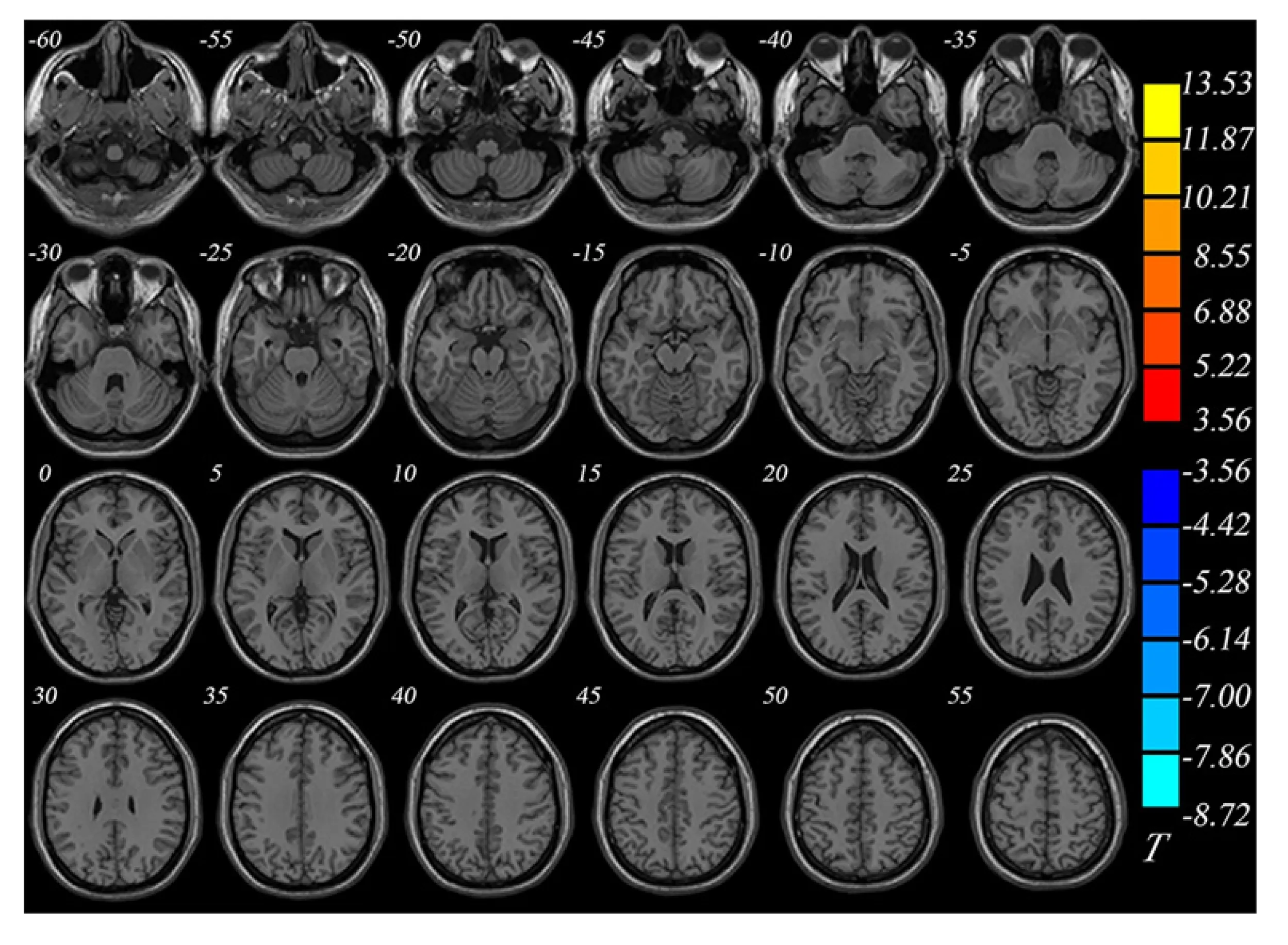
Figure 5 Comparison of gray matter volumes between the unipolar depression group and bipolar depression group (Brainnet viewer).
CONCLUSION
In conclusion, UD patients showed smaller GMVs than the controls in the right fusiform gyrus, left inferior occipital gyrus, left paracentral lobule, right superior and inferior temporal gyri, and right posterior lobe of the cerebellum, and larger GMVs than the controls in the left posterior central gyrus and left middle frontal gyrus. There were no differences in GMV between patients with BD and those with UD. Although different patterns of change were observed, the present study does not suggest VBM as a supplementary technique for the differential diagnosis of UD and BD, and additional studies with a larger sample size are necessary. Classification techniques based on machine learning could be explored[26].
ARTICLE HIGHLIGHTS
Research background
Previous studies using voxel-based morphometry (VBM) revealed changes in gray matter volume (GMV) patients with depression.
Research motivation
The differences of GMV using VBM between bipolar disorder (BD) and unipolar depression (UD) are less known.
Research objectives
To analyze the whole-brain GMV data of patients with untreated UD and BD compared with healthy controls.
Research methods
Patients with BD, those with UD, and non-depressive controls were enrolled to further analyze the brain images.
Research results
There were differences in GMV between UD patients and controls, as well as between BD patients and controls. There were no differences in GMV between UD and BD patients.
Research conclusions
BM might have a low value for differentiating between UD and BD. However, patients with UD and BD had different patterns of changes in GMV when compared with healthy controls.
Research perspectives
Classification techniques based on machine learning could be explored.
 World Journal of Clinical Cases2021年6期
- World Journal of Clinical Cases的其它文章
- Interactive platform for peer review: A proposal to improve the current peer review system
- Animal models of cathartic colon
- New indicators in evaluation of hemolysis, elevated liver enzymes,and low platelet syndrome: A case-control study
- Analysis of hospitalization costs related to fall injuries in elderly patients
- Effect of alprostadil in the treatment of intensive care unit patients with acute renal injury
- Etomidate vs propofol in coronary heart disease patients undergoing major noncardiac surgery: A randomized clinical trial
