囊性包虫病所致过敏性休克中IgE、IgG1与T细胞激活的关系
2020-08-27 20:01李杰戴庆亚力·亚森
中国医药导报 2020年19期
李杰 戴庆 亚力·亚森

[摘要] 目的 探索囊性包蟲病所致过敏性休克过程中特异性变应原与T细胞激活的关系,为临床治疗提供新思路。 方法 采集2015年1月~2017年12月在新疆医科大学第一附属医院行囊性包虫病内囊摘除术30例患者(研究组)血清,其中术中发生过敏性休克患者作为过敏性休克组(10例),未发生过敏性休克患者作为无过敏性休克组(20例);另选同期健康体检者血样作为对照组(30名)。分析各组人群血清免疫球蛋白(Ig)E与IgG1水平,检测各组人群血清中白细胞介素(IL)-10、IL-4、IL-2与γ干扰素(IFN-γ)表达量,并进行相关性分析。结果 过敏性休克组术中IgE和IgG1表达量高于无过敏性休克组,差异有统计学意义(P < 0.05)。术后1周各组人群血清中IgE和IgG1表达量比较,差异无统计学意义(P > 0.05)。术前研究组血清IL-4与IL-10表达量高于对照组,差异有统计学意义(P < 0.05)。过敏性休克组术中血清IL-4、IL-10低于无过敏性休克组,IFN-γ、IL-2高于无过敏性休克组,差异有统计学意义(P < 0.05)。术后1周过敏性休克组血清IL-4、IL-10表达量高于无过敏性休克组和对照组,差异有统计学意义(P < 0.05)。IgE与IL-2、IFN-γ呈正相关(r = 0.96、0.68,P < 0.05),IgG1与IL-2、IFN-γ呈正相关(r = 0.60、0.79,P < 0.05)。 结论 过敏性休克发生主要是Th、Th2型细胞失衡,其中Th1型在过敏性休克中占主导作用,其激活与IgE和IgG1相关。
[关键词] 囊性包虫病;过敏性休克;Th1;Th2
[中图分类号] R532.32 [文献标识码] A [文章编号] 1673-7210(2020)07(a)-0093-04
[Abstract] Objective To explore the relationship between specific allergens and T cell activation during anaphylactic shock caused by cystic echinococcosis, and to provide new ideas for clinical treatment. Methods Serum was collected from 30 patients (research group) who were underwent cystic hydatid cystectomy in the First Affiliated Hospital of Xinjiang Medical University from January 2015 to December 2017. The patients with anaphylactic shock during the operation were taken as the anaphylactic shock group (10 cases), while patients without anaphylactic shock were taken as the non-anaphylactic shock group (20 cases), and the blood samples of healthy examinees from the same period were selected as the control group (30 people). The levels of serum immunoglobulin (Ig) E and IgG1 in each group were analyzed, while the expression levels of interleukin (IL)-10, IL-4, IL-2 and γ interferon (IFN-γ) in the serum of each group were detected, and correlation analysis was performed. Results The expression levels of IgE and IgG1 during the operation in the anaphylactic shock group were higher than those in the non-anaphylactic shock group, and the differences were statistically significant (P < 0.05). There were no statistically significant differences in serum IgE and IgG1 expression levels among the groups after 1 week of operation (P > 0.05). The expression levels of serum IL-4 and IL-10 before the operation in the study group were higher than those in the control group, and the differences were statistically significant (P < 0.05). The serum IL-4 and IL-10 during the operation in the anaphylactic shock group were lower than those in the non-anaphylactic shock group, while the IFN-γ and IL-2 were higher than those in the non-anaphylactic shock group, and the differences were statistically significant (P < 0.05). The serum IL-4 and IL-10 expression levels of the anaphylactic shock group were higher than those of the non-anaphylactic shock group and the control group after 1 week of operation, and the differences were statistically significant (P < 0.05). IgE was positively correlated with IL-2 and IFN-γ (r = 0.96, 0.68, P < 0.05), while IgG1 was positively correlated with IL-2 and IFN-γ (r = 0.60, 0.79, P < 0.05). Conclusion As long as anaphylactic shock occurs, Th1 and Th2 type cells are imbalanced. Th1 type plays a leading role in anaphylactic shock, and its activation is related to IgE and IgG1.
[Key words] Cystic echinococcosis; Anaphylactic shock; Th1; Th2
包虫病(echinococcosis)是棘球绦虫寄生于宿主体内所导致的一种严重人畜共患病,疾病呈世界性分布[1-3]。人体感染包虫后,幼虫在人体内形成囊性包虫,导致囊性包虫病。目前,全世界约有3百万囊型包虫病(Eg)或泡型包虫病(Em)患者[4-5],囊性包虫病所致过敏性休克(anaphylactic shock,AS)是患者死亡的重要并发症。前期研究发现[6-8],无论术前术后,AS患者免疫球蛋白(Ig)E,IgG和IgG1均显著升高,且术前IgG和IgG1水平升高是AS重要的风险因子。同时,Th1、Th2型细胞在过敏性休克中具有相反效应,这两种细胞失衡在疾病发生过程中具有重要意义[9-10]。Th1、Th2型细胞在疾病的不同时期出现,AS发生时Th1型较Th2型发挥的作用更为明显[10-11],但具体机制尚不明确。本研究通过不同抗体与细胞类型的关系,分析疾病发生发展过程中特异性抗体与细胞类型的关系。
1 资料与方法
1.1 一般资料
选取2015年1月~2017年12月在新疆医科大学第一附属医院明确诊断为Eg需手术治疗的患者30例作为研究组,围术期发生过敏性休克患者作为过敏性休克组(10例),其中男6例,女4例;年龄35~62岁,平均(44.23±2.54)岁;将未发生过敏性休克患者作为无过敏性休克组(20例),其中男11例,女9例;年龄33~66岁,平均(45.19±2.68)岁。另选取同期的健康体检人群作为对照组(30名),其中男18名,女12名;年龄32~60岁,平均(44.78±3.98)岁。三组人群一般资料比较,差异无统计学意义(P > 0.05),具有可比性。
过敏性休克诊断标准:采用基于Sampson等[12]略作修改的过敏性休克诊断标准[13]:①暴露于已知过敏原后(几分钟到数小时),皮肤点刺测试呈阴性(即皮肤划痕症阴性)Eg患者;②囊液外溢导致血压急剧下降至<80/50 mmHg(1 mmHg=0.133 kPa)(收缩压较基础血压下降>30%即认为发生休克);③在血压下降前或同时,常有一些与过敏相关的症状发生,如皮肤潮红、瘙痒,继以广泛的荨麻疹和/或血管神经性水肿。
1.2 方法
1.2.1 标本采集 健康体检者于体检时及1周后各留取1次晨起空腹血样。患者于入院后次日早上空腹采血,过敏性休克组分别于发生过敏时、术后1周进行采血;无过敏性休克组于手术摘除包虫时、术后1周进行采血。每次采血量均为5 mL,收集血样静置0.5 h,3000 r/min离心10 min,离心半径1.5 cm。收集血清,置于-80℃冰箱保存备用。
1.2.2 IgE和IgG1检测 按照IgE(北京润泽康生物科技有限公司,SXRIGE)和IgG1(北京中冉旭升科技发展有限公司,D710352-0096)试剂盒操作指南进行检测,主要包括:①加载试剂;②定标,标本检测;③记录数据。
1.2.3 白细胞介素(IL)-2、IL-4、IL-10以及γ干扰素(IFN-γ)检测 按照IL-2(上海莱兹生物科技有限公司,5020000501)、IL-4(上海莱兹生物科技有限公司,5020000801)、IL-10(广州旋风生物科技有限公司,2275-IL-095)及IFN-γ(上海莱兹生物科技有限公司,5020003701)试剂盒操作。主要包括:①标准曲线的建立;②加样;③每孔加入一抗常温放置45 min;④洗板5次,拍干;⑤加显色液放置20 min;⑥加终止液,450 nm处读取吸光度值。在标准曲线上查出样品吸光度值所对应的样品浓度。
1.3 统计学方法
采用SPSS 22.0软件对所得数据进行统计分析。计量资料以均数±标准差(x±s)表示,多样本均数比较采用单因素方差分析,两两间均数的多重比较采用Scheffe检验,方差不齐时采用Dunnett-T或Dunnett-C检验。通过线性回归方法进行相关性分析。以P < 0.05为差异有统计学意义。
2 结果
2.1 各组人群血清IgE和IgG1水平比较
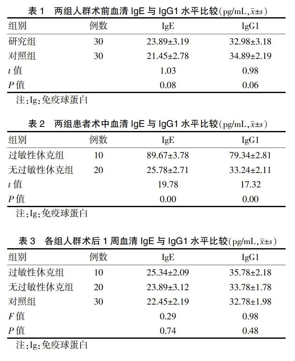
术前,研究组和对照组血清IgE和IgG1比较,差异无统计学意义(P > 0.05)。見表1。过敏性休克组术中IgE和IgG1表达量高于无过敏性休克组,差异有统计学意义(P < 0.05)。见表2。术后1周各组人群血清中IgE和IgG1表达量比较,差异无统计学意义(P > 0.05)。见表3。
2.2 各组人群血清IL-2、IL-4、IL-10及IFN-γ表达量比较
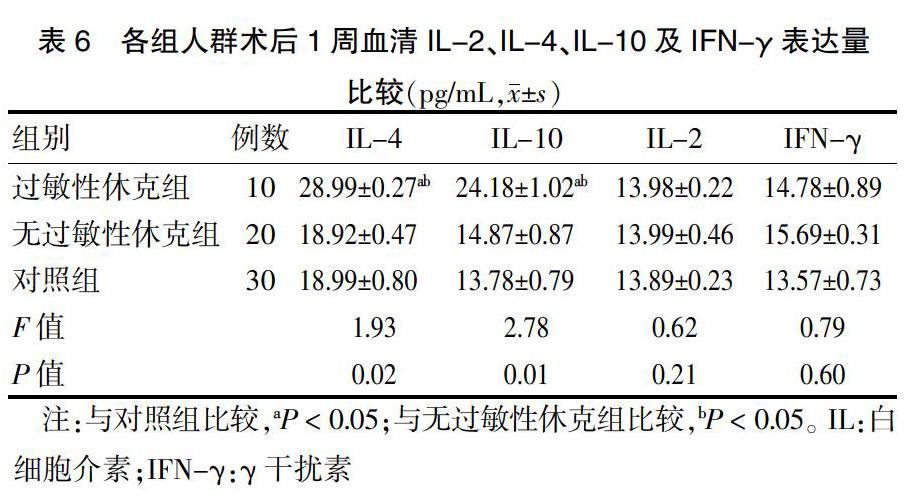
术前,研究组血清IL-4与IL-10表达量高于对照组,差异有统计学意义(P < 0.05)。见表4。过敏性休克组术中血清IL-4、IL-10低于无过敏性休克组,IFN-γ、IL-2高于无过敏性休克组,差异有统计学意义(P < 0.05)。见表5。术后1周过敏性休克组血清IL-4、IL-10表达量高于无过敏性休克组和对照组,差异有统计学意义(P < 0.05)。见表6。
2.3 相关性分析
IgE与IL-2、IFN-γ呈正相关(r = 0.96、0.68,P < 0.05),IgG1与IL-2、IFN-γ呈正相关(r = 0.60、0.79,P < 0.05),IgE、IgG1与IL-4、IL-10无相关性(P > 0.05)。
3 讨论
Eg导致的过敏性休克是包虫病的严重并发症[14-16],具体机制不明确。前期研究发现,囊液含有导致过敏性休克发生的特异性抗原,能与IgE特异性结合,提示IgE在过敏性休克发生过程中具有重要作用[14,17]。术前IgG和IgG1水平升高是过敏性休克的重要风险因子[18],主要是由于囊破裂会募集IgG2和IgG3同型抗体到囊包[19]。在过敏性休克发生时Th1型细胞占据主导作用,而在后期则是Th2型细胞发挥作用[20]。本研究通过对纳入人群血清IgE、IgG1含量及Th1、Th2型细胞相关因子进行比较分析发现,术中过敏性休克组血清IgE、IgG1表达量高于无过敏性休克组,提示在过敏性休克发生过程中IgE和IgG1是疾病发生的危险因素。术中过敏性休克组IL-4、IL-10低于无过敏性休克组,且IFN-γ与IL-2高于无过敏性休克组;研究组IL-2、IL-4、IL-10与IFN-γ表达量均高于对照组。提示,在过敏性休克发生过程中主要是通过Th1型细胞分泌炎性因子发挥作用,Th2型细胞在疾病发生过程中一直有参与,但在过敏性休克发生过程中不占主导作用。相关性分析显示,IgE、IgG1与IL-2、IFN-γ呈正相关,与IL-4、IL-10无相关性,进一步证实在炎症发生过程中主要是Th1型发挥作用。提示,Eg患者由于虫体在体内形成包囊,激活自身免疫系统,主要是激活Th2型细胞。Th2型细胞产生IL-4、IL-10等细胞因子的表达升高,升高的IL-4可对Th2型细胞具有稳定作用,使机体在没有特异性过敏原刺激下维持在平衡状态,机体中IgE、IgG1表达量处于生理平衡状态。而在手术过程中由于囊液溢出,其中的特异型抗原激活Th1型细胞,产生的IFN-γ激活Th1型细胞的生成,并且IFN-γ可消除IL-4对Th2型细胞的稳定作用,使Th2型细胞转变为Th1型细胞。Th1型细胞激活使细胞分泌IgE、IgG1,在生理情况下IgE和IgG1处于平衡状态,但在术中随着Th1、Th2型细胞失衡,导致IgE、IgG1在体内表达失衡,引起过敏性休克的发生。
综上,在Eg过敏性休克发生过程中主要是包囊的破裂,囊液中的特异性变应原引起体内IgE、IgG1表达增加,促进Th1型细胞增加,Th2型细胞受到抑制,引起过敏反应。
[参考文献]
[1] 李亦梅.包虫病所致过敏性休克患者免疫学保护性因素的研究[D].乌鲁木齐:新疆医科大学,2011.
[2] Thompson RC. The taxonomy,phylogeny and transmission of Echinococcus. Exp Parasitol [J]. Exp Parasitol,2008, 119(4):439-446.
[3] Moro P,Schantz PM. Echinococcosis:a review [J]. Int J Infect Dis,2009,13(2):125-133.
[4] Nunnari G,Pinzone MR,Gruttadauria S,et al. Hepatic ech-inococcosis:clinical and therapeutic aspects [J]. World Gastroenterol,2012,18(13):1448-1458.
[5] Adel-Patient K,Bernard H,Ah-Leung S,et al. Peanut- and cow′s milk-specific IgE,Th2 cells and local anaphylactic reaction are induced in Balb/c mice orally sensitized with cholera toxin [J]. Allergy,2015,60(5):658-664.
[6] Li S,Qian R,Wang S,et al. Identification of IgE and IgG1 specific antigens in Echinococcus granulosus cyst fluid [J]. Braz J Med Biol Res,2017,50(7):e6071.
[7] 鄭宏,徐志新,杨戈雄,等.感染细粒棘球蚴绵羊诱发过敏性休克期间IgG、IgG1、IgE抗体水平的探讨[J].中国寄生虫学与寄生虫病杂志,2003,21:42-45.
[8] Zhou X,Wang W,Cui F,et al. Extracellular vesicles derived from Echinococcus granulosus hydatid cyst fluid from patients: isolation,characterization and evaluation of immunomodulatory functions on T cells [J]. Int J Parasitol,2019,49(13/14):1029-1037.
[9] Yue X,Li H,Tang J,et al. Rapid and label-free screening of echinococcosis serum profiles through surface-enhanced Raman spectroscopy [J]. Anal Bioanal Chem,2019, 25(3):161-168.
[10] Moghaddam SM,Picot S,Ahmadpour E. Interactions between hydatid cyst and regulated cell death may provide new therapeutic opportunities [J]. Parasite,2019,26(1):70.
[11] Bayraktar MR,Mehmet N,Durmaz R. Th1 and Th2 inducing cytokines in Cystic echinococcosis [J]. Turkiye Parazitol Derg,2005,29(3):167-170.
[12] SamPson HA,Munoz-Furlong A,Bock SA,et al. SymPosium on the definition and management of anaphylaxis:Summary report [J]. J Allergy Clin Immunol,2005,115(3):584-591.
[13] 谷美龄,李亦梅,郑宏.囊型包虫病致过敏性休克患者IFN-γ和IL-4的表达及其意义[J].中国病原生物学杂志,2011,6(3):34-38.
[14] Li Y,Zheng H,Gu M,et al. Comparisons of serum total IgE,IgG,and IgG1 levels in patients with and without echinococcosis-induced anaphylactic shock [J]. Am J Trop Med Hyg,2012,87(1):104-108.
[15] Siracusano A,Margutti P,Delunardo F,et al. Molecular cross-talk in host-parasite relationships:he intriguing immunomodulatory role of Echinococcus antigen B in cystic echinococcosis [J]. Int J Parasitol,2008,38(12):1371-1376.
[16] Almond RJ,Flanagan BF,Antonopoulos A,et al. Differential immunogenicity and allergenicity of native and recombinant human lactoferrins:Role of glycosylation [J]. Eur J Immunol,2013,43(1):170-181.
[17] Zhang Q,Ye J,Zheng H. Dexamethasone attenuates ech-inococcosis-induced allergic reactions via regulatory T cells in mice [J]. BMC Immunology,2016,17(1):4.
[18] Furuhashi K,Chua YL,Wong KHS,et al. Priming with high and low respiratory allergen dose induces differential CD4+ T helper type 2 cells and IgE/IgG1 antibody responses in mice [J]. Immunology,2017,151(2):227.
[19] Turquetineves A,Otte M,Schwartz C,et al. The Extracellular Domains of IgG1 and T Cell-Derived IL-4/IL-13 Are Critical for the Polyclonal Memory IgE Response In Vivo [J]. PLoS Biol,2015,13(11):112-119.
[20] Dematteis S,Baz A,Rottenberg M,et al. Antibody and Th1/Th2-type responses in BALB/c mice inoculated with live or dead Echinococcus granulosus protoscoleces [J]. J Helminthol,2010,21(1):19-26.
(收稿日期:2019-11-29)