Comparative Identification between Kadsura coccinea and Kadsura heteroclite
Xincheng QU, Qimin HU,Yunfeng HUANG, Liping DOU
1. Guangxi Institute of Traditional Chinese Medicine, Nanning 530022, China; 2. Guangxi Key Laboratory of Traditional Chinese Medicine Quality Standards, Nanning 530022, China; 3. First Affiliated Hospital of Guangxi University of Chinese Medicine, Nanning 530022, China
Abstract [Objectives] The purpose was to provide scientific basis for the identification and application of Kadsura coccinea and its chief adulterant Kadsura heteroclite. [Methods] The botanical characteristics, traits and microscopic morphology of the two plants were compared. [Results] K. coccinea was clearly distinguished from K. heteroclite in botanical characteristics, traits and microscopic morphology. [Conclusions] This study is of great significance to the identification and development of K. coccinea.
Key words Kadsura coccinea, Kadsura heteroclite, Botanical characteristic, Trait identification, Microscopic identification
1 Introduction
Kadsuracoccinea(Lem.) A. C. Smith (K.chinensisHance ex Benth;K.hainanensisMerr.), also known as Fantuanteng, Dazuan, Jichangfeng, Fengshateng, Jiufantuan, Guoshan Longteng, Guoshanfeng, Xueteng, Choufantuan, Feihong Nanwuweizi, Da Fantuanteng, Houye Wuweizi and Daye Zuangufeng, is usually used as medicine with its roots and canes, with effects of promoting qi circulation, relieving pain, removing stasis and promoting blood circulation. In Yao ethnic group of Guangxi,K.coccineais often used to treat gastric an duodenal ulcers, chronic gastritis, acute gastroenteritis, rheumatic arthralgia, traumatic injuries, fractures, dysmenorrhea, abdominal pain due to postpartum stasis, hernia,etc[1-4]. Its chief adulterant isKadsuraheteroclite. In this paper, the botanical characteristics, traits and microscopic morphology ofK.coccineaandK.heteroclitewere studied to provide a scientific basis for their identification, development and application.
2 Materials and Methods
2.1MaterialsK.coccineawas collected in Napo, Guangxi in June, 2015, andK.heteroclitewas collected in Jinxiu, Guangxi in July, 2015. They were identified by Professor Lai Maoxiang from Guangxi Institute of Traditional Chinese Medicine. The reagents used in the experiment were of analytical grade.
Ermatoky0416 rotary slicer (Japan), AK-400A swing mill (Zhejiang), Ni-U Nikon advanced biological microscope and video imaging device (Japan).
2.2 MethodsThe botanical characteristics, traits and microscopic morphology ofK.coccineaandK.heteroclitewere identified according to the methods ofChinesePharmacopoeia. For microscopic inspection, the roots and canes ofK.coccineaandK.heteroclitewere prepared into paraffin sections, and the powder ofK.coccineaandK.heteroclitewas passed through No.5 sieve and prepared into powder sections. The sections were observed and photographed under a microscope.
3 Results
3.1Characteristicsoforiginalplants
3.1.1K.coccinea. Woody vine, glabrous. Leaf: leathery, oblong to ovate-lanceolate, 7-18 cm long, 3-8 cm wide, blunt or short-acuminate in apex, broadly wedge-shaped or nearly round in base, entire, with 6-7 lateral veins on each side, and unapparent in vein network. Petiole: 1.0-2.5 cm long. Flower: solitary in leaf axil, binate for rare cases, dioecious. Male flower: perianth red, 10-16 segments, elliptical in the largest petal of the middle round, 2.0-2.5 cm long, about 14 mm wide, thickened obviously in the innermost three segments, fleshy. Torus: long conical, 7-10 mm long, with 1-20 subulate branching appendages in the apex. Androecium: ellipsoidal or subglobose, 6-7 mm in diameter, with 14-48 stamens. Filament: surrounded by two anther chambers at the top. Pedicel: 1-4 cm long. Female flower: similar in perianth with that of male flower, short subulate in style, without shield-shaped stigma at the top, oblong in carpel, with 50-80 pistils, 5-10 mm long in pedicel. Aggregate fruit is nearly spherical, red or dark purple, and 6-10 cm or larger in diameter. Small berries are obovate, up to 4 cm long, leathery in exocarp, without apparent seeds. Seeds are heart-shaped or ovate-heart-shaped, 1.0-1.5 cm long, and 0.8-1.0 cm wide. Flowering period lasts from April to July, and fruiting period lasts from July to November.
3.1.2K.heteroclite. Woody vine, glabrous. Cane: brown, with ellipsoidal pits. Stem: thick in cork layer, longitudinally split. Leaf: oval-shaped to broadly oval-shaped, 6-15 cm long, 3-7 cm wide, acuminate or shape in apex, broadly wedge-shaped or nearly obtuse in base, entire or with detached small serrations in the upper half, 7-11 lateral veins on each side, apparent in network of veins. Petiole: 0.6-2.5 cm long. Flower, solitary in leaf axil, dioecious, white or light yellow in perianth, 11-15 petals, smaller in outer and inner rounds, biggest in the middle round, oval to obovate, 8-16 mm long, and 5-12 mm wide. Male flower: torus oval, elongated cylindrical in apex, conical protruding from the stamen group. Androecium: ellipsoidal, 6-7 mm long, about 5 mm in diameter, with 50-65 stamens. Stamen: 0.8-1.8 mm long. Filaments and anther septa are connected to form a nearly wide flat square. The top of the septum is horizontally oblong. The length of anther chamber is about as long as that of stamen. The filaments are extremely short. Pedicel: 3-20 mm long, with several bracteoles. Gynoecia: nearly spherical, 6-8 mm in diameter, with 30-55 pistils. Ovary: oblong-ovate. There is shield-shaped stigma on the top of style. Pedicel: 3-30 mm long. Aggregate fruit is nearly spherical, and 2.5-4.0 cm in diameter. Mature carpel: obovate, 10-22 mm long, leathery without apparent seeds when it is dry, 2-3 seeds, 4-5 seeds for rare cases, oblong kidney-shaped, 5-6 mm long and 3-5 mm wide. Flowering period lasts from May to August, and fruiting period lasts from August to December[5].
3.2Traitidentification
3.2.1K.coccinea. Root: cylindrical, 0.5-3.0 cm in diameter; dark brown or taupe in surface, rough, with elliptical pores and transverse cracks, easy to separate between bark and xylem; tough in texture, difficult to break. Cross section: bark thick, granular, gray-brown or tan, with pale yellow dots scattered; xylem yellowish white or brownish yellow, with many fine holes. Cane: cylindrical, 0.6-3.0 cm in diameter, gray-brown or tan in surface, with irregular cracks for rough cases, with white lenticels for fine cases, cork flaky, bends breaking into horizontal grooves; tough in texture, difficult to break. Cross section: fibrous; bark thick, brownish black, easy to peel, with taste of raw guava, less residue; xylem tan to light brown, radial holes (vessels) visible, with dark brown pith in the center. It smells slightly fragrant and tastes slightly sweet.
3.2.2K.heteroclite. Root: cylindrical, slightly curved, 0.5-3.0 cm in diameter; gray-brown to tan in surface, with longitudinal wrinkles and horizontal cracks; hard in texture, difficult to break. Cross section: bark thick, gray-brown or yellowish brown, with yellow dots; xylem gray-yellow or light yellow, with many fine holes. Cane, cylindrical, 0.6-5.0 cm in diameter. Vine: dark brown, with obvious deep vertical stripes, with punctate lenticels. Old stem: cork thick, massive vertical cracks, soft in texture, greasy, with long horizontal lenticels, dark tan where cork peels off; tough in texture, hard to break. Cross section: bark tan, slightly fibrous; xylem brown or pale yellow, with numerous vessels; pith brown. It smells slightly fragrant and tastes astringent[6].
3.3Microscopicidentification
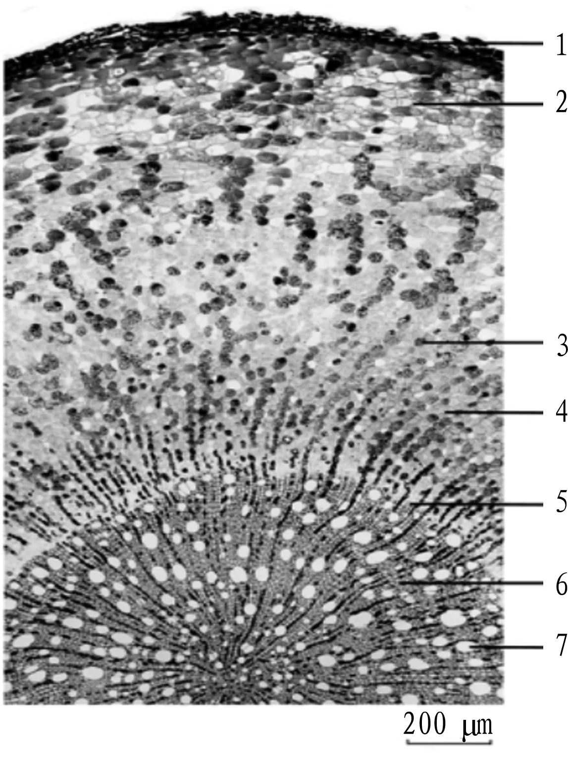
3.3.1K.coccinea. (i) Root cross section. Cork layer: numerous layers of cells, oblate, brownish yellow. Bark layer: broad, cells round, oblong or irregular, secretory cells and a few crystal-embedded fibers scattered. Phloem: bast fibers scattered, calcium oxalate cubes embedded in the outer wall of a single fiber or fiber bundle to form crystal-embedded fibers. Cambium: loop. Xylem: vessel 20-80 μm in diameter, 1-3 rows of cells in xylem ray, containing dark brown matter. The parenchyma cells contain starch granules (Fig.1).
(ii) Stem cross section. Cork layer: numerous layers of cells, oblate, with brown-yellow substance in the cell cavity. Bark layer: numerous layers of cells, suborbicular, oblong or irregular, secretory cells and crystal-embedded cells scattered, pericyclic fibers distributed intermittently in a loop. Phloem: thick, sieve cells suborbicular, phloem rays extending to the bark, bast fibers arranged into 5-10 layers, fiber wall thick, bast fiber cells containing calcium oxalate cubes to form crystal sheath fibers, calcium oxalate cubic crystals 5-16 μm in diameter. Xylem: vessels distributed singly or radially, 25-128 μm in diameter; ray cells 1-4 layers, rectangular, most containing brown-yellow substance. Pith: parenchyma cells round or suborbicular, some shrinking to form voids (Fig.2).
(iii) Powder. Powder: brownish yellow. Fiber: bundled or separated, yellowish brown, sharpened or obtuse at both ends. Lignocellulose: cell wall straight and thin, cell cavity large, with linear or herringbone pit, 18-28 μm. Bast fiber: cell wall thick, some cell wall containing calcium oxalate cubic crystals to form crystal sheath fibers, 25-38 μm in diameter. Eleocyte: mostly broken, suborbicular for intact ones, containing round yellow oil droplets. Grit cell: rectangular, approximately square or irregular, cell wall thick, mostly containing calcium oxalate cubic crystals, 33-51 μm in diameter. Cork cell: brownish yellow, polygonal. Starch granule: single or multiple, semi-circular, oval, triangular or beak-shaped, 5-25 μm in diameter. Vessel: yellowish brown, bevelled or threaded, 30-43 μm in diameter (Fig.3).
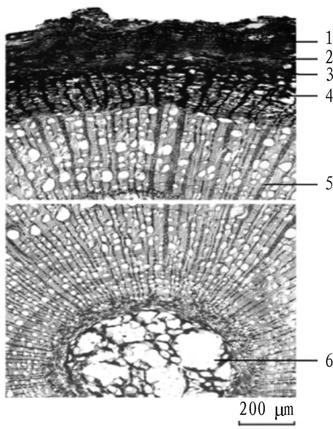
3.3.2K.heteroclite. (i) Root cross section. Cork layer: numerous layers of cells, rectangular, brownish yellow. Cork cambium: 1-2 layers of cells, rectangular, closely arranged. Phelloderm: 3-4 layers of cells. Bark layer: numerous layers of cells, secretory cells and a few of crystal-embedded fibers scattered. Phloem: with secretory cells, bast fiber singular, calcium oxalate cubes embedded in the outer wall of fibers or fiber bundle to form crystal-embedded fibers. Cambium: loop. Xylem: vessel 22-140 μm, 1-4 layers of cells in xylem ray, containing dark brown secretions. The parenchyma cells contain starch granules (Fig.4).
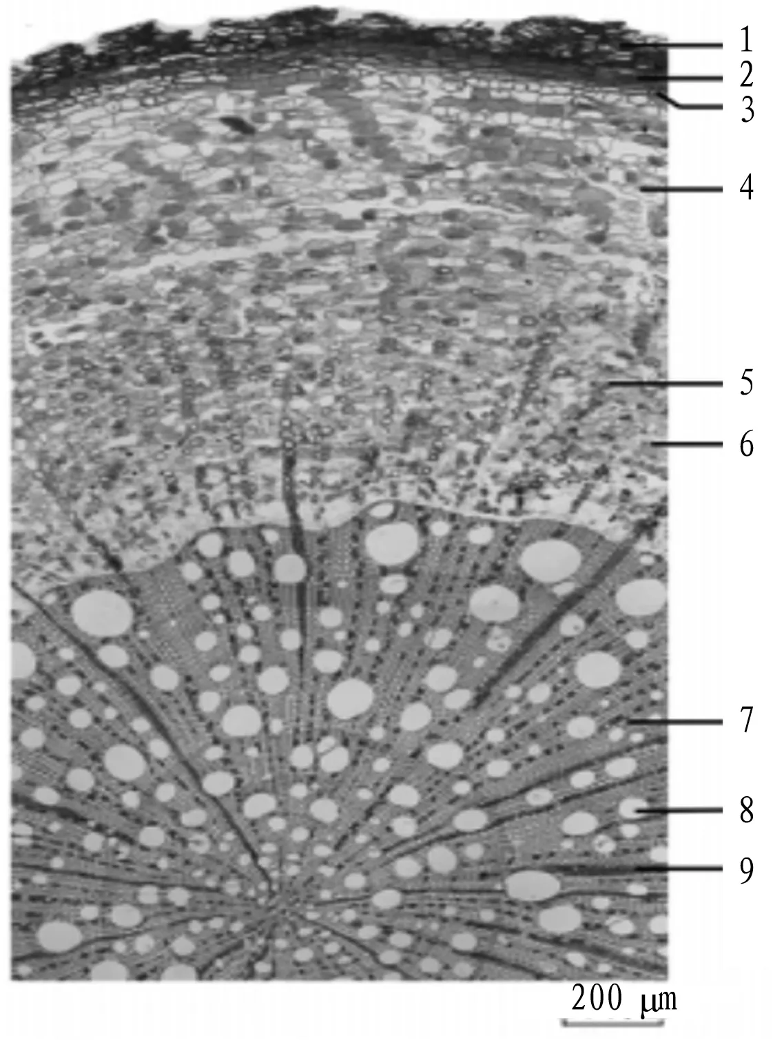
(ii) Stem cross section. Cork layer: 6-13 layers of cells, approximately square. Bark layer: thin, secretory cells and grit cells scattered, grit cells oval or irregular, wall thick, cell cavity small, pores and grooves unapparent, most containing calcium oxalate cubes; pericyclic fiber bundle mostly connecting to grit cells in a ring, fiber cell thin, lignified. Phloem layer: cells suborbicular, some containing brownish-yellow secretions; bast fibers single or connected, and small cubic crystals of calcium oxalate embedded at the edges to form crystal-embedded fibers. Xylem: vessels mostly scattered, 25-86 μm in diameter, xylem ray consisting of 1-3 layers of cells, some containing brown secretions. Pith: parenchyma cells suborbicular, pale yellow, thin-walled, some shrinking, crystal-contained cells distributed (Fig.5).
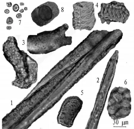
(iii) Powder. Powder: tan. Fiber, more, bundled or separated. Bast fiber: long spindle, 18-36 μm in diameter, buckler-shaped or broken at the ends, 2-5 fibers aggregating, mostly incomplete, outer layer of secondary wall embedded with a large number of calcium oxalate cubic crystals, cubic crystal 2-8 μm in diameter. Lignocellulose: fusiform, blunt pointed or flat at the end, wall thickness 5-8 μm, with oblique linear or oblique cross-shaped pit, 22-34 μm in diameter. Grit cell: off-white, approximately square, branched or irregular, wall extremely thick, pore and groove unapparent, cell cavity small or invisible, 36-78 μm in diameter, interior and edge embedded with a large number of calcium oxalate cubic crystals, cubic crystal 3-9 μm. Vessel: perforated or trapezoidal, mostly broken, 36-78 μm in diameter. Xylem parenchyma: pale yellow, nearly rectangular, 56-100 μm long, 35-58 μm wide, with circular pits. Cork cell: pale yellow, polygonal on the surface. Starch granule: single or multiple, round, oval or subtrianguslar, 5-16 μm in diameter (Fig.6).

Note: 1. cork; 2. bark; 3. bast fiber; 4. phloem; 5. cambium; 6. xylem; 7. vessel.
Fig.1 Morphology of root cross section ofKadsuracoccinea

Note: 1. cork; 2. pericyclic fiber; 3. bark; 4. phloem; 5. xylem; 6. pith.
Fig.2 Morphology of stem cross section ofKadsuracoccinea

Note: 1. bast fiber; 2. lignocellulose; 3. eleocyte; 4. grit cell; 5. cork cell; 6. starch granule; 7. vessel.
Fig.3 Morphology of powder ofKadsuracoccinea

Note: 1. cork; 2. phellogen; 3. phelloderm; 4. bark; 5. phloem; 6. bast fiber; 7. xylem; 8. vessel; 9. xylem ray.
Fig.4 Morphology of root cross section ofKadsuraheteroclite

Note: 1. cork; 2. bark; 3. pericyclic fiber; 4. phloem; 5. xylem; 6. vessel; 7. pith.
Fig.5 Morphology of stem cross section ofKadsuraheteroclite

Note: 1. bast fiber; 2. lignocellulose; 3. grit cell; 4. vessel fragment; 5. xylem parenchyma; 6. cork cell; 7. starch granule; 8. eleocyte.
Fig.6 Morphology of powder ofKadsuraheteroclite
4 Conclusions
The above research shows thatK.coccineais clearly distinguished fromK.heteroclitein botanical characteristics, traits and microscopic morphology. In terms of botanical characteristics, the leaves ofK.coccineaare obtuse or short acuminate in apex and broadly wedge-shaped or suborbicular in base, with 6-7 lateral veins in each side and unapparent vein network; the perianth segments are red; and the seeds are heart-shaped or ovate-heart-shaped, 1.0-1.5 cm long and 0.8-1.0 cm wide. The leaves ofK.heterocliteare serrulate in the upper edge, with 7-11 lateral veins on each side and apparent vein network; the perianth segments are white or pale yellow; and the seeds are oblong-kidney-shaped, 5-6 mm long and 3-5 mm wide. In terms of traits, the roots ofK.coccineaare easy to separate between bark and xylem, and their cross section has thick and granular bark; the canes are brownish yellow to grayish black, and the cross section is coarsely fibrous, with thick and easily-peeled bark; and it has a taste of raw guava and little residue. The old stem ofK.heteroclitehas thick and soft cork layer and feels slippery when touched, and the cross section is slightly fibrous. From the perspective of microscopic morphology, the cork cells in the root cross section ofK.coccineaare oblate, the bark layer is thicker, and the xylem vessels have a diameter of 20-80 μm; the pericyclic fiber bundles in the stem cross section are intermittently distributed in a loop, the xylem vessels are distributed singly or in series, with a diameter of 25-128 μm; and the vessels in the powder section are mainly pitted vessels and threaded vessels. The cork cells in the root cross section ofK.heterocliteare rectangular, the phellogen is visible, the xylem vessels are mostly scattered, with a diameter of 22-140 μm; the cork cells in the stem cross section are approximately square, the pericyclic fiber bundles are mostly connected to the grit cells in a ring shape, and crystal-embedded cells are scattered in the pith; and the vessels in the powder section are mainly pitted vessels and threaded vessels. The research results have certain significance for the identification and development ofK.coccinea.
- Medicinal Plant的其它文章
- Application of Chaihu plus Longgu Muli Decoction in Treatment of Physical and Mental Diseases
- Study on Pharmacological Effects of Calycosin in Astragali Radix
- Advances in Research on Treatment of Heart Failure with Yangxinshi Tablet
- Advances in Chemical Constituents and Pharmacological Activity of Pholidota spp.
- Treatment of Arthralgia Syndrome from Zang and Fu
- Effects of Zingber mioga Aqueous Extract on Hepatic Anti-alcoholism in Mice

