
Reappraisal of Serridentinus gobiensis Osborn &Granger and Miomastodon tongxinensis Chen:the validity of Miomastodon
2020-05-07 10:28:42WANGShiQiZHANGXiaoXiaoLIChunXiao
古脊椎动物学报(中英文) 2020年2期
WANG Shi-Qi ZHANG Xiao-Xiao,3,4 LI Chun-Xiao,3
(1 Key Laboratory of Vertebrate Evolution and Human Origins of Chinese Academy of Sciences, Institute of Vertebrate Paleontology and Paleoanthropology, Chinese Academy of Sciences Beijing 100044)
(2 CAS Center for Excellence in Life and Paleoenvironment Beijing 100044 wangshiqi@ivpp.ac.cn)
(3 University of Chinese Academy of Sciences Beijing 100049)
(4 Tianjin Natural History Museum Tianjin 300201)
Abstract The elephantimorph proboscideans,Serridentinus gobiensis Osborn &Granger,1932,and Miomastodon tongxinensis Chen,1978,from the Middle Miocene of northern China,were revised as Zygolophodon gobiensis (Osborn &Granger,1932).However,their phylogenetic positions are still being debated because of their intermediate morphology between the typical bunodont (Gomphotheriidae) and zygodont (Mammutidae) elephantimorphs.In the present paper,we compare their dental and mandibular morphology with that of the Eurasian Z.turicensis,Gomphotherium subtapiroideum,and G.tassyi,as well as the North American Mio.merriami and G.productum.It appears that S.gobiensis and Mio.tongxinensis share with Mio.merriami the slightly more bunodont molar morphology than that of Z.turicensis,e.g.,the thicker enamel,thicker pretrite crescentoids,higher interlophid enamel pillars in buccal view,and the narrower contour majorly caused by the narrower posttrite half loph(id)s.S. gobiensis and Mio. merriami also possess an“erected oval cross-sectioned mandibular tusk”,in which the cross-section is mediolaterally compressed (dorsoventral diameter being larger than the mediolateral one).Whereas,in Z.turicensis and G.productum,the mandibular tusk is“laid oval cross-sectioned”,in which the cross-section is dorsoventrally compressed (dorsoventral diameter is smaller than the mediolateral one).Therefore,it is reasonable to revive the genus Miomastodon Osborn,1922,which contains the species that were previously attributed to Zygolophodon,but they have relatively bunodont molar morphology (i.e.,the robust type of the Z.turicensis group).The mandibular tusk with erected oval cross-section seems to be a synapomorphy of Miomastodon species.Furthermore,the molar morphology of G.subtapiroideum and G. tassyi also exhibits intermediate status between the typical bunodonts and zygodonts.However,the mandibular symphysis of G.subtapiroideum and G. tassyi is stronger than that of Miomastodon,and the mandibular tusk is pyriform cross-sectioned.The validity of Miomastodon and G.subtapiroideum/tassyi obscures the boundary between the Gomphotheriidae and Mammutidae,and suggests that the evolutions of the Gomphotheriidae and Mammutidae are deeply involved in with each other,rather than straightforwardly detached.This phenomenon has been revealed by a collagen sequence analysis among Notiomastodon,Mammut,and extant elephants,which should be further studied.
Key words North China;Middle Miocene;proboscidean,Gomphotheriidae,Mammutidae
SerridentinusOsborn,1923,was established based onMastodon productusCope,1875,and several North AmericanMastodonspecies.Later,Osborn and his colleagues expanded the extent ofSerridentinus,and published several Eurasian species,e.g.,S.gobiensisOsborn &Granger,1932,from Tunggur,northern China.The holotype is a right hemimandible carrying the moderately to deeply worn m2 and the erupting m3,with complete mandibular symphysis and lower tusks.MiomastodonOsborn,1922,was established mainly based onMastodon merriamiOsborn,1921,which he regarded as the first“trueMastodon”in North America.He also broadened the nameMiomastodonto Eurasian species,e.g.,Mio.deperetiOsborn,1936.Chen (1978) reported another Eurasian species,Mio.tongxinensisfrom Tongxin,northern China,which is represented by a pair of deeply worn lower and upper third molars.
Many debates occurred over the validity ofSerridentinus gobiensisandMiomastodon tongxinensissubsequently.Tobien (1972) did not agree with the validity of the genusSerridentinus,and synonymized most of itsspecies withGomphotherium.However,he considered that the molar ofS.gobiensisdisplays the zygodont pattern (Tobien,1972).Tassy(1985) thought thatS.gobiensisbelongs to the robust type ofZygolophodon.Later,Tobien et al.(1988) attributed all the Chinese samples from Middle or early Late Miocene with more or less zygodont features into the single species,Z.gobiensis,includingMio.tongxinensis.Tobien (1996) further considered thatZ.gobiensisbelongs to theZ.turicensisgroup.However,Z.gobiensissensu Tobien et al.(1988) bears much variation and spans quite a long geological period.Therefore,further study ofZ.gobiensissensu Tobien et al.(1988)should be carried out,and the key problem is the taxonomy ofS.gobiensis.
1 Material and methods
1.1 Institutional abbreviations
AMNH and FAMNH,American Museum of Natural History,New York,USA;HMV,Hezheng Paleozoological Museum,Hezheng,China;IVPP,Institute of Vertebrate Paleontology and Paleoanthropology,Chinese Academy of Sciences,Beijing,China;MNHN,Muséum National d'Histoire Naturelle,Paris,France;NHMW,Naturhistorisches Museum Wien,Vienna,Austria;NMB,Naturhistorisches Museum Basel,Basel,Switzerland;SNSB-BSPG,Staatliche Naturwissenschaftliche Sammlungen Bayerns -Bayerische Staatssammlung für Paläontologie und Geologie,Munich,Germany;THP,Tianjin Natural History Museum,Tianjin,China.
1.2 Materials and terminology
The material described in the present work is housed in AMNH and IVPP.The comparative material is housed in AMNH,IVPP,HMV,MNHN,NHMW,NMB,SNSB-BSPG and THP.Others were illustrated in previous publications (Cope,1873;Lortet and Chantre,1878;Schlesinger,1917;Osborn,1921,1922,1929,1936;Borissiak,1936;Lehmann,1950;Tassy,1977,2013,2014;Madden and Storer,1985;Chen,1988;Göhlich,2010;Lofgren and Anand,2011;Wang et al.,2017).
The terminology of the occlusal structure of gomphotheriid molars and mandible follows Tassy (2013,2014;Fig.1A) with several modifications.Specifically,we used the terms pretrite central conule and pretrite crescentoid for different crown elements,which Osborn (1936) had already dealt with (he used conule or serration for pretrite central conule and spur or crest for crescentoid,see Osborn,1936:393).A pretrite crescentoid (green color) is a thick or thin enamel projection that originates from the mesial or distal side of the pretrite main cusp(id) and runs to the base of the loph(id)s (Fig.1D,green color).The proximal end of a crescentoid merges closely with the attached main cusp(id) without a sulcus between them.Whereas,a pretrite central conule (blue color) is a round,bulbous cone (Fig.1E,blue color),or subdivided enamel cones that are either arranged in a line or serrated (Fig.1A,posterior pretrite central conule 2).It should be noted that the boundary between crescentoids and central conule might be vague after moderate or deep wear.Pretrite central conules rise directedly from the base of the interloph(id)or the anterior cingulum(id),with a sulcus that clearly separates the adjacent loph(id).Pretrite crescentoids are lacking in the most typical bunodonts (i.e.,Gomphotherium angustidens) and pretrite central conule are absent in typical zygodonts (i.e.,Mammut borsoni).However,in some cases,the two elements coexist (Fig.1A).A short and thick pretrite crescentoid derives from the peak of the main cusp(id),it is followed by a single or subdivided pretrite central conule(s) that is separated from the pretrite crescentoid by a sulcus,e.g.,inGomphotherium productum.
1.3 Measurements,specimen illustrations,and data analyses
Mandibular and cheek teeth measurements follow Tassy (2013,2014;Fig.1B,C).Measurements were preferentially obtained using callipers (in mm).If not available,photos and 3D digital models were also used,and the data were obtained using the public software Image J (V 1.48) (Schneider et al.,2012).
Illustrations of mandibles and molars in the present article were based on photos taken by a Nikon D7 100 camera equipped with an AF-S NIKKOR 18−300 mm optical zoom lenses,or snapshots of 3D digital models generated by a handheld Artec Spider 3-dimensional scanner.
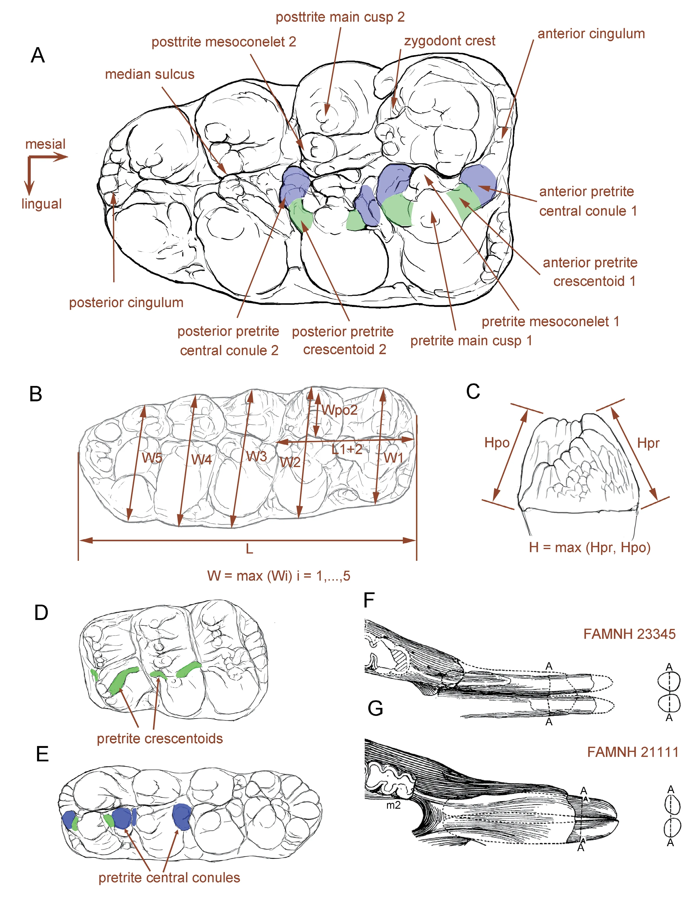
Fig.1 Terminology and measurements of mastodont molars
Zygodont taxa are characterized by the remarkable widening of their cheek teeth,for more efficient bolus compression (Königswald,2016),particularly by the widening of the posttrite half loph(id)s.Thus,in our cheek teeth measurement protocol,in addition to the general measurements as described in Tassy (2014),we measured the posttrite width of the second loph(id),from the median sulcus to the summit of the posttrite main cusp(id) (Wpo2;Fig.1B).For comparison of biometric data between taxa with different size,the raw data should be normalized.For the posttrite width of the second loph(id) (Wpo2),we used the width of the second loph(id) (W2) for normalization,i.e.Wpo2/W2 (Fig.1B).For maximal width(W),we measured the length of the first two loph(id)s (L1+2) rather than the whole length for normalization,i.e.W/L1+2.This treatment is advantageous for comparing M1-M3 or m1-m3 in the same plot.In the present paper,we only compared the lower cheek teeth because the upper cheek teeth are very rare in our concerning material.We figured both width vs.length of m3 and Wpo2/W2 vs.W/L1+2.
For the mandibular tusk measurements,we measured the dorsoventral (Ddv) and mediolateral diameters (Dml),and figured Dml vs.Ddv for the mandibular tusk comparison.For comparing the mandibular symphysis,we measured the length of the symphysis (Ls),maximal width of the symphysis (Ws),and length of the cheek tooth row (Lt) (after Tassy,2013,measurements 2,11,and 24,respectively).We figured Ls/Lt vs.Ws/Lt,which means the relative length and relative width of mandibular symphysis,respectively.
2 Systematic paleontology

Type speciesMiomastodon merriami(Osborn,1921).
Referred speciesMiomastodon tongxinensisChen,1978,Miomastodon gobiensis(Osborn &Granger,1932),Miomastodon metachinjiensis(Osborn,1929).
DiagnosisLongirostrine trilophodont elephantimorph with narrow and moderately elongated symphysis;mandibular tusks straight,parallel-protruding,close to each other,and with an“erected oval cross-section”(Ddv>Dml);posttrite half lophids moderately subdivided,mesiodistally compressed,and lacking posttrite central conules;pretrite crescentoids thicker than those ofZygolophodon turicensis,especially for lower molars;pretrite central conules usually present on the first and second interlophids of lower molars.
Differs from the other members of the Mammutidae (Losodokodon,Eozygodon,Zygolophodon,andMammut,Sinomammut)in the relatively bunodont characters of the cheek teeth,in the slightly chevroned and alternatively positioned half lophid of m3;also differs fromMammutin the relatively longer symphysis (Ls/Lt>1,see Fig.5D and Table S3) and mandibular tusks.Differs fromGomphotherium subtapiroideumandG.tassyiin weaker mandibular symphysis (Ws/Lt<0.4,see Fig.5D and Table S3);in mandibular tusks with oval rather than pyriform cross-section.Differs from the other longirostrine trilophodont gomphotheriid taxa in the more zygodont characters of the cheek teeth.

(Figs.2A−D,4A−C;Tables 1,2)
Serridentinus gobiensisOsborn and Granger,1932
Zygolophodon gobiensis(Osborn &Granger,1932) Tobien,1972,p.176
Zygolophodon gobiensis(Osborn &Granger,1932) Tassy,1985,p.511
partimZygolophodon gobiensis(Osborn &Granger,1932) Tobien et al.,1988,p.146−156,figs.39,41,43,44
nonZygolophodon nemonguensisChow and Chang,1961
nonZygolophodon gromovaeDubrovo,1970
nonZygolophodon jiningensisChow and Chang,1974
nonZygolophodon chinjiensis(Osborn,1929) Chow et al.,1978
Type specimenAMNH 26461,a right hemimandible carrying mandibular symphysis with a pair of lower tusks.The tooth row consists of the vacancy of m1,moderately worn m2,and erupting m3.
Type locality and horizon6.4 km (4 miles) northwest to the Wolf Camp (Zhunwuguer locality),Tunggur Formation,Tamuqin Fauna,~MN7/8,see below (Osborn and Granger,1932;Wang et al.,2003;Qiu et al.,2013).
OccurrenceTunggur region,Nei Mongol,China,late Middle Miocene.
Referred materialAMNH 26476,a right m3 with partial mandibular fragments (see Tobien et al.,1988:fig.41).
Differential diagnosesMiomastodonwith relatively large size (~120% m3 length ofMio.merriami) and relatively longer symphysis.Differs fromMio.merriamiin the longer mandibular symphysis and in possessing the pretrite central conules in the first and second interlophids of the lower molars;differs fromMio.tongxinensisin the larger dimensions and in the narrower m3 with a strong fourth lophid;differs fromMio.metachinjiensisin the relatively stronger pretrite central conules,in the greater subdivision of the posttrite half lophids,and in the smaller m3 dimensions.
RemarksThe type hemimandible ofMiomastodon gobiensisOsborn &Granger,1932 was discovered by Andrews,the head of the Central Asiatic Expedition 1930,from an isolated hill north of the Wolf Camp (Osborn and Granger,1932),Middle Miocene Tunggur Formation.In recent publications,this locality was recognized as“Zhunwuguer”(Wang et al.,2003).Based on Spoch (1929),Mio.gobiensiswas recovered from the upmost cross-bedded sandstones which overlies a thick layer of limestones.It possibly coexisted with a very derivedPlatybelodon(more derived than the type species ofPl.grangeri),and the layer also produces the Tamuqing small mammal assemblage,which is the youngest small mammal assemblage in the Tunggur Formation,with the approximate age of 11.8 Ma (Wang et al.,2003;Qiu et al.,2013).
Descriptions(see Osborn and Granger,1932:11;Tobien et al.,1988:147,149) In AMNH 26461 (Fig.2),the left corpus and ramus are broken while the other parts are complete.In dorsal view (Fig.2A) the ramus is thin and plate-like.The condyloid process of the mandible is oval and its long axis is transversely oriented.The shape of mandibular corpus looks like a triangle of which the acute angle is anteriorly oriented.The m1 alveolus was healing,leaving a relatively large vacancy.The mid-axes of the m2 and m3 are not in line.The symphysis is moderately elongated.It is relatively narrow and the distal part is only slightly widened.The symphyseal trough is deep and the two interalveolar crests border each side of the symphyseal trough.The posterior end of the symphysis is almost in line with the anterior end of the m1 alveolus.The distal end of the symphysis displays an obtuse angle with an anteriorly oriented apex.In lateral view (Fig.2B),the ramus is high,and the dorsally protruded condyloid process is much higher than the hook-like coronoid process.These two processes are linked by a smoothly curved mandibular notch.The masseter fossa is trapezoidal.The angular process is rather blunt,but slightly ventrally bulging.The anterior end of the mandibular corpus is slightly thicker than its posterior end.The symphysis stretches slightly ventrally,and the ventral border of the mandible is nearly straight.The anterior mental foramen is large,and is distant from the tooth row,but the posterior mental foramina are absent.In rostral view(Fig.2C),the symphyseal trough is smooth and deep.It dips down rostrally and shows two medially oblique edges at the distal end.The anterior mental foramen is relatively rounded and faces anteriorly.The ascending ramus is high and thin,and the large mandibular condyle is slightly medially oblique.
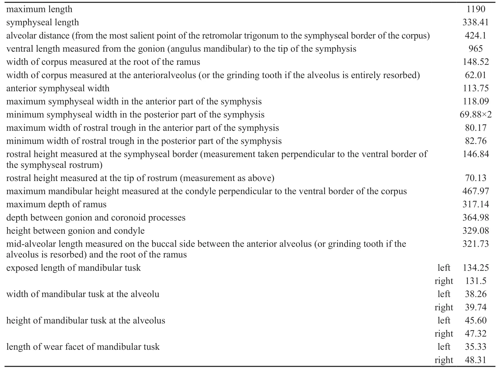
Table 1 Mandibular and tusk measurements of Miomastodon gobiensis* (mm)
The mandibular tusks (Fig.2A−D) are simply rod-like.They are straight,closely apposed,with a relatively short exposed length (~130 mm,precise measurements see Table 1).The cross-section is oval and its maximal axis is slightly dorsolateral-ventromedial oriented.The dorsoventral diameter is larger than the mediolateral diameter,which we call the“erected oval cross-section”(the ratio of Ddv/Dml >1,see Table S2,Fig.2D).The main wear facet(Fig.2C) is also oval shaped.It obliquely cuts the rostrodorsal end of the tusk.Another small facet (Fig.2C) is just beneath the main facet.It is perpendicular to the longitudinal axis of the tooth.

Fig.2 Type mandible of Miomastodon gobiensis,AMNH 26461,from the Zhunwuguer locality,Tunggur region,Nei Mongol,in dorsal (A),lateral (B),and rostral (C) views,and the contour of the cross-section of mandibular tusks,in rostral view (D)
The m2 is moderately worn (Fig.3A−C).In occlusal view,the width of lophids increases from anterior to posterior.The first pretrite half lophid is trifoliate with a small mesoconelet and nearly symmetrical anterior and posterior crescentoids.The crescentoids are thick,and a small and bulbous posterior central conule is present.The first posttrite half lophid is slightly mesiodistally compressed and the sulcus between the main cusp and the mesoconelets is clear.The first interlophid is relatively mesiodistally wide and is blocked by the pretrite central conules.A large conule is present at the entoflexid.The second lophid is similar to the first one.The anterior and posterior crescentoids are smaller than those of the first lophid.The second posterior pretrite central conule is also present.The posttrite half lophid displays a tendency for subdivision and mesiodistal compression,and the separation of the main cuspid and the mesoconelet can be observed.The second interlophid is also mesiodistally wide.The third pretrite half lophid possesses a small anterior pretrite crescentoid and a small anterior pretrite central conule,but the posterior pretrite crescentoid is unclear.The posttrite half lophid is more strongly subdivided than the anterior two,showing a series of serrated conelets,and the separation of the main cuspid and the mesoconelet cannot be observed.Cingulids are present on the anterior and posterior margins of the tooth and two large conelets arise on the center of the posterior cingulid.In buccal view (Fig.3B),the lophids are“∩-shaped”.The first interlophid is“U-shaped”and the second interlophid is“V-shaped”.The two interlophids are stuffed up by the central conules and crescentoids due to deep wear.In lingual view (Fig.3C),the lophids are“Λ-shaped”and the interlophids are“V-shaped”.
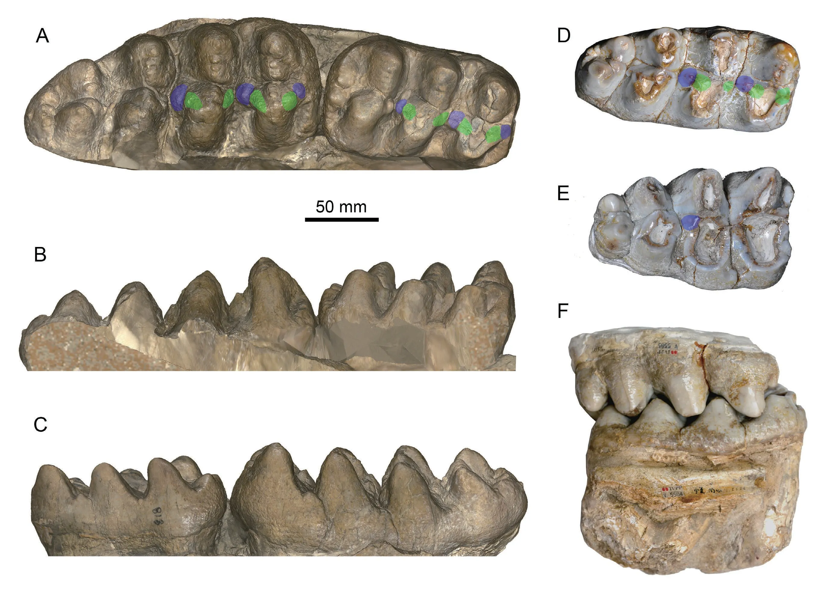
Fig.3 Cheek teeth of Miomastodon gobiensis and Mio.tongxinensis
The m3 (Fig.3A−C) is erupting from the first three lophids.However,the covering boney plate has been removed and the entire tooth is exposed.In occlusal view (Fig.3A),this tooth has four lophids plus a strong posterior cingulid and is long leaf-shaped.Lophids 2 and 3 display a slight tendency for alternative position of half lophids and the posterior three lophids are slightly chevroned.The first pretrite half lophids has weak but subdivided mesoconelets.The pretrite anterior and posterior crescentoids are strong,and the anterior and posterior pretrite central conule rises from the anterior cingulid and the first interlophid,respectively.The first posttrite half lophid has a strong main cuspid and a subdivided mesoconelet.The anterior zygodont crest is present and the posterior one is absent.The second pretrite half lophid also has a small and subdivided mesoconelet.The anterior and posterior pretrite crescentoids are weak,the anterior pretrite central conule is almost absent,and the posterior one is relatively strong.The posttrite half lophid is composed of a line of small conelets,and the main cuspid and the mesoconelet are indistinguishable.The third pretrite half lophid possesses a main cuspid that is equivalent to the mesoconelet,the anterior pretrite crescentoid is very weak,and the other accessory structures are absent.The third posttrite half lophid is composed of four equal-sized conelets,and the main cuspid and the mesoconelets are indistinguishable.The third interlophid is mesiodistally open.The fourth lophid is almost the same as the third one,but much smaller.Cingulids are present on the anterior and posterior ends of the teeth.In buccal view (Fig.3B),the lophids are“∩-shaped”.The first interlophid is“U-shaped”and the second and third interlophids are“V-shaped”.The anterior two interlophids are half stuffed up by the central conules and crescentoids (central conules reach the half height of the interlophids).In lingual view (Fig.3C),like the m2,the lophids are“Λ-shaped”and the interlophids are“V-shape”.

(Fig.3D−F;Table 2)
partimZygolophodongobiensis(Osborn &Granger,1932) Tobien et al.,1988,p.146−156,figs.39,41,43,44
Type specimenIVPP V 5584,a deeply worn right m3 (Chen,1978:pl.II,fig.2).
Type locality and horizonZhangenbao Formation in the Tongxin region,Ningxia.Late Early or early Middle Miocene.
Referred specimenIVPP V 5585,a deeply worn right M3 (Chen,1978:pl.II,fig.1),probably belonging to the same individual as the type m3
Differential diagnosesMiomastodonwith small dimensions (~85% m3 length ofMio.merriamiand~75% ofMio.gobiensisandMio.metachinjiensis,precise measurements see Table 2).Differs from the otherMiomastodonspecies in the smaller size,mostly because of the less developed fourth loph(id);also differs fromMio.merriamiin the presence of the first and second posterior pretrite central conules of the lower molars.
RemarksChen (1978) reportedMiomastodon tongxinensis,represented by a pair of lower and upper third molars that are deeply worn.The size is fairly small,therefore might represent a senile female individual.The material was collected from Gujiazhuangzi,a local village of Tongxin region,where the Early to Middle Miocene Zhangenbao Formation is well exposed and produces abundant fossil mammals.The Zhangenbao Formation consists of five fluviolacustrine sedimental circles,and the first and second circles represent the late Early Miocene and the third to fifth represent the early Middle Miocene.The precise locality ofMio.tongxinensisis unclear,and there are two possibilities:one is from the second circle at Miaoerling,which is characterized by the ferrygineous sandstones attached on the specimens;and another one is from the fourth circle at Shataigou,the nearest fossil locality to the Gujiazhuangzi village.
Descriptions(see Chen,1978:103,104;Tobien et al.,1988:152,153) IVPP V 5584 is a deeply worn right m3 (Fig.3D).In occlusal view,the tooth is long leaf-shaped and is relatively narrow.The second lophid is slightly wider than the other lophids.The lophids 2 and 3 display a slight tendency for alternative position of half lophids and the posterior three lophids are slightly chevroned.The first pretrite half lophid is trifoliate.The enamel walls of the posterior pretrite crescentoid and central conule are connected to each other,only showing a distal inflated enamel loop.The posttrite half lophid shows mesiodistally compression and subdivision.The main cuspid is strongly oblique.Fairly weak anterior and posterior zygodont crests are present.The first interlophid is mesiodistally wide.The second pretrite half lophid is similar to the first one.The posterior pretrite central conule is strong,linked to the pretrite lophid by the thick posterior pretrite crescentoid.The second posttrite half lophid is also mesiodistally compressed,but it is more deeply worn than the first half lophid.The second interlophid is also mesiodistally wide.The anterior and posterior crescentoids of the third pretrite half lophid are weak,and only a small posterior pretrite central conule is present.The posttrite half lophid shows distinct separation of the main cuspid and the mesoconelet.The fourth pretrite half lophid has a strong mesoconelet that is anteriorly positioned and the posttrite half lophid is small.Cingulids are present on the anterior and posterior margins of the tooth.
IVPP V 5585 is a deeply worn right M3 (Fig.3E) that very possibly belongs to the same individual as V 5584,as they are perfectly matched (Fig.3F).In occlusal view,it is wide with a triangular shape.The posterior two lophs are chevroned.The first two pretrite half lophs are so deeply worn showing two oval or subcircular enamel rings.However,the posterior pretrite central conule of the second loph is prominent.The first two posttrite half lophs are mesiodistally compressed,and form two transversely elongated enamel rings.The anterior two interlophs are mesiodistally narrow,relative to those of the m3.On the third loph,the pretrite half loph lacks a posterior crescentoid and a central conule,and the posttrite half loph shows a clear groove between the main cusp and the mesoconelet.The fourth loph is very small,but the pretrite half loph possesses an anterior central conule and a mesoconelets.The posterior cingulum is almost absent.
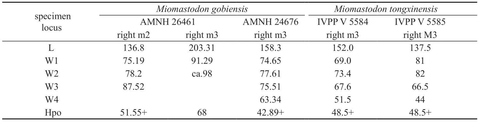
Table 2 Cheek tooth measurements of Miomastodon* (mm)
3 Results and discussion
3.1 Molar comparisons
3.1.1 Comparison toZygolophodon turicensis
Tobien et al.(1988) allocated most of the specimens from the middle to early Late Miocene of China showing less or more zygodont characters into one species,Zygolophodon gobiensis,includingSerridentinus gobiensisOsborn &Granger,1932,andMiomastodon tongxinensisChen,1978.Tobien et al.(1988:155) stated that the type mandible ofZ.gobiensis(i.e.,S.gobiensisOsborn &Granger,1932) belongs to the robust or primitive morph-type.Other specimens,e.g.,Z.gromovaeDubrovo,1970,Z.(Turicius)nemonguensisChow &Chang,1961,andZ.jiningensisChow &Chang,1974,were attributed to the gracile or advanced morph-type ofZ.gobiensis(Tobien et al.,1988).The two morph-types were also recognized in the European sample ofZ.turicensis(Tobien,1975;Tassy,1985).The gracile type ofZygolophodonis indisputable,as the type specimen ofZ.turicensisis absolutely the gracile type (see below).However,the robust type ofZygolophodonwas always confused with various bunodont taxa,and thus gave rise to much controversy.Tobien et al.(1988) interpreted the two morph-types as a result of functional partition.However,the morphological distinction between the two types is clearly shown on the unworn specimens,and functional partition in a single species is untenable.
The type specimen ofZygolophodon turicensisis a left m2 from Elgg,Switzerland,Middle Miocene (Fig.4F-H).It shows a high grade of zygodonty.In occlusal view (Fig.4F),the posttrite half lophids are highly compressed;and the conelets are strongly subdivided,arranging in line as a sharp edge.The anterior and posterior crescentoids are sharp and slender,and no anterior and posterior central conules rise from the interlophids.In buccal and lingual views (Fig.4G,H),the lophids are Λ-shaped preserving a sharp peak.In buccal view(Fig.4G),the interlophids exhibit high and deep V-shaped notches.These features are typical in the gracile type ofZ.turicensis.

Fig.4 Molars of the comparative species of the relevant taxa
The molars of the type specimen ofSerridentinus gobiensis,as the other robust type ofZygolophodon,display more bunodont morphology thanZ.turicensis.In occlusal view(Fig.3A),the posttrite half lophids are moderately subdivided,the separation of the posttrite main cuspid and the corresponding mesoconelet is unclear,and these arranged posttrite conelets are relatively blunt rather than sharp.The pretrite mesoconelets are more bulbous than those of the typicalZ.turicensis.The anterior and posterior crescentoids are thicker than those of the typicalZ.turicensis.More importantly,in the lophids 1 and 2,a round and bulbous posterior pretrite central conule is present at the distal end of the corresponding posterior pretrite crescentoid.This element is absolutely absent in the type m2 ofZ.turicensis.As a result,in buccal view (Fig.3B),in the unworn teeth,the interlophids are half stuffed up by enamel pillars (crescentoids and central conules),showing shallow interlophid notches,and in buccal and lingual views (Fig.3B,C),the lophids display blunt peaks.Furthermore,the lophids 2 and 3 exhibit slight alternative positions and slight chevroned patterns,which is very uncommon in the gracileZ.turicensisand the other mammutid taxa.Similar morphology is also shown inMiomastodon tongxinensis(Fig.3D,E).Therefore,a better solution is to restrict the genus nameZygolophodononly to the gracile morph-type,and attribute the robust type,e.g.,S.gobiensisandMio.tongxinensis,to another genus.
3.1.2 Comparison toMiomastodon merriami
Osborn (1921) reportedMastodon merriami,a pair of lower m3 and portions of two upper tusks from Virgin Valley Formation,Nevada,USA.Subsequently,Osborn (1922)established the new genusMiomastodonfor hisMastodon merriami,that is,Mio.merriami(Osborn,1921).Osborn (1936) attributedMio.proavusfrom Pawnee Creek,Colorado,USA,published by Frick (1933),toMio.merriami,and placed the hypodigm ofMastodon proavusCope,1873 (a P4 and a fragment of an M1) toSerridentinus.However,later researchers often synonymizedMastodon merriamiOsborn,1921,andMastodon proavusCope,1873,withZ.proavus(Cope,1873) (Madden and Storer,1985;Lofgren and Anand,2011).Although the specific name“proavusCope,1873”was published earlier than“merriamiOsborn,1921”,the characters ofMastodon proavusare obscure,and its hypodigm was possibly heterogeneous(Madden and Storer,1985).The M1 ofMastodon proavusis to a large degree incomplete,and was presumed to belong to a bunodont taxon;and the P4,more possibly belonging to a zygodont taxon,possesses very little morphological information for defining a species.Here we regardMastodon proavusCope,1873,as nomen dubium.
The type m3 ofMastodon merriamiOsborn,1921 (Fig.4A-C),although moderately to deeply worn,processes thicker enamel walls than that of typicalZygolophodon turicensis.The posttrite half lophids are also mesiodistally compressed,leaving mesiodistally wide interlophids,but the posttrite half lophids are relatively blunt.Although the posterior pretrite central conules seem to be absent,the anterior and posterior pretrite crescentoids are remarkably thicker than those ofZ.turicensis.Therefore,likeSerridentinus gobiensisandMiomastodon tongxinensis,the molar morphology ofMastodon merriamiis more bunodont than that ofZ.turicensis.As a result,the genusMiomastodonseems to be valid and should be revived for those untypical“Zygolophodon”with more or less bunodont molar morphology,such asMio.merriami,Mio.gobiensis,andMio.tongxinensis.Here,we regard each of these species as valid taxa (see the differential diagnosis).
3.1.3 Comparison toGomphotherium productumand the other relatedGomphotheriumspecies
Miomastodon gobiensiswas originally attributed toSerridentinusOsborn,1923.AlthoughSerridentinushas been synonymized withGomphotherium,it is necessary to compareMiomastodon gobiensiswith species that had previously been assigned intoSerridentinus.
The type species ofSerridentinuswasMastodon productusCope,1875,which was established based on an incomplete mandible (AMNH 14383) from Santa Fé,New Mexico,USA.Osborn (1923) transferred this species to his newly established genusSerridentinus,i.e.,S.productus(Cope,1875),along with several North American species.Tobien (1972)synonymizedSerridentinuswithGomphotherium,and attributed almost all of the North American species into one speciesG.productum.It should be mentioned that Tobien’sG.productumis a complex that contains more than one species.For simplicity,we compareMiomastodon gobiensisonly with the type specimen ofMastodon productusCope,1875.InG.productum(Fig.4L-N),the posttrite main cuspid and mesoconelet are clearly separated,but they are not subdivided.The anterior and posterior crescentoids are short or absent,and the anterior and posterior central conules are strong and even subdivided as serration.The interlophids are relatively mesiodistally narrower than those ofMio.gobiensis.In buccal view(Fig.4M),the interlophids are nearly fully stuffed up by enamel pillars rather than half stuffed up as inMio.gobiensis.The interlophids are rather narrow.Generally speaking,the molar morphology ofG.productumis rather bunodont,and that ofMio.gobiensisis intermediate between the bunodontG.productumand the zygodontZ.turicensis.
Osborn (1936) also attributed several EurasianTrilophodon(=Gomphotherium) species intoSerridentinus.The most important one is perhapsMastodon angustidensformasubtapiroidesSchlesinger,1917,from Eibiswald,Austria,which is nowadays referred to asGomphotherium subtapiroideum(Schlesinger,1917).Göhlich (2010) published abundant material ofG.subtapiroideumfrom Sandelzhausen,and Wang et al.(2017) published a new species,G.tassyi,of which the tooth morphology is closely related toG.subtapiroideum.
In the lower molars ofGomphotherium subtapiroideumandG.tassyi(Fig.4O-T),in occlusial view (Fig.4O,R),the anterior and posterior crescentoids are present,with moderately developed (sometimes subdivided) posterior central conules.This feature is similar to that ofMiomastodon gobiensis.The posttrite half lophids are moderately mesiodistally compressed,leaving relatively mesiodistally wide interlophids,also similar to those ofMio.gobiensis.However,the posttrite main cuspid and mesoconelet are less well subdivided than those ofMio.gobiensis.Alternative position and chevron structure are absent inG.subtapiroideumandG.tassyi,either.In buccal view (Fig.4P,S),the interlophids are half stuffed up by the enamel pillars,as are those ofMio.gobiensis.Generally speaking,the molar ofMio.gobiensisresembles that ofG.subtapiroideumandG.tassyimore than the other gomphotheriid and mammutid species.
3.1.4 Biometric comparison of molars
The m3s of the typical zygodont (MammutandZygolophodon,Fig.5A) and bunodont taxa (Gomphotherium productumandG.angustidens,Fig.5A) are clearly separated in the length-width plot.However,samples ofMiomastodonandG.subtapiroideum/tassyiare scattered along the boundary between the ranges of zygodont and bunodont taxa (Fig.5A) and overlap with each other.In the plot of W/L1+2-Wpo2/W2 (Fig.5B),which enables the comparison of the m1-3 in the same panel,the sample of the zygodont taxa(Fig.5B) is clustered at the top right of the panel and that of theG.angustidens(Fig.5B)at the bottom left.However,the samples ofMiomastodon,G.subtapiroideum/tassyi,and ofG.productum(Fig.5B) are located between the ranges of zygodont andG.angustidenssamples,with slight overlaps at their boundary.Nevertheless,the samples ofMiomastodon,G.subtapiroideum/tassyi,andG.productumcannot be separated from each other (Fig.5B).The biometric comparison indicates that the width of theMiomastodonlower molar is intermediate between the typical zygodont and bunodont taxa,which is mainly contributed by the posttrite half lophid width.However,the biometric data ofMiomastodoncannot be separated fromG.subtapiroideum/tassyiandG.productum.
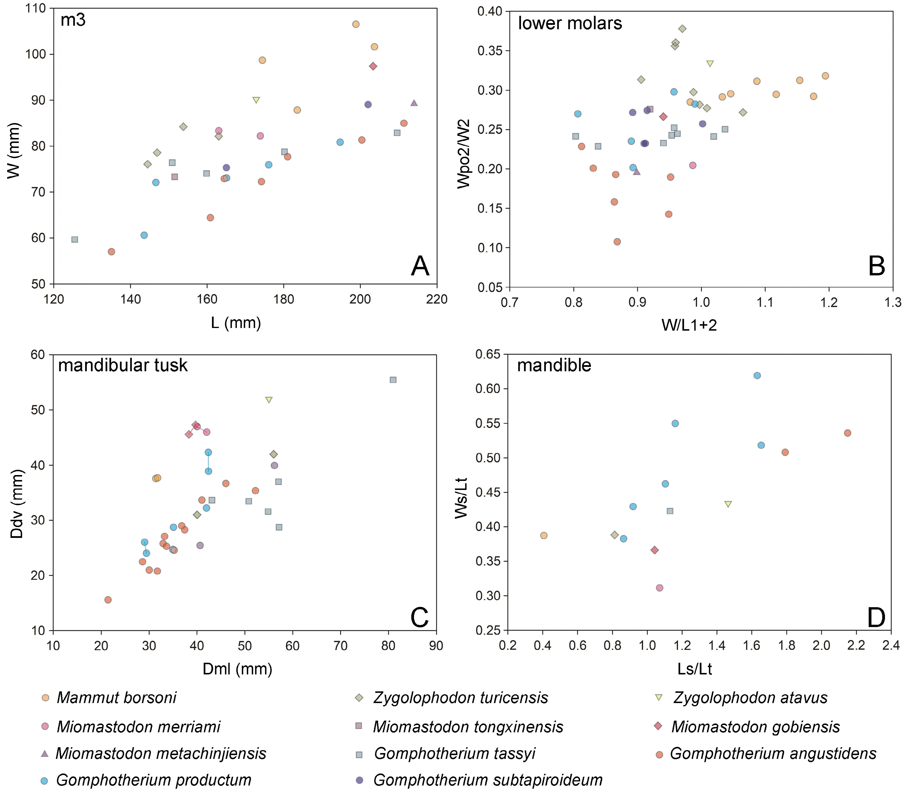
Fig.5 Scatter plots of bunodont and zygodont cheek teeth and mandibular tusks of the relevant taxa
3.2 Comparison of the mandible of Miomastodon gobiensis to the other taxa
The mandible ofMiomastodon merriamihas been reported by Frick (1933) from Pawnee Creek,Colorado,and by Madden and Storer (1985) from Wood Mountain,Canada.A prominent difference betweenMio.merriamiandMio.gobiensisis that the mandibular symphysis ofMio.merriamiis shorter.The other features are very similar,including the slightly ventrally deflected mandibular symphysis,and the slightly dorsally bent mandibular tusk.Perhaps the most important feature shared withMio.gobiensisis that the mandibular tusk is erected oval (see below).
The mandible ofZygolophodon turicensiswas rarely reported,except for a nearly complete mandible from Freising,Germany.However,the specimen had been ruined during World War II (Göhlich,1998).Based on the descriptions and illustrations in the previous publications (see Osborn 1936:fig.657;Lehmann,1950:pl.14,fig.26),the mandibular tusk is laid oval cross-sectioned,unlike the erected oval cross-sectioned lower tusks inMiomastodon gobiensis.The laid oval cross-section of lower tusk is also known inZ.turicensisfrom Simorre,France (Tassy,1977).The mandibular symphysis of the FreisingZ.turicensisis moderately elongated as that ofMio.gobiensis(Fig.5D).In lateral view,the mandibular symphysis of the FreisingZ.turicensisis likewise slightly ventrally declined,but the ramus is lower than those ofMio.gobiensis.
The mandible ofMiomastodon gobiensisshared several features withGomphotherium productum(Fig.6A,B).Both possess long and rod-like mandibular tusks,and both have a moderately elongated mandibular symphysis.However,inG.productum,the cross-section of the mandibular tusk is also laid oval (Fig.1G),as inZ.turicensis.In lateral view (Fig.6A),the mandibular tusk ofG.productumis more dorsally bent than that ofMio.gobiensis.In dorsal view (Fig.6B),the mandibular symphysis ofG.productumis slightly wider than that ofMio.gobiensis.
The mandibular and tusk morphology ofGomphotherium subtapiroideumandG.tassyiis apparently distinct from those ofMiomastodon gobiensis.InG.subtapiroideumandG.tassyi,the cross-section of mandibular tusk ispyriform with a clear dorsal groove.Furthermore,the mandibular symphysis ofG.tassyiseems to be thicker and longer,and more ventrally inclined than that ofMio.gobiensis(Fig.6C,D).
In the ancestral taxa of both the Gomphotheriidae and Mammutidae,for example,in the primitive mammutidEozygodon,in theGomphotherium annectensgroup,and inG.angustidens,the mandibular tusk is pyriform cross-sectioned with a strong or weak longitudinal dorsal groove,which is a plesiomorphy of elephantimorphs.This pyriform cross-sectioned mandibular tusk is also present inG.subtapiroideumandG.tassyi.In the derived taxa,for example,in the mammutidMiomastodon,Zygolophodonand gomphotheriidG.productum,the mandibular tusk is oval or circular.However,the cross-section of the lower tusk ofMiomastodondiffers fromZygolophodonandG.productumin having a more derived feature,the erected oval cross-section.This difference is clearly observed in the biometric plot (Fig.5C),in which the samples ofMio.gobiensisandMio.merriamispecies positions at the top left corner.This feature,the erected oval cross-sectioned lower tusk,is possibly an autapomorphy of the genusMiomastodon,which should be further attested in otherMiomastodonspecies.

Fig.6 Mandibles of Gomphotherium productum and G.tassyi, in comparison with Miomastodon gobiensis
In biometric comparison of mandibular symphysis (Fig.5D),Miomastodon gobiensisis positioned at the somewhat left of the bottom,indicating a very narrow and relatively short symphysis;whileMammut borsoni,being positioned at the leftmost and relatively lower part of the panel,has a shortest symphysis among all,and the symphysis is slightly wider than that ofMiomastodon.The sample ofZygolophodon turicensisis closer toMammut borsonithan toMiomastodon.Gomphotherium angustidenshas the longest symphysis amongst them all,being at the right of the panel.Zygolophodon atavusandG.tassyihave mandibular symphysis that is intermediated betweenMiomastodonandG.angustidens.WhereasG.productumshows large variation in the biometric data,but generally,the symphysis ofG.productumis relative wide.
3.3 Summary of morphological differences among Miomastodon gobiensis,Mio.merriami,Zygolophodon turicensis,Gomphotherium pruductum,and G.subtapiroideum
We integrate the key morphological points amongMiomastodongobiensis,Mio.merriami,Zygolophodon turicensis,Gomphotherium pruductum,andG.subtapiroideum/tassyiin Table 3.Based on the morphological comparison of the mandible and lower dentition,it can be inferred that the mandibular and lower dentition morphology ofMio.gobiensisandMio.merriamiis betweenZygolophodon turicensisand severalGomphotheriumspecies,e.g.G.productumandG.subtapiroideum/tassyi.For lower molar morphology,Mio.gobiensisandMio.merriamiresembleG.subtapiroideum/tassyimore than the other taxa,and they display intermediate morphology between the typical zygodont taxonZ.turicensisand the relatively bunodont taxonG.pruductum(the more typical bunodont taxa includesG.angustidensandG.annectens).In regards to mandibular morphology,Mio.gobiensisresembles that ofZ.turicensisand the typical specimens ofG.productum;however,seems more derived than that ofG.subtapiroideum/tassyi.Furthermore,Mio.merriamipossesses a shorter mandibular symphysis thanMio.gobiensis.One possible interpretation is that the length of the mandibular symphysis represents sexual dimorphism.This hypothesis cannot be verified,unless the two types of mandible are recovered from the same locality in the future.Another interpretation is thatMio.merriamiandMio.gobiensisrepresent distinct evolutionary directions.The former shortened their mandibular symphysis and tusks faster than the latter.Nevertheless,the similar tooth morphology strongly indicates thatSerridentinus gobiensiscould be attributed intoMiomastodon.Furthermore,the presence ofMiomastodon,as well asG.subtapiroideum/tassyiobscures the boundary of the Gomphotheriidae and Mammutidae.To further address this problem,it should be traced back to the early differentiation of the two families possibly during the Oligocene.As we know,the Oligocene elephantimorphPalaeomastodonalso displays intermediate cheek tooth morphology between zygodonts and bunodonts,and was once considered an ancestor of the Mammutidae(Osborn,1936).A collagen sequence phylogeny (Buckley et al.,2019) clustersNotiomastodon platensis(what we considered as gomphotheres) not with extant elephantids (believed to be derived from gomphotheres),but withMammut americanum,which totally conflicts with the most common opinion of proboscidean evolution.It evokes the following speculation that gene introgression might occur between gomphotheres and mammutids.

Table 3 Comparison of mandible and dentition among selected species of Zygolophodon,Miomastodon,and Gomphotherium
3.4 Discussion of other related taxa
3.4.1Serridentinus metachinjiensisOsborn,1929,andSerridentinus chinjiensisOsborn,1929
Serridentinus metachinjiensisOsborn,1929,was represented by a fragmentary right hemimandible with m2 and m3 (AMNH 19414) (Fig.4D,E).The type locality is Chinji Bungalow,Pakistan,the Middle Miocene Chinji Formation.Tobien (1972) retained it withinGomphotherium,whereas Tassy (1983,1985) transferred it toZygolophodon.The molar morphology ofS.metachinjiensisis similar to that ofMiomastodon gobiensis.InS.metachinjiensis(Fig.4D,E),the pretrite crescentoids are thinner and central conules are smaller than those ofMio.gobiensis.However,the posttrite main cuspids and mesoconelets are slightly more bulbous and less subdivided than those ofMio.gobiensis,although these differences are minor.Serridentinus metachinjiensismight also represent a species that is similar toGomphotherium subtapiroideum.However,the presence of slight alternative position and chevron of lophids leads us to revise it asMio.metachinjiensis.Serridentinus chinjiensisOsborn,1929 was represented by an incomplete right M3 (AMNH 19447) (Fig.4I-K).It was also discovered from the Chinji Bungalow,the Chinji Formation,but the horizon seems slightly lower than that ofS.metachinjiensis.Tassy (1983) synonymizedS.chinjiensisOsborn,1929,withZ.metachinjiensis(Osborn,1929).Although fragmentary,the M3 exhibits typical zygodont features,such as the presence of clear pretrite crescentoids without central conules,highly subdivided posttrite half lophs with a sharp row of conelet summits (Fig.4I),and the deep V-shaped interlophid notches in lateral view (Fig.4J,K).Therefore,we would rather placeS.chinjiensisOsborn,1929,intoZygolophodon,and keep the validity of the specific name,Z.chinjiensis(Osborn,1929).
3.4.2Zygolophodon(?)junggarensisChen,1988
Chen (1988) erected this species based on fragmented upper jaws from the north of the Junggar Basin,China,Middle Miocene Halamagai Formation.Based on the plate and the description (Chen,1988:pl.4,fig.3),it displays intermediate molar morphology between bunodonts and zygodonts.The posttrite lophs are moderately subdivided,the pretrite mesoconelets are bulbous,and pretrite crescentoids seem to be relatively thick.Therefore,this species should be attributed toMiomastodon.Further comparison is unavailable because few lower cheek teeth were known inMiomastodon.Here we temporary referred it toMiomastodonsp.However,the Junggar sample might be close toMio.tongxinensis.
3.4.3Mastodon atavusBorissiak,1936
This species is represented by a nearly complete skeleton from the Early Miocene of Turgai,Kazakhstan.However,it lacks the braincase and upper jaws.Originally,Borissiak(1936) considered it to be closely related toGomphotherium angustidens.Tassy (1985) and Tobien (1996) referred it toZygolophodon atavus.The mandible is well preserved.The mandibular symphysis is slightly ventrally inclined and the mandibular tusks are rod-like with a ventrally inclined wear facet at the tip.However,the mandibular symphysis seems much longer than that ofZ.turicensis(Borissiak,1936:pl.1,figs.3,4;also see Fig.5D).The crosssection of the mandibular tusks is nearly circular,but with a slightly larger Dml than Ddv (laid oval;Fig.6C;55×52 mm).Unfortunately,the molars are fairly deeply worn,so that the crown morphology is not well-known (Borissiak,1936:pl.2,figs.1,2).The dimensions of the m3,and the width-length ratio of the first two lophids also falls into the ranges ofZygolophodon(Fig.5A,B).Therefore,we retain this species to beZ.atavus,following Tassy (1985) and Tobien (1996).
4 Conclusions
In the present paper,Serridentinus gobiensisOsborn &Granger,1932,Miomastodon tongxinensisChen,1978,and several related taxa are reevaluated,based on dental and mandibular features.We conclude that the genusMiomastodonOsborn,1922,should be revived,including at least the following species:Mio.merriami(Osborn,1921),Mio.tongxinensisChen,1978,Mio.gobiensis(Osborn &Granger,1932),andMio.metachinjiensis(Osborn,1929).The mandibular tusk ofMiomastodonis oval cross-sectioned,and the dorsoventral diameter is larger than the mediolateral diameter,which can be regarded as a generic autapomorphy (only known inMio.merriamiandMio.gobiensis).Miomastodon,as a mammutid,possesses more bunodont cheek teeth than the contemporaryZygolophodon.However,the molar morphology ofMiomastodonis similar to that ofGomphotherium subtapiroideum/tassyi,which possesses stronger mandibular symphysis and pyriform cross-sectioned lower tusk.The presence ofMiomastodonandG.subtapiroideum/tassyistrongly suggests an involved evolutionary pattern existing between the Gomphotheriidae and Mammutidae.
AcknowledgmentsWe thank Chen Guan-Fang,IVPP,China,for her extensive advice on this work.We thank J.Meng,AMNH,USA,P.Tassy,MNHN,France,U.Göhlich,NHMW,Austria,L.Costeur,NMB,Switzerland,G.Rössner,SNSB-BSPG,Germany,R.Second and G.Corner,University of Nebraska State Museum of Natural History,USA,for allowing us access to the various taxa of proboscideans in the collection.We thank the two reviewers D.Mothé and Ji X P for their great advice on the original manuscript.We thank Y.O’Connor for the improvement of English.This work was supported by the Chinese Academy of Sciences(grant nos.XDB26000000,XDA20070203,QYZDY-SSW-DQC022,GJHZ1885),the National Natural Science Foundation of China (grant nos.41872001,41430102),and Special Research Program of Basic Science and Technology of the Ministry of Science and Technology (grant no.2015FY310100).
Supplementary material can be found at the website of Vertebrate PalAsiatica (http://english.ivpp.cas.cn/sp/PalAsiatica/vp_list/) in Vol.58,Issue 2.
- 古脊椎动物学报(中英文)的其它文章
- Isotopic (C,O) variations of fossil enamel bioapatite caused by different preparation and measurement protocols:a case study of Gigantopithecus fauna
- A Late Miocene Huerzelerimys (Rodentia:Muridae) skull from Hezheng,Gansu,China
- A new species of Scleropages (Osteoglossidae,Osteoglossomorpha)from the Eocene of Guangdong,China
- A redescription of the Silurian Sinogaleaspis shankouensis(Galeaspida,stem-Gnathostomata) from Jiangxi,China