Subclavian lmpella 5.0 to the rescue in a non-ST elevation myocardial infarction patient requiring unprotected left main rotablation:A case report
Vasileios Panoulas,María Monteagudo-Vela,Konstantinos Kalogeras,Andre Simon
Abstract BACKGROUND Often in patients with significant three-vessel or left main disease there is coexistent significant peripheral disease rendering them poor candidates for percutaneous left ventricular support during revascularization.Evidence on the management of such cases is limited.CASE SUMMARY We describe a case of such a patient with critical distal left main disease and chronically occluded right coronary artery who presented with chest pain and a non-ST elevation myocardial infarction and had significantly impaired left ventricular function.With the aid of our cardiothoracic surgeons a cut down subclavian Impella 5.0 was inserted and high risk rotablation percutaneous coronary intervention carried out successfully.CONCLUSION This case highlights the need for cross-specialty collaborations in such high-risk cases were alternative access is needed for insertion of large bore mechanical circulatory support devices.
Key words: Impella;Subclavian;Rotablation;Left main;Percutaneous coronary intervention;Case report
INTRODUCTION
In patients with stable coronary disease,the use of Impella for high risk percutaneous coronary intervention(PCI)has been associated with improved mid-term outcomes compared to intra-aortic balloon pump[1,2].Data from the large retrospective evaluation of the USpella registry[3]support the feasibility,safety,and hemodynamic usefulness of Impella device for the treatment of unprotected left main interventions using the percutaneous 2.5 and CP Impellas.However,often in these high-risk patients,the iliofemoral disease is so extensive that does not allow percutaneous peripheral arterial interventions.Limited reports exist on the management of such patients that require alternative access for mechanical circulatory support.
CASE PRESENTATION
We present a case of a well-functioning 71-year-old gentleman who was originally admitted with chest pain via the primary PCI pathway.
On admission he had a blood pressure of 145/85 mmHg with regular pulse and a soft ejection systolic murmur.His lung auscultation revealed bibasal crepitations and he had pitting oedema to his mid shins.He was saturating on air at 94%.
His electrocardiogram showed transient anteroseptal ST elevation and a small troponin rise of 400 ng/L.
His left ventricular(LV)function was severely impaired with ejection fraction of 30%,inferior partial scar and hypokinesia elsewhere.He also had mild aortic stenosis.
He had extensive peripheral arterial disease(PAD)with external iliac diameters of 3.5 mm bilaterally,previous aortic stent,endovascular aneurysm repair for abdominal aortic aneurysm(Figure 1A),right carotid endarterectomy,old right basal ganglia ischaemic infarct,hypertension,hypercholesterolaemia and smoking.
FINAL DIAGNOSIS
Coronary angiogram performed immediately on admission(Figure 1B)showed a tight calcific distal left main stem(LMS)bifurcation,tight proximal calcific left circumflex and significant calcific mid left anterior descending lesions.Right coronary artery was chronically occluded proximally and collateralized by the left system.By the end of the diagnostic procedure his chest pain had settled and the patient was discussed with the on-call surgeon.
In essence this patient presented with a non-ST elevation myocardial infarction with critical distal calcific left main disease,which was his last remaining conduit as his right coronary artery was a chronic total occlusion.

Figure 1 Heavily diseased peripheral and coronary vascular trees. A:3D reconstructions of abdominal and iliofemoral arterial systems showing previous abdominal aneurysm,and endovascular aneurysm repair alongside the extensive ilio-femoral disease;B:Initial coronary angiogram demonstrating tight distal left main stem and proximal left circumflex calcific disease.The right coronary artery is a chronic total occlusion.
TREATMENT
The heart team agreed on urgent coronary artery bypass grafting.However,over the next couple of days,while completing his pre-surgical work-up(including carotid doppers,deptartmental echocardiogram and lung function tests),he developed recurrent transient STE chest pains with troponin rise up to 7000 ng/L,pulmonary oedema and impending cardiogenic shock with LV deterioration to 15%.
At that stage,an urgent decision was made by the heart team for high-risk PCI using Impella 5.0 supportviathe subclavian access,under general anaesthesia.The subclavian artery was dissected and exposed.A 10-mm silver-coated Dacron graft was anastomosed to the subclavian artery and a 5.0 Impella was placed successfully in the LV(Figure 2).
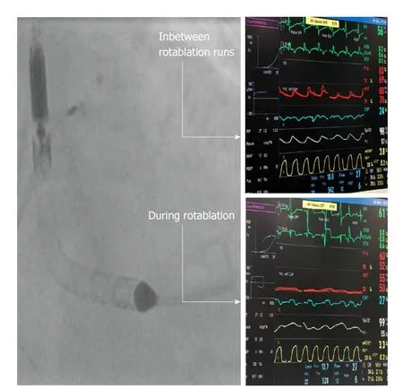
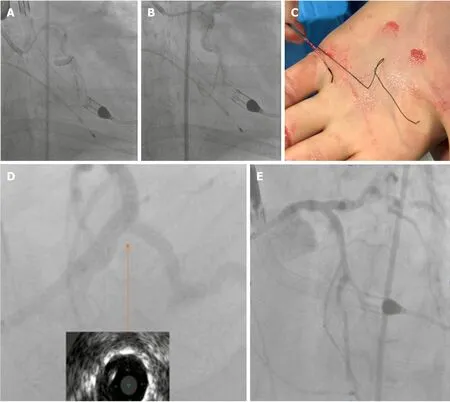
Subsequently using the right femoral access and an 8F EBU 3.5 guidecather the left main was initially rotablated with 1.75 mm burr.The loss of pulsatility during the runs was prominent,however mean arterial pressure was sustained at about 55 mmHg due to the presence of the 5.0 Impella(Figure 3).Despite a couple of complications and equipment failures,including localized LMS dissection balloon entrapment on coronary wire and undeployed stent dislodgement,a good result was obtained with reverse culotte LMS bifurcation stenting and targeted PCI of mid left anterior descending and proximal left circumflex lesions(Figure 4).
OUTCOME AND FOLLOW-UP
Following his successful LMS bifurcation rotablation PCI the patients was extubated the following day and the Impella 5.0 explanted 5 d later.He made an excellent recovery and was discharged home 10 d later.On one-year follow up the patient was doing remarkably well and his LV had recovered fully,with a current ejection fraction of 55%.He is fully compliant with his heart failure and antiplatelet regime.He still has mild aortic stenosis for which he will be kept under surveillance on an annual basis.

Figure 2 A 10-mm silver-coated Dacron graft was anastomosed to the subclavian artery and an lmpella 5.0 was inserted.
DISCUSSION
This case illustrates the importance of cross-specialty collaboration in overcoming challenges in coronary revascularization of high-risk patients with prohibited iliofemoral access for percutaneous mechanical circulatory support devices.
The co-existence of PAD and cardiovascular disease(CVD)is common with nearly 45% of patients with PAD suffering from simultaneous cardiovascular disease[4].Recently the first use of intravascular lithotripsy(IVL)to treat vascular disease using the Shockwave IVL device(Shockwave Medical Inc)in iliofemoral arteries for modification of calcified plaque in an attempt to facilitate percutaneous Impella CP implantation was described[5].Furthermore a fully percutaneous transaxillary approach for implantation of Impella CP was been described and is feasible[6,7].When it comes,however,to implantation of an Impella 5.0 percutaneous options are limited(e.g.transcaval[8]),whereas surgical axillary cut-down has been established as a safe technique[9].
An alternative approach to tackling this case may have been the use of Impella 5.0 to stabilize the patient prior to off-pump CABG.However,our surgical team felt that in view of the ongoing ischaemic symptoms,the surgical risk would have been prohibitive and an immediate treatment with PCI would be the preferred option.
CONCLUSION
Our case report suggests that the use of surgical cut down to facilitate last remaining conduit high risk PCI in unstable patients with poor left ventricular function is feasible and safe.(1)Impella 5.0 provides a high level of support,which allows the operators to optimize their revascularization techniques and overcome,without stress,complications that may occur during high risk left main PCI(last conduit);(2)The Impella 5.0 can be inserted using subclavian cut down in cases with peripheral vascular access not amenable to large bore access.The transcaval access technique,even though promising for Impella 5.0,has yet to be widely adopted;and(3)Despite the emergence of IVL often in patients with very extensive disease IVL and peripheral angioplasty may not be feasible and a cross-specialty collaboration is needed to facilitate use of alternative access for mechanical circulatory support.

Figure 3 During rotablation runs with the 1.75 mm burr the pulsatility was lost,however,the mean arterial pressure was maintained due to the presence of the lmpella.

Figure 4 After a stormy procedure a good angiographic and intravenous ultrasound result was obtained.A:Support issues despite use of guideliner leading on to stent dismounting off its balloon undeployed after it got trapped on a calcific bend;B:Localised small perforation/dissection at the distal left main;C:Extreme left circumflex tortuosity causing deformation of Choice PT XS coronary wire and balloon trapping on the wire;D:Spider view and intravenous ultrasound in distal left main stem showing good stent expansion and apposition;E:LAO cranial view showing good final left main stem Culotte result.
 World Journal of Cardiology2020年4期
- World Journal of Cardiology的其它文章
- Autonomic laterality in caloric vestibular stimulation
- Comparative assessment of clinical profile and outcomes after primary percutaneous coronary intervention in young patients with single vs multivessel disease
- Do age-associated changes of voltage-gated sodium channel isoforms expressed in the mammalian heart predispose the elderly to atrial fibrillation?
- Role of gut microbiota in cardiovascular diseases
