Cutaneous ciliated cyst on the anterior neck in young women:A case report
Yon Hee Kim,Jihyoun Lee
Yon Hee Kim,Department of Pathology,Soonchunhyang University Seoul Hospital,Seoul 04401,South Korea
Jihyoun Lee,Department of Surgery,Soonchunhyang University Seoul Hospital,Seoul 04401,South Korea
Abstract BACKGROUND A cutaneous ciliated cyst(CCC)is a rare,benign tumor in young female adults,which is usually found on the lower extremities.CASE SUMMARY We found an uncommon location of CCC in the anterolateral cervical area and reviewed the literature.A 20-year-old female complained of a well-defined,painless,palpable mass that started several years ago.The mass was tense and movable and located at the anterolateral aspect of the neck.Imaging showed a non-enhancing round mass.Surgical excision biopsy was performed,and the cystic mass was revealed to be a CCC.CONCLUSION The rare location of CCC can be found in anterior neck area,which should be another diagnostic option for mass on anterior neck.
Key Words:Head and neck neoplasms;Female;Young adult;Subcutaneous mass;Mixed tumor;Mullerian;Case report
INTRODUCTION
Morphologic evaluation or physical examination is helpful in the differential diagnosis of benign skin lesions in the cervical region.However,if the lesion is located deep in the subcutaneous area and does not have pathognomonic findings,such as central punctum of the epidermoid cyst,the differential diagnosis can be challenging and requires further imaging modalities.Otherwise,the location of the mass can be associated with its origin.Congenital neck masses can be classified by their location.Thyroglossal duct cyst,cervical clefts,and teratomas are mostly located at the midline,and branchial cleft anomalies,lymph nodes,and thyroid lesions are mostly located in the lateral neck[1].
Cutaneous ciliated cyst(CCC)was first reported in 1978 by Farmer and Helwig.It is a rare benign tumor of Mullerian heterotopias,and it has been reported as frequent in young females after puberty[2].The most common location is the lower extremities.There have been serial case reports that reported unusual locations of CCC,such as the back[2],abdominal wall[3],or scalp[4].It rarely occurs in male patients,and the locations reported are in the perineum,inguinal,and shoulder areas.
To our knowledge,there are limited reports of CCC that develop in the anterolateral neck area.We report our experience and summarize previous literature.
CASE PRESENTATION
Chief complaints
A 20-year-old female patient visited the clinic with a palpable nodule in her right anterior neck area.
History of present illness
The mass was first noticed a few years previous,and she could barely remember,but it had not grown since then.
History of past illness,personal and family history
She did not have any specific medical history,including congenital anomaly.However,she had a family history of papillary thyroid cancer of her father.
Physical examination
The mass was located in the subcutaneous tissue of the mediolateral area of the anterior neck,close to the right head portion of the clavicle.It was movable and tense and not accompanied by pain,fever,or redness.No specific abnormality was seen on her thyroid gland and midline cervical area.
Laboratory examinations
Ultrasonography showed a well-demarcated low echoic round mass,measuring 2.2 cm× 1.5 cm.The complete blood count showed that the white blood cell count was 4500/mm3,hemoglobin 13.9 g/dL,hematocrit 42.8%,and platelet count 264000 /µL.Neutrophil showed 55.82% in differential count,and lymphocyte showed 32.45%,monocyte 9.32%,eosinophil 1.70%,and basophil 0.71%.Blood chemistry results,including aspartate aminotransferase,alanine aminotransferase,total bilirubin,alkaline phosphatase,uric acid,gamma guanosine triphosphate,and lactate dehydrogenase,were within normal limits.Serum β-human chorionic gonadotropin detected to be less than 1.20 mIU/mL.
Imaging examinations
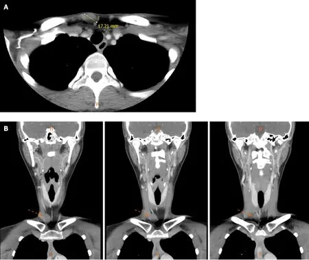
Computed tomography(CT)was performed for the differential diagnosis and spatial evaluation.Enhanced CT imaging showed a non-enhancing well-defined ovoid mass in the right paramidline supraclavicular fossa that measured 2.2 cm × 1 cm × 1.5 cm(Figure 1A and B),and the differential diagnosis from the image was dermoid or epidermoid cyst.
FINAL DIAGNOSIS
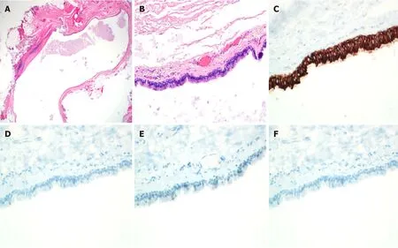
She had undergone an excisional biopsy for therapeutic and diagnostic purposes.In the gross examination,the lesion was an ovoid-shaped,solid,soft mass,and thin fibrous capsules surrounded the entire lesion.The microscopic findings revealed a well-defined and thin-walled,ovoid,unilocular cystic lesion lined by ciliated pseudostratified cuboidal to columnar epithelium showing focal intraluminal papillary projections in the subcutaneous tissue.The luminal surface of the cystic lesion was filled with amorphous proteinous materials without keratinous and solid mass-like lesion.The outer surface of the cystic lesion was completely covered with thin collagenous tissue without smooth muscle bundles.It also showed focal chronic inflammatory change with no lymphoid follicles or other components.The peritumoral subcutaneous tissue revealed no thyroid,cartilaginous,or skin appendageal tissue(Figure 2).
TREARMENT
After complete excision of the lesion,there was no development of seroma or wound infection.
OUTCOME AND FOLLOW-UP
After six months of follow-up,there was no evidence of local recurrence.
DISCUSSION
The location of CCC is mostly around the perineum or lower extremities,and is also reported in the abdominal wall,back,fingertips,and scalp.We reviewed the literature available of full-text files using PubMed search between 1978 and 2019(Table 1).Most of the clinical features were palpable cystic masses remaining indolent for months to decades,except one report of a rapid growth two months after two years from its first notice[5].The case reports included atients of various races,including African,Caucasian,and Asian.When the mass was ruptured during operation,the cystic contents showed clear serous or yellowish fluid materials.Limited evidence of longterm follow-up was found.Rosset al[6]reported a 36-year-old female patient with CCC on the dorsal aspect of the foot,found indolent since the patient’s late teens.Santoset al[7]reported a 35-year-old male patient who had CCC in his right perineum;they reported no evidence of recurrence or malignant change during follow-up.
CCC developed in male patients was found in a few reports[7-11];the locations were the scalp,perineum,and back.There has been a debate whether it is a persistent Mullerian cyst or ciliated metaplasia in the eccrine cyst lining[12].Because CCC has been regarded as Mullerian in origin,in addition to the morphologic evaluation under hematoxylin and eosin stain,there were reports of the use of estrogen receptor(ER),progesterone receptor,WT1,or PAX-8 immunostaining[13].Like other studies,the results of the immunohistochemical staining for ER,S-100,and desmin in this patient were negative[9].The choice of the diagnostic modality of a neck mass is sometimes challenging,especially if the mass is cystic nature.Cystic lesions on the lateral neck in young adult can have malignant potential,such as metastatic thyroid cancer[14,15].Fine needle aspiration cytology(FNAC)can be useful in solid lesions or metastatic thyroid cancer,but the diagnostic yield might not be sufficient for a definite diagnosis in cystic masses.Moreover,FNAC can be performed for the diagnosis of thyroglossal duct cysts[16]or epidermal cysts.However,it might cause an infection that interferes with the subsequent surgical procedure.

Table 1 Literature review in clinical features of cutaneous ciliated cyst
CONCLUSION
The rare location of this case should be helpful to diagnostic decision of masses on the lateral neck in young female adults.

Figure 1 Enhanced computed tomography of the neck.A:Non-enhancing round mass on the subcutaneous area in the anterior neck area in axial view;B:Serial images of coronal section.

Figure 2 Pathologic findings reveal a well-defined ovoid unilocular cystic lesion lined by ciliated pseudostratified cuboidal to columnar epithelium.A and B:Focal intraluminal papillary projections in the subcutaneous tissue(Hematoxylin and eosin stain,× 400);C:The lining epithelium reveals immunoreactive cytokeratin(cytokeratin,× 400);D-F:It reveals negative findings for immunostainings(D:Estrogen receptor,× 400;E:Desmin,× 400;F:S-100,×400).
 World Journal of Clinical Cases2020年19期
- World Journal of Clinical Cases的其它文章
- Role of monoclonal antibody drugs in the treatment of COVID-19
- Review of simulation model for education of point-of-care ultrasound using easy-to-make tools
- Liver injury in COVID-19:A minireview
- Transanal minimally invasive surgery vs endoscopic mucosal resection for rectal benign tumors and rectal carcinoids:A retrospective analysis
- Impact of mTOR gene polymorphisms and gene-tea interaction on susceptibility to tuberculosis
- Establishment and validation of a nomogram to predict the risk of ovarian metastasis in gastric cancer:Based on a large cohort
