Application of persulfate in low-temperature atmospheric-pressure plasma jet for enhanced treatment of onychomycosis
Qunxia ZHANG (张群霞), Cao FANG (方草), Zhu CHEN (陈祝) and Qing HUANG (黄青)
1 Key Laboratory of High Magnetic Field and Ion Beam Physical Biology, Institute of Technical Biology and Agriculture Engineering, Hefei Institutes of Physical Science, Chinese Academy of Sciences, Hefei 230031, People’s Republic of China
2 University of Science and Technology of China, Hefei 230026, People’s Republic of China
3 Anhui Jianzhu University, Hefei 230601, People’s Republic of China
Abstract Fungal infection of human nails,or onychomycosis,affects 10%of the world’s adult population,but current therapies have various drawbacks. In this work, we employed a self-made lowtemperature plasma(LTP)device,namely,an atmospheric-pressure plasma jet(APPJ)device to treat the nails infected with Trichophyton rubrum(T.rubrum)with the aid of persulfate solution.We found that persulfate solution had a promoting effect on plasma treatment of onychomycosis.With addition of sodium persulfate, the APPJ therapy could cure onychomycosis after several times of treatment. As such, this work has demonstrated a novel and effective approach which makes good use of LTP technique in the treatment of onychomycosis.
Keywords: low-temperature plasma (LTP), persulfate, onychomycosis, Trichophyton rubrum
1. Introduction
Onychomycosis is the most common nail disease caused by fungal infection, accounting for about 50% of the nail diseases. Onychomycosis is caused by dermatophytes, non-dermatophytes, and yeasts with a prevalence of approximately 2%-13% worldwide [1, 2]. Among fungal strains associated with onychomycosis,Trichophyton rubrum and Trichophyton mentagrophyte are two of the most important infectious pathogen, accounting for 80% of all dermatophytoses in humans [3]. Although onychomycosis is not fatal, it is widespread in human fungal infections.Typical symptoms of onychomycosis include discoloration, thickening and increased fragility of the nail, which may lead to nail detachment in the later stage of disease. These symptoms are
4 These authors are contributed equally to this work.not only related to personal aesthetics, but also may cause serious consequences such as pain and mobility problems[4].
Plasma is an ionized gas that can be produced using high voltages applied to a variety of gas source components,including argon,helium,nitrogen,compressed air,or a mixed gas. Low-temperature plasma (LTP) can generate free radicals, excited molecules, charged particles and ultraviolet photons [5], and can effectively inactivate microorganisms.Several research groups have studied the biomedical effects of different room-temperature plasmas on cells, tissues, and animals. Many studies have shown that atmospheric-pressure room-temperature plasma (ARTP) has strong inactivation effects on bacteria [6-8], fungi [9-11], biofilms [12-14],viruses [15], spores [16, 17], etc. Nowadays, atmosphericpressure-LTP or ARTP has been widely used in biomedical fields such as tumor treatment [18], coagulation [19], wound healing [20], and dermatological treatment [21].
Persulfate is a strong oxidant with a high redox potential of 2.01 V[22],and it has been applied in the emerging field of advanced oxidation technology [23]. Persulfate has high stability and can be activated by heat, ultrasonic, ultraviolet or vacuum UV irradiation, activated carbon, alkaline pH,hydrazine,phenol and transition metals or zero-valent iron to produce sulfate radicals (SO4−·) with even higher redox potential (2.6-3.1 V) [24, 25], which is equivalent to the redox potential of the commonly used ·OH with strong oxidation(2.8 V).Moreover,the lifetime of SO4−·is much longer than that of hydroxyl radicals(·OH),and so SO4−·can exhibit higher selectivity and reactivity [26]. Actually, due to this property, persulfate has been adopted to degrade organic pollutants [23, 27] and antibiotics [28].
In this work, therefore, we applied atmospheric-pressure plasma jet (APPJ) at ambient temperature to treat onychomycosis, and especially, we applied persulfate in the treatment, to test if persulfate could treat fungus with improved treatment efficiency. The fungus we chose, T. rubrum, is the main pathogen known for fungal infection of human nails.In recent years, researchers have carried out a series of investigation for inactivation of T. rubrum using plasma technology. Daeschlein et al inactivated several clinical fungi C.albicans, T. rubrum, T. interdigitale and M. canis using an argon atmospheric-pressure LTP jet device,and observed the different antibacterial effects [29]. Heinlin et al effectively inactivated T.rubrum and M.canis using LTP,indicating that low-temperature atmospheric-pressure plasma has broad application prospects in the treatment of skin fungal infections [30]. In another study, Shapourzadeh et al treated T.rubrum spore suspension with LTP for 90 s,120 s,150 s,and 180 s, respectively, and their results showed that the growth of T. rubrum was inhibited by 62%-91% [31]. However, for treatment of onychomycosis, it still remains a challenge to inactivate T. rubrum in nails in an effective way. Our idea was thus to make use of an effective additive in the LTP treatment to assist the killing of fungi.Therefore, to facilitate the plasma treatment of onychomycosis, in this work, we proposed the use of persulfate in the APPJ treatment of onychomycosis. As a result, we showed that our APPJ treatment could activate persulfate to form SO4−· and so the efficiency of inactivation of T. rubrum could be greatly improved. After several times of persulfate and plasma treatment,T.rubrum in nails could be completely inactivated,and so onychomycosis could be cured.
2. Materials and methods
2.1. Chemicals
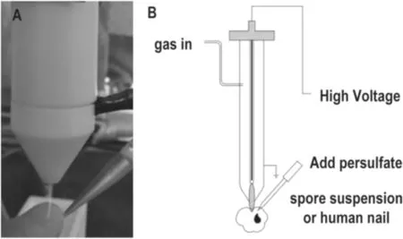
Figure 1.Photograph(A)and schematic plot(B)of the He plasma jet device applied to human nail infected with T. rubrum.
Peptone and agar for Sabouraud dextrose agar (SDA) media preparation and potassium hydroxide (KOH) were obtained from Sangon Biotech Co., Ltd, Shanghai. Glucose for SDA media preparation, sodium persulfate (Na2S2O8), sodium chloride (NaCl), ethanol, tert-butyl alcohol (TBA), salicylic acid, acetonitrile, methanol and citric acid were purchased from Sinopharm Chemical Reagent Co., Ltd, China.LIVE/DEADTMBacLightTMBacterial Viability Kit (SYTO 9/PI) was supplied by Invitrogen, America. 5,5-Dimethyl-1-pyrroline N-oxide (DMPO) was purchased from Macklin Biochemical Co., Ltd, Shanghai. Rhodamine B (RhB) was purchased from Aladdin biochemical technology Co., Ltd,Shanghai. Acetic acid was purchased from Shanghai Titan Scientific Co.,Ltd,China.Sodium persulfate was prepared in ultrapure water. All chemicals were of high-purity and thus used without further purification.
2.2. Low-temperature APPJ device
The photograph and schematic diagram of the self-made APPJ device used in the experiment are shown in figure 1.Briefly,a 15 cm long stainless steel electrode was placed in a quartz tube with the upper end of the anode connected to an AC power supply. The peak voltage, current and frequency were about 9.7 kV, 2.8 mA and 9.6 kHz, respectively. The power was 12 W according to our previous [32]. So for the plasma-jet irradiated area with diameter circa 1 mm, the power distribution per area was estimated about 3.8 W mm−2.The gas source for APPJ used in the experiment was helium,and the gas flow rate was 1 l min−1. The working gas was introduced into the tube at side 2 min before plasma initiation in order to exclude the air in the tube. When the sample was irradiated by the APPJ,the distance between the jet outlet and the sample was 5 mm.
2.3. Fungal strain and culture conditions
The standard strain of T. rubrum was purchased from the Institute of Dermatology, Chinese Academy of Medical Sciences (Nanjing). T. rubrum was cultured on SDA and incubated at 28°C for 7 d or until sporulation. The culture was covered with 5 ml of 0.9% sterile saline, and T. rubrum was scraped from the medium with an inoculating loop and counted with a hemocytometer to adjust to a final concentration of 106CFU ml−1.
2.4. Establishment of T. rubrum in vitro infection nail model
A model of onychomycosis was established based on the method described by Peres et al with some modifications[33].The nails of healthy adult volunteers were collected with no history of fungal diseases and nail diseases, and no nail cosmetics such as nail polish were used. The donors did not use antifungal drugs systematically or locally for the latest 3 months. Before the experiment, the nails of the same size were placed in a centrifuge tube, soaked in sterile water, and centrifuged at a speed of 6000 rpm min−1three times to remove dirt and breeding microorganisms on the deck surface. After that, the nails were washed three times with 75%ethanol solution, and finally washed three times with sterile water to wash away the residual ethanol solution on the nail surface.The cleaned nails were placed in a clean bench to be completely dry, and irradiated with ultraviolet light for one hour. After that, the nails were placed in a 12-well plate and co-cultured with 1 ml of T.rubrum suspension,and incubated at 28°C for one month until the nails were infected with T. rubrum.
2.5. APPJ treatment of T. rubrum spore suspension
During the plasma treatment, the solution samples were placed approximately 5 mm from the exit of the jet. The prepared spore suspension solutions of 160 and 40 μl of different concentrations of sodium persulfate solution/sterile were pipetted into selected wells of 96-well microtiter plates,and treated with the APPJ for 0 min, 1 min, 2 min, 3 min,4 min, 5 min, respectively. All the experiments were performed in triplicate. After the treatment, the treated bacterial solutions were diluted 100 times, and then 10 μl of diluted suspension was taken from the diluted solution and cultured on SDA and incubated for 5 d at 28°C until the colonies were formed. To observe the fungal inactivation directly, we also employed fluorescence microscopy with SYTO 9/PI staining method. The SYTO 9 green fluorescence label was used to identify the living cells while the PI red fluorescence label(which can only transfuse through a damaged cell membrane)was used to distinguish the damaged cells.With adding 0.1 M sodium persulfate into the spore suspension,the samples were then stained with SYTO 9/PI. After staining for 15 min in a dark room,all the samples were then washed using 2 ml 0.9%sterile physiological saline, repeated three times. Then the plasma-treated samples were immediately examined using fluorescence microscopy(Thermo Fisher Scientific EVOSTM)for direction observation of T. rubrum inactivation.
2.6. APPJ treatment of onychomycosis
During the plasma treatment, the infected nail samples were placed approximately 5 mm from the plasma jet exit and the treatment time of all nail samples was maintained for about different durations(3-5 min).The nail samples were placed in a 24-well plate and each well was added with 200 μl of sterile water or 0.02 M sodium persulfate solution. After the plasma treatment, the added sterile water/sodium persulfate solution was aspirated, and the treated nails were washed three times with sterile water.After the nails were completely dried,they were placed in SDA and cultured at 28°C. The plasma treatment was performed every 2 d for a total of three treatment times. We found that 5 min treatment time was enough for the antimycotic effect on T.rubrum.After three treatment sessions, the nails were then cut into two lengthwise pieces and treated with 20% potassium hydroxide (KOH) as prepared for the method of direct microscopy. Besides, the method of culture recovery was also applied for verification and comparison.

Figure 2.Summary of the processes of preparation and plasma treatment.
2.6.1. Evaluation of the therapeutic efficacy. The therapeutic effect can be evaluated using both the fungal culture approach and fungal direct microscopy approach. For the method of culture recovery, the procedure is illustrated in figure 2. In this approach,each nail was cut into 10 small pieces along the cross section after the plasma treatment. The nail fragments were then cultured on SDA and incubated for 2 weeks at 28°C. The effect of treatment was evaluated based on the number of culture positives in the tested nail fragments [34].
2.6.2. Observation of the hypha with light microscope.Besides,the infection of nails can also be examined by direct microscopy [35]. In this experiment, we used 20% KOH treatment of nails for direct microscopy. The nails were scraped and placed on the glass slide, then a drop of 20%KOH was added onto the nails, and then the nails were covered with the coverslips. KOH could dissolve the nails keratin, and the hypha could be observed under the light microscope (Qibu Biotechnology XDS-3KY).
2.7. Detection of the sulfate radical in the plasma-treated PS solutions
2.7.1. Electron spin resonance (ESR) measurement. ESR spectroscopy can probe the generation of·O2−,·OH and SO4−·in the reaction systems [36-38]. In order to confirm the sulfate radical during the plasma processing, we also conducted ESR experiment in which DMPO was used to trap the short-lived radicals at ambient temperature. In the experiment, ultrapure water of 400 μl, sodium persulfate solution of 0.1 M 100 μl and 5,5-Dimethyl-1-pyrroline N-oxide (DMPO) of 0.1 mol l−1500 μl were pipetted into selected wells of 24-well plates,and treated with the APPJ for 5 min.After the plasma treatment,the samples were measured by ESR equipment (Bruker EMX plus 10/12, equipped with Oxford ESR910 liquid helium cryostat).
2.7.2. Estimation of the contribution from SO4−·in the plasma treatment. For the determination of the contribution of SO4−·,an appropriate strategy is to eliminate·OH contribution using a chemical scavenger selective to ·OH. In this way,people have employed tert-butyl alcohol (TBA) as the selective ·OH scavenger to discriminate the contributions of·OH and SO4−·in the overall pollutant degradation[39].So in our experiment, we employed the similar approach, and used rhodamine B(RhB)to see its color change during the plasma treatment so as to estimate the APPJ treatment effect on the solution. During the APPJ treatment, the solution samples were placed approximately 5 mm from the exit of the jet.The prepared RhB solution of 400 μl mixed with 100 μl water or 0.1 M sodium persulfate solution was pipetted into a selected well in the 24-well plates, and treated with the APPJ for 1-5 min. All the experiments were performed in triplicate.After the treatment, the samples were detected by a UV-vis spectrometer (SHIMADZU UV-2550).
2.7.3.Detection of hydroxyl radical with salicylic acid(SA). A total of 0.4 ml salicylic acid solution (500 mg l−1) dissolved in H2O was added with 0.1 ml H2O and then the mixture was exposed to APPJ under the same plasma conditions as applied for T. rubrum treatment. After plasma treatment, the samples were detected using high performance liquid chromatography(HPLC, Shimadzu, Japan), and the HPLC instrument was equipped with an ultraviolet detector and a C18 column (5 μm, 4.6 × 250 mm2). The HPLC eluent was prepared using 10% (v/v) HPLC-grade acetonitrile, 10% methanol,0.03 M citric acid, 0.3 M acetic acid and water, and the flow rate was set at 1.0 ml min−1[40, 41].
3. Results and discussion
3.1. Effect of sodium persulfate solution combined with APPJ on T. rubrum spore suspension
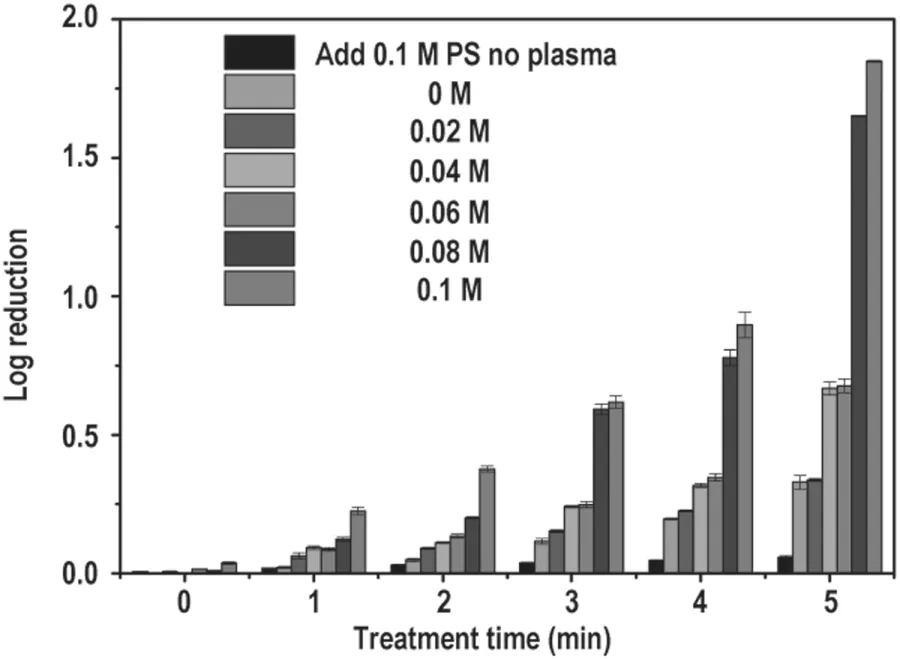
Figure 3.Comparison of T. rubrum log reduction after exposure to APPJ for 0, 1, 2, 3, 4, 5 min for different sodium persulfate concentrations (0, 0.02, 0.04, 0.06, 0.08, 0.1 M), respectively. The measurements were made after the samples were cultured on SDA and incubated statically for 5 d at 28 °C. Each experiment was repeated for three times. (PS: sodium persulfate).
After incubation for 5 d the number of colonies was counted,and the inactivation effect was quantitatively calculated. The survival rate for T. rubrum with or without the presence of sodium persulfate was measured depending on the treatment time and sodium persulfate concentration. The results are shown in figure 3. The log reduction for T. rubrum without sodium persulfate after He-plasma treatment for 3 min was 0.12, which increased to 0.33 with increasing the treatment time. With the presence of 0.06 M sodium persulfate in the fugal spore suspension, the log reduction for T. rubrum after treatment for 3 and 5 min was 0.25 and 0.68, respectively.By adding different concentrations of sodium persulfate(0-0.1 M) into the spore suspension, we found that the sterilization effect was gradually increased as the concentration of the sodium persulfate solution increased. The results indicate that in the presence of persulfate, T. rubrum showed 98.6%growth inhibition under the given discharge conditions.Figure 4 shows the fluorescent images of labeled T. rubrum under the fluorescence microscope, confirming that plasma plus persulfate adding could achieve the best killing of T. rubrum. This fluorescence-acquired result is consistent with the result obtained from the CFU method.
3.2.Observation of nail infection model under light microscope
After the nail fragments were co-cultured with T. rubrum for one month,the nails were placed under an optical microscope for observation. The results are shown in figure 5. The nails that were not infected with T. rubrum had a smooth surface and a clear outline; while for the T. rubrum-infected nails, if they were infected with a large amount of T. rubrum on the surface, the T. rubrum hyphae invaded the nails, and so the nail outline was blurred.
3.3.Therapeutic effect of sodium persulfate solution combined with APPJ on onychomycosis

Figure 4.Fluorescent images for evaluation inactivated effect of T. rubrum spore suspension after exposure to APPJ by using SYTO 9/PI dye. A: the negative control group; B: plasma after 5 min; C: plasma + 0.1 M sodium persulfate after 5 min plasma treatment.

Figure 5.The upper-panel shows the optical microscope images of uninfected and infected intact nails with T.rubrum,respectively.The low-panel shows the images of uninfected and infected scraped nail fragment, respectively. The nail scraps were soaked in 20% KOH,then visualized under white light (original magnification: ×25).
We found that plasma treatment once or twice did not show complete growth inhibition of T.rubrum,so we chose a total of three times for the plasma treatment. T. rubrum-infected nails were treated with APPJ every two days after each plasma treatment. After the three times of plasma treatment,the nails were cultured on SDA and directly examined with 20% KOH fungal microscopy. The fungal culture and KOH fungal microscopy are the gold standard for the detection of onychomycosis [42]. The results for the therapeutic effect of sodium persulfate solution combined with APPJ on onychomycosis are shown in figure 6 and table 1. The results show that the addition of sodium persulfate without plasma treatment had negligible therapeutic effect on onychomycosis,and the sole plasma treatment effect was not enough to kill all the fungal cells, while adding persulfate had a synergistic effect on plasma sterilization, which promoted curing onychomycosis significantly. Figure 7 shows that the infected nails became cloudy on gross appearance,and as the treatment time increased, the appearance of white turbidity became less and less until it was healed.
3.4. Contribution from sulfate radical in the plasma treatment
Qualitatively, we could detect the ·OH and SO4−· radicals by ESR spectroscopy, and the results are displayed in figure 8,which confirms the existence of both DMPO-·OH and DMPO-SO4−· in the plasma treatment. The ESR spectra exhibit the four- and six-line signals, which are the characteristic peaks of DMPO-·OH and DMPO-SO4−· adducts,respectively [43]. To be noted, there is no or negligible of sulfate radical without PS presence or before the plasma treatment.
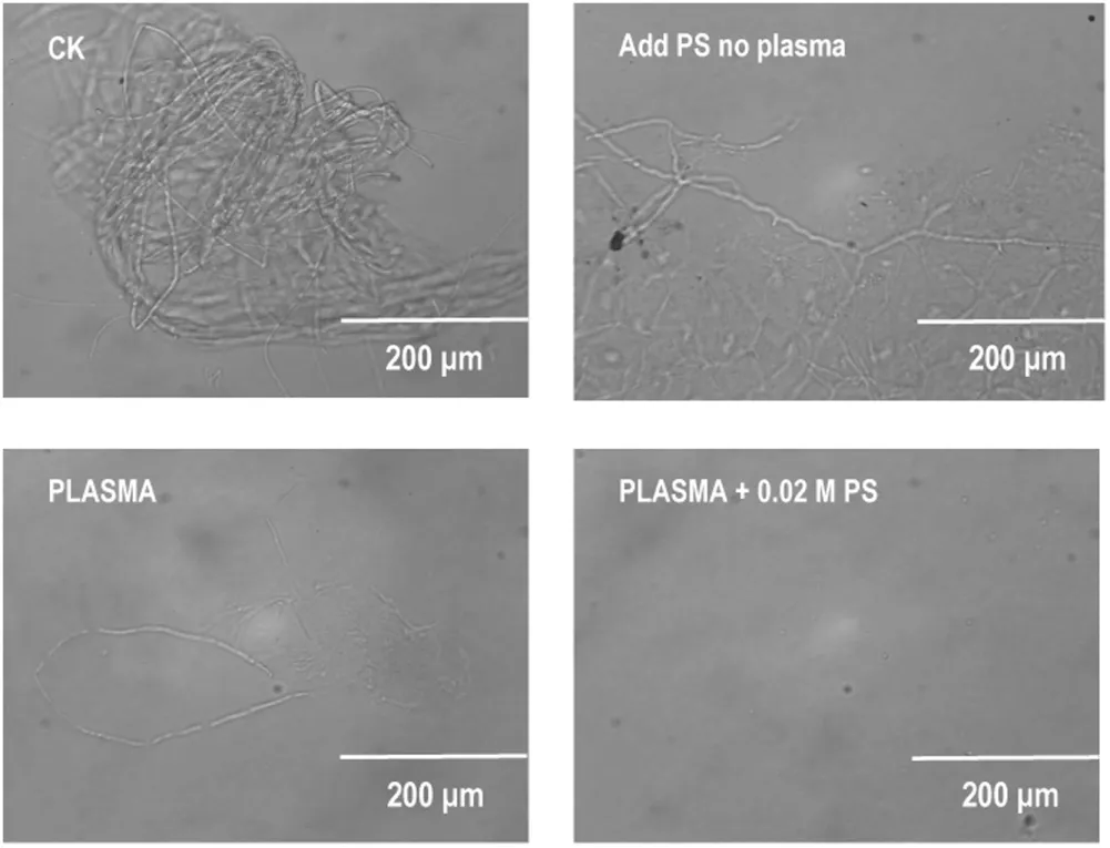
Figure 6.Fungal direct microscopy for evaluation of efficacy of APPJ combined with persulfate solution.The four nail scrap groups were the negative control group,the PS group,the plasma group,and the plasma plus sodium persulfate(0.02 M)group,respectively.The nail scraps were obtained by soaking the nails in 20% KOH,visualized under white light (original magnification: ×25).(PS: sodium persulfate).
Since there were both sulfate and hydroxyl radicals in the solution, and with hydroxly radical activating PS to form sulfate radical in the solution,so relatively,we could estimate the contribution of SO4−· during the plasma treatment using the method reported previously [44], in which the relative contributions of ·OH and SO4−· can be determined by comparing the difference in the removal efficiencies with and without ·OH scavenger addition. Figure 9 shows the result,from which we could estimate that the activated persulfate radical was over 90% (the contribution of SO4−·/the contributions of SO4−· and ·OH) for the plasma treatment for 5 min.
To further estimate the concentration of sulfate radical,because we found no literature for a quantitative measurement, we thus took an approximate approach by estimating the concentration of hydroxyl radical under the same condition using HPLC method. As a result, we found 5 min APPJ treatment of water could produce hydroxyl radical with concentration about 1.9 mM in the solution.Considering that the solution contained majority of sulfate radical as shown by figure 9, if we assume 90% hydroxyl radical was used up to activate PS to form sulfate radical,so we can estimate that the sulfate radical concentration was about 1.7 mM. Although itis a very rough estimation,it confirms anyway that the role of sulfate radical cannot be neglected in the treatment.

Table 1.Culture recovery: evaluation of therapeutic efficacy.

Figure 7.Photographs of infected nails. The four groups were the negative control group, the PS group, the plasma group, and the plasma plus sodium persulfate (0.02 M) group, respectively. These photographs showed that the therapeutic effect of APPJ combined with sodium persulfate on onychomycosis. (PS: sodium persulfate).

Figure 8.ESR signals of aqueous dispersion for DMPO- ·OH and DMPO- in the sterile water and the plasma plus persulfate adding to the system. The measurements were carried out with magnetic field of 3350 G, sweep width of 100 G, microwave frequency of 9.396 799 GHz,modulation frequency of 100 kHz,and power of 5.02 mW. (PS: sodium persulfate).
The results presented in this work clearly show that sodium persulfate solution can indeed promote plasma to inactivate T. rubrum and effectively treat onychomycosis. In recent years, scientists have conducted extensive research on the antibacterial properties of plasma. Table 2 lists some published antimicrobial results using plasma on different microorganisms as reported by other groups as well some results due to our work.Xiong et al used a helium plasma jet,a surface microdischarge plasma device, and a floating electrode dielectric barrier discharge to treat model nails with either E.coli bacteria or T. rubrum fungus applied to the nail surface. The results showed that all the three plasma devices could significantly kill bacteria and fungi on bovine hoof slices(human nail replacement model)[45].Lipner et al used a low temperature plasma to treat onychomycosis, and most patients were satisfied with the treatment [46]. In this work,we used He APPJ to inactivate T. rubrum spore suspension with the aid of persulfate additive. We tested this discharge device to inactivate E.coli for comparison, and the bactericidal effect was remarkable and comparable with the results of other research groups in the absence of sodium persulfate,while in the presence of sodium persulfate, the sterilization efficiency can be significantly improved (table 2). In this work, we have also showed that the indirect treatment (i.e.adding sterile water or sodium persulfate solution during treatment) was more efficient for dealing with the infected nails,because direct treatment(without adding any solutions)could only kill fungi on the surface of the nail, whereas indirect treatment could not only allow the plasma produced active species to penetrate the nail fungi, but also potentiate the effect on plasma treatment of onychomycosis by activating persulfate.
In this work, we showed that onychomycosis could be cured after three times of plasma plus persulfate treatment.Although the therapeutic effect was based on the in vitro onychomycosis model, we understand that the treatment should also depend on the degree of the infection and some other specific circumstances of the patients. Besides, onychomycosis can be caused by a variety of fungi, such as,Trichophyton rubrum and Trichophyton mentagrophyte. In comparison of varied pathogens, Trichophyton rubrum (T.rubrum) represents the most clinically important specie.Therefore, here we only chose T. rubrum in this research for the conception proof.Of course,in real cases,we may have to consider other fungi that may cause onychomycosis, including Trichophyton mentagrophyte as well.
4. Concluding remarks
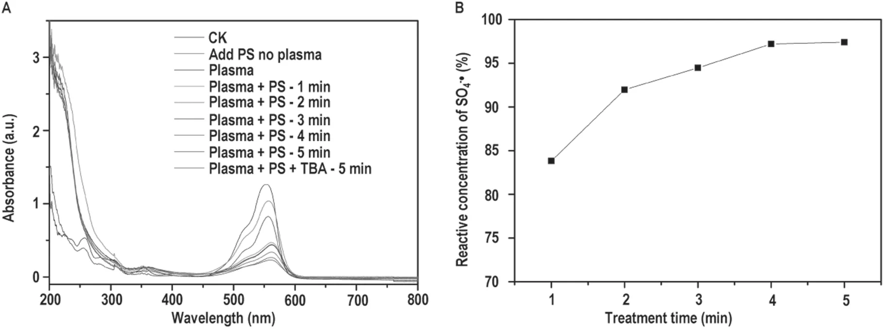
Figure 9.(A) UV-Vis absorption spectra of RhB. The RhB characteristic absorption peaks in the visible region of 553 nm (PS: sodium persulfate; TBA: tert-butyl alcohol), (B) The relative percentage concentration of sulfate radical after the plasma treatment for 1-5 min.
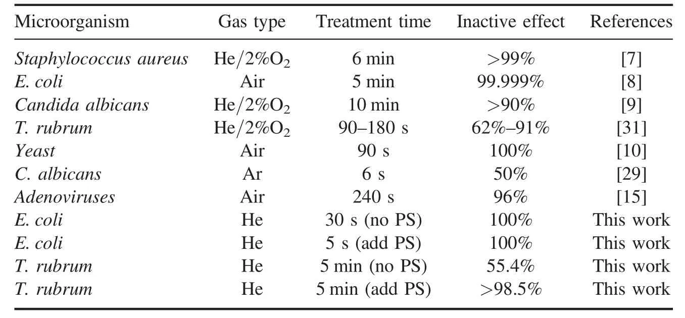
Table 2.Antimicrobial results using plasma on different microorganisms (PS: sodium persulfate).
Plasma treatment of human onychomycosis has potential clinical application prospects. Compared with other methods for treating onychomycosis, plasma treatment is relatively safe,simple,inexpensive and effective.In this experiment,we added sterile water or sodium persulfate solution to the nails during plasma treatment of onychomycosis, and investigated the effect and the mechanism involved in the treatment of onychomycosis. Using the APPJ-persulfate approach, the plasma could not only kill the fungi with its produced oxygen reactive species, but also activate persulfate to form longerlived reactive sulfate radical SO4−· (the lifetime of SO4−· is much longer than that of the hydroxyl radical (·OH)), which thus improved significantly the efficiency of killing T.rubrum and the treatment of onychomycosis. Therefore, this work may provide a more effective clinical application of LTP to treat human onychomycosis.
Acknowledgments
We would like to thank Mr Chuankai Xia and Dr Chunjun Yang for providing Trichophyton rubrum. A portion of this work(ESR measurement)was performed with assistant of Dr Wei Tong on the Steady High Magnetic Field Facilities,High Magnetic Field Laboratory, CAS. This work is supported by National Natural Science Foundation of China (Nos.11635013 and 11775272).
 Plasma Science and Technology2020年2期
- Plasma Science and Technology的其它文章
- Analysis of the characteristics of the plasma of an RF driven ion source for a neutral beam injector
- Synthesis of vertical graphene nanowalls by cracking n-dodecane using RF inductivelycoupled plasma-enhanced chemical vapor deposition
- Magnetic field enhanced radio frequency ion source and its application for Siincorporation diamond-like carbon film preparation
- Microwave frequency downshift in the timevarying collision plasma
- Comparison of Reynolds average Navier-Stokes turbulence models in numerical simulations of the DC arc plasma torch
- Effects of fast ions produced by ICRF heating on the pressure at EAST
