Elderly male with blurry vision
Anthony Maselli, Sarah E. Frasure
Department of Emergency Medicine, Brigham and Women's Hospital, Boston, Massachusetts 02115, USA
Dear editor,
We present a case of an elderly gentleman who was punched in the face and subsequently diagnosed by an emergency physician with acute angle closure glaucoma secondary to a posterior lens dislocation.Slit lamp evaluation, ocular ultrasound, and computed tomography (CT) imaging, can identify a dislocated lens. A rare complication of a lens dislocation includes secondary acute angle closure glaucoma. Treatment of lens dislocation includes prosthetic lens placement or reattachment in cases of a partial lens dislocation.
CASE
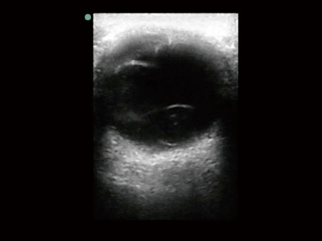
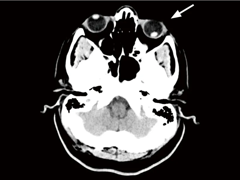
A 71-year-old male with a history of hypertension and glaucoma presented to the emergency department(ED) with pain and decreased vision in the left eye after being punched in the face three weeks prior while on vacation. He denied diplopia, headache, neck pain,or rhinorrhea. His vital signs were unremarkable. His physical exam was notable for a fixed, dilated left pupil to 7 mm with scleral injection. Intraocular pressure was measured at 14 mmHg OD and 56 mmHg OS. Extraocular movements were intact. A point-of-care ultrasound(Figure 1) and CT scan (Figure 2) were performed.
The patient was diagnosed with acute angle closure glaucoma secondary to a posterior lens dislocation.The patient was seen by the ophthalmology service. In an attempt to reduce the intraocular pressure, he was provided with an oral carbonic anhydrase inhibitor,latanoprost, brimonidine, and timolol eye drops. Delayed surgical repair of the lens was scheduled.
DISCUSSION

Figure 1. Point-of-care ultrasound of the patient’s left eye performed with a linear transducer (4-12 MHz) in the transverse position demonstrates a dislocated lens (arrow). The dislocated lens is identified as a round structure with a hyperechoic rim; it is visualized in the posterior aspect of the vitreous chamber.

Figure 2. Axial view of the patient’s head on computed tomography(CT) imaging demonstrates a complete lens dislocation in the left eye(arrow).
Lens dislocation occurs when the lens detaches from the ciliary bodies. Risk factors include blunt ocular trauma, Marfan syndrome, Weill-Marchesani syndrome,sulfite oxidase deficiency, and homocystinuria.[1-3]Slit lamp evaluation, ocular ultrasound, and CT imaging, can detect a dislocated lens. Treatment of lens dislocation involves prosthetic lens placement or re-attachment in cases of a partial lens dislocation.[1,4]A unusual complication of a lens dislocation includes secondary acute angle closure glaucoma; displacement of the lens causes narrowing of the anterior chamber angle and pupillary block. Emergent ophthalmology consultation and immediate reduction in intraocular pressure is paramount in cases of acute angle closure glaucoma secondary to lens dislocation.
Funding:None.
Ethical approval:Not needed.
Conflicts of interest:The authors declare that there are no conflicts of interest relevant to the content of the article.
Contributors:AM proposed the study and wrote the first draft.All authors read and approved the final version of the paper.
 World journal of emergency medicine2019年1期
- World journal of emergency medicine的其它文章
- Instructions for Authors
- Hemodynamic improvement using methylene blue after calcium channel blocker overdose
- Intussusception in an infant with two non-diagnostic abdominal ultrasound studies
- Assessment of clinical dehydration using point of care ultrasound for pediatric patients in rural Panama
- Prehospital response to respiratory distress by the public ambulance system in a Ukrainian city
- Establishment of trauma registry at Queen Elizabeth Central Hospital (QECH), Blantyre, Malawi and mapping of high risk geographic areas for trauma
