Subungual Exostosis Misdiagnosed as Subungual Wart
Yu-Ning Zhang, Jing-Yu Ren, Shu-Ping Guo, Hong-Ye Liu,∗
1Mathematics and Statistics Faculty of Central South University, Changsha, Hunan 410012, China, 2Department of Dermatology,First Hospital of Shanxi Medical University, Taiyuan, Shanxi 030001, China.
Introduction
Subungual exostosis (SE) is a clinically rare benign bone tumor that develops underneath or beside the nail bed.SE can occur in any finger or toe,but it is most common at the distal phalanx of the hallux, accounting for about 70%-80% of cases.1Patients generally have no subjective symptoms and often seek medical care for secondary infection or nail plate deformation. Because of its special location, SE is liable to be misdiagnosed as a subungual wart or other nail-related lesion. We herein report a case of SE in a patient,who was misdiagnosed as a subungual wart, in order to improve the knowledge of clinician to SE.
Case report
An 11-year-old girl presented to the First Hospital of Shanxi Medical University on August 2018 because of an eight-month history of nail deformation of the left hallux.A pea-sized gray-yellow subungual neoplasm had appeared on the distal phalanx of left hallux eight months ago.The patient denied any known triggers for the lesion and was otherwise asymptomatic. The neoplasm slowly got enlarged,leading to nail plate lifting and onycholysis.She subsequently developed pain and discomfort while walking.She visited a local hospital,and a subungual wart was diagnosed. Three cycles of cryotherapy were ineffective. The patient had no known drug or food allergy and no other systemic diseases.
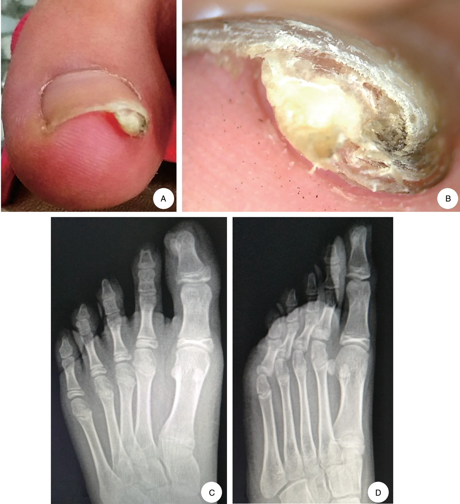
Physical examination revealed a well-demarcated,grayyellowverrucousneoplasmmeasuringabout1.0cm×0.8cm at the distal phalanx of the left hallux. The lesion was stiff, and the nail plate was lifted and separated(Fig.1A).Dermoscopic examination revealed a gray-yellow hyperkeratotic subungual nodule without vascular structures. Except for the periphery, the surface was basically smooth (Fig. 1B). Imageological examination revealed an osteophyte connected to the distal phalanx of the left hallux(Fig.1C and 1D).A diagnosis of SE was made based on the above results.
The patient was transferred to the orthopedics department for surgical resection, and complete removal of the neoplasm was effective.
Discussion
SE is a benign bone tumor that usually involves the distal toe, and was first reported by Dupuytren in 1817.2It commonly occurs in children and teenagers.The pathogenesis of SE remains incompletely understood, but it is generally believed to be associated with trauma,infection,and genetic factors.3The most common clinical manifestation is a firm,slowly progressing exogenous nodule at the distal aspect of the toe or finger. Obvious subjective symptoms are generally absent,but nail plate deformation may develop,and swelling and pain may occur because of mechanical stimulation or secondary infection. Imaging findings reveal an osseous tumor originating from the distal phalanx of the toe or finger, which has a normal bone structure with no osteolytic destruction or periosteal reaction.
Radiographs are very useful for a definitive diagnosis of SE. Dermoscopy is a recently developed technique for examining skin abnormalities;it is easy to perform and has been used in the diagnosis and differential diagnosis of various skin lesions.To the best ofourknowledge,however,no report to date has described the dermoscopic findings of SE.In the present case,dermoscopic examination showed a well-demarcated,gray-yellow hyperkeratotic nodule with a smooth surface and no vascular structures.

Figure 1. Clinical presentation and laboratory examination results of the lesion. A: A well-demarcated, gray-yellow verrucous neoplasm of 1.0cm×0.8cm was located at the distal phalanx of the left hallux.The nail plate was lifted and separated.B:Dermoscopic examination showed a gray-yellow hyperkeratotic subungual nodule without vascular structures.Except for the periphery,the surface was basically smooth.C and D:Radiographs revealed an osteophyte connected to the distal phalanx of the left hallux,which had a normal bone structure with no osteolytic destruction or periosteal reaction. Posterior-anterior (C) and oblique (D) radiographs of the same lesion.
SE should be distinguished from a subungual wart,paronychia, osteochondroma, and pyogenic granuloma.4The above findings differ from multiple papillary hyperplasia in common warts. The present case suggests that dermoscopic examination may have high clinical value in the differential diagnosis of SE, but the dermoscopic characteristics of this disease are still unclear,and a large sample is needed for observation and summary.
In our case,the diagnosis of SE was confirmed based on the medical history, clinical manifestations, and dermoscopic and X-ray examination findings.Surgical resection was effective.
- 国际皮肤性病学杂志的其它文章
- Solid-Cystic Hidradenoma
- Arteriosclerosis Obliterans Presenting as Multiple Leg Ulcers: A Case Report
- Interstitial Mycosis Fungoides with Systemic Sclerosis-Like Features: A Case Report
- Nasal Type Extranodal NK/T-Cell Lymphoma Presenting with Unilateral Facial Erythemas,Nodules, and Necrosis
- Lentigines within Nevus Depigmentosus
- Severe Port Wine Stain with Significant Nodules and Alveolar Bone Invasion Leading to Restricted Mouth Opening

