Study on the Inhibitory Effect of Scopoletin on Proliferation and Metastasis of Breast Cancer Cells
Xiaoyan LIU Juan LI Zhiqiang MEI

Abstract [Objectives] This study was conducted to investigate the effect of scopoletin on the proliferation of breast cancer cell lines.
[Methods] Cell viability, gene expression and cell cycle were measured by cell culture, RTPCR and flow cytometry. The effects of different concentrations of scopoletin on proliferation and cell cycle of breast cancer cells were compared.
[Results] After a certain period of time, the breast cancer cells showed different degrees of apoptosis, and the growth cycle was arrested in the G1 phase, which promoted the downregulation of three genes in breast cancer metastasis.
[Conclusions] The study proves that scopoletin inhibits the proliferation and metastasis of breast cancer cells in a concentration and timedependent manner.
Key words Scopoletin; Breast cancer; Inhibition; Study
Breast cancer is one of the most common malignant tumors in women. Although the current surgery and radiotherapy and chemotherapy can improve the survival rate of patients, there are still some patients with recurrence and metastasis, especially the complications such as hair loss and thoracic deformity caused by breast surgery and radiotherapy and chemotherapy, which affect the quality of life of women. It is of great significance to seek effective and lowtoxic drugs for the prevention and treatment of breast cancer. Recent studies have found that scopoletin has a significant antitumor effect on a variety of malignant tumors. Scopoletin is widely found in a variety of plants, and the content is relatively higher in seeds of Melia azedarach Linn., leaves of Artemisia capillaris Thunb., stems and leaves of Artemisia annua Linn. and whole plant of Arabidopsis thaliana. Studies have shown that scopoletin has the effects of lowering blood fat and blood pressure[1-2], resisting inflammation[3-4], protecting liver[5-6], enhancing memory activity[7-8]and resisting tumor[9]. In this study, the effects of scopoletin on cell proliferation and metastasis in human breast cancer cells were investigated, so as to explore its possible mechanism.
Materials and Methods
Materials
Human breast cancer cell line MDAMB435 was purchased from Shanghai Institute of Biochemistry and Cell Biology, Chinese Academy of Sciences. Scopoletin was purchased from Shanghai Yuanye Biotechnology Co., Ltd., prepared into a 100× stock solution with dimethyl sulfoxide (purchased from sigma) and stored in a refrigerator at -20 ℃. Other reagents and instruments used in this experiment included RNeasy Mini Kit (Cat No: 74104, Qiagen), MAXIMASC ultrapure water purifier, DMEM medium (Gibco), and MTT purchased from Beijing Huarui Biological Pharmaceutical Co., Ltd. and stored at 4 ℃ in dark place.
Methods
Cell culture
The breast cancer cell line MDAMB435 was cultured with a high glucose DMEM (10% fetal bovine serum) medium in a carbon dioxide incubator at 37 ℃ with 5% CO2 and saturated humidity. The medium was changed every 2-3 d, and the cells were subjected to digestion and passage with 0.25% trypsin and 0.02% EDTA. When the cells were in the logarithmic growth phase, they were digested and centrifuged for collection.
Cell grouping
Cells in the logarithmic growth phase were adjusted to a concentration of 5×10⁴ cells/ml and inoculated in 96wellplates. 100 μl of cell suspension was added to each well. After 24 hof culture, the experimental groups were separately added with scopoletin to a final concentration of 0, 100, 250 and 500 μg/ml, respectively. Another well was added with the culture medium containing no cells, and used as a blank control group for zero adjustment. Each group was provided with 3 duplicate wells.
Detection of cell activity
After 24, 48 and 72 h of treatment with scopoletin, 20 μl of MTT (5 mg/ml) was added, and 4 h later, the supernatant was discarded. DMSO was added according to 200 μl/well, followed by shaking well for 10 min. The absorbance (A value) of each well was determined with a fullwavelength microplate reader (wavelength 570 nm), and the cell growth inhibition rate was calculated according to Cell growth inhibition rate (%)=(1-Aexperimental group/Acontrol)×100%.
Flow cytometry analysis of cell cycle
The breast cancer cells in the logarithmic growth phase were inoculated into 6well plates, and after 24 h of culture, scopoletin was added to the culture medium to a final concentration of 100, 250 and 500 μg/ml, respectively, followed by 48 h of culture (the control group was added with complete medium). The cells were washed and digested with PBS, and then centrifuged. 3.5 ml of anhydrous ethanol was added to fix the cells for 30 min. Then, 200 μl of PBS and 2 μl of RNase (0.25 mg/ml) were added and incubated at 37 ℃ for 30 min. Staining was performed with 0.5 ml of 50 μg/ml for 30 min, and the cells were filtered (300 μm nylon mesh) into an EP tube, and subjected to cell analysis on an EPICS flow cytometer (Beckman-Coulter).
Expression of genes related to breast cancer metastasis
The total cellular RNA was extracted using the RNeasy mini kit (Cat No: 74104, Qiagen) 6 h after the treatment with scopoletin, and the RNA was measured using an ND1000 UV/visible spectrophotometer (NanoDrop, USA). In a 10 μl RT reaction system, 2 μl of 5×RT buffer, 1 μl of dNTPs, 0.5 μl of random primer, 0.5 μl of Rev. Ace (purchased from TOYOBO), 0.25 μl of Super RI, 0.25 μl of RTenhancer, 2.25 μl RNase free water and 3.25μl of RNA (150 ng/μl) were added. The specific procedures were as follows: 30 ℃ for 10 min, 42 ℃ for 30 min, 99 ℃ for 5 min, and finally 4 ℃ for 5 min, and the product was incubated at 16 ℃. After the synthesis, the cDNA was diluted by adding 40 μl of ddH2O to serve as a template for quantitative PCR (qPCR), and the 18SRNA gene was used as an internal control. In a 10 μl reaction system, 5 μl of 2× PCR probe mix, 0.02 μl of probe, 1 μl of primer, 2 μl of H2O and 2 μl of cDNA were mixed, and 40 cycles of amplification were performed in a StepOne plus Thermocycler. Realtime quantitative PCR was performed to detect mRNA expression levels of EMTrelated genes. The primers of related genes were as follows: Vimentin gene upstream primer: TGGTCTAACGGTTTCCCCTA, and downstream primer: GACCTCGGAGCGAGAGTG, Zeb1 gene upstream primer: TGACTATCAAAAGGAAGTCAATGG, and downstream primer: GTGCAGGAGGGACCTCTTTA, and Ncadherin upstream primer: TGGGAAATATAGACAAGCTGGAA, and downstream primer: CTGTTATGTTGAGCTCCTCACTGT. The internal reference gene Q18s used the upstream primer: GCAATTATTCCCCATGAACG, and downstream primer: GGGACTTAATCAACGCAAGC.
Experimental data processing
The results were expressed as mean±standard deviation. The qtest was used for intragroup and intergroup comparison, in which P<0.05 was considered as having statistical significance. The calculation software was SPSS13.0 for windows.
Results and Analysis
Morphological observation
Under the inverted microscope, the breast cancer cells in the control group grew vigorously in a fusiform reticular pattern with neat edges, and the cells bestrewed the whole culture dish. After 24, 48, and 72 h of treatment with the scopoletin, the cells turned round to different degrees with the treatment time, and the nuclei were broken into circular bodies of different sizes. Some of the cells had cell membrane detached and suspended, and cell debris can be seen.
Detection of cell activity by MTT assay method
The inhibition rate of cell growth in each group was significantly different from that in the untreated group (P<0.05). After 24 h of treatment with different concentrations of scopoletin, the 100, 250 and 500 μg/ml treatment groups showed the inhibition rate of breast cancer cell lines of 22.62%±0.33%*, 32.23%±0.11%* and 41.41%±0.22%*, respectively. After 48 h, the inhibition rates of breast cancer cell lines at 100, 250 and 500 μg/ml were 25.52%±0.28%*, 38.43%±1.25%* and 52.41%±1.22%*, respectively. After 72 h, the inhibition rates of breast cancer cell lines at 100, 250 and 500 μg/ml were 32.72%±0.28%*, 68.63%±0.36%* and 70.53%±1.15%*, respectively. Scopoletin had a significant inhibitory effect on breast cancer cells, and its inhibitory effect was enhanced with the increase of the concentration of scopoletin.
Cell cycle results from flow cytometry analysis
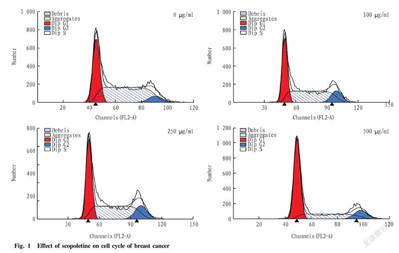
The experimental results showed that with the increase of concentration, most of the breast cancer cells were arrested in the G1 phase. In the 500 μg/ml group, 60.12%±3.13%, 27.14%±1.25%and 12.74%±0.19% of the cells stopped in the G1 phase, S phase and G2 phase, respectively, while only 35.83%±0.21% of the cells in the control group stopped in the G1 phase, and the cells stopped in the S phase and G2 phase accounted for 57.15%±2.13% and 7.03%±0.18%, respectively. The differences were significant.
Effects of scopoletin on the expression of breast cancer metastasisrelated genes
The mRNA expression of the three genes was analyzed by realtime PCR. The results showed that with the concentration of scopoletin increasing, the mRNA expression levels of VIM, Zeb1 and Ncad decreased in a dosedependent manner. It can be seen that scopoletin can inhibit the expression of the three genes, thereby inhibiting the metastasis of breast cancer cells.
Discussion
Epithelialmesenchymal transition (EMT) refers to the biological process by which epithelial cells are transformed into interstitial phenotype cells. It plays an important role in embryonic development, cancer metastasis, drug resistance and various fibrotic diseases. Its main features are the reduction of cell adhesion molecule expression and the conversion of cytokeratin cytoskeleton into vimentinbased cytoskeleton with the characteristics of mesenchymal cells in morphology. Through EMT, epithelial cells lose their epithelial phenotype such as attachment to the basement membrane, and obtain higher antiapoptotic, invasive and migratory capabilities. EMT is an important biological process for the migration and invasion of epithelialderived malignant cells. VIM, Zeb1 and Ncad are the three most important genes for cancer cell metastasis. These three genes can promote the transformation of epithelial cells into mesenchymal cells, which is to promote the occurrence of EMT. Through the effects of different concentrations of scopoletin on breast cancer cells, it can be found that the expression of these three genes was also decreased with the concentration increasing, indicating that the drug has an inhibitory effect on the metastasis of breast cancer cells.
Its antitumor mechanism may be inhibition of cell cycle arrest and induction of apoptosis and differentiation of tumor cells through inhibition of tumor cell signaling. More reports are about the antitumor activity of scopoletin on T lymphoma cells and prostate cancer PC3 cells[9-10]. Intensive research has shown that this drug is found to be beneficial for reducing intracellular protein content and acid phosphatase activity. At the same time, scopoletin also has an antiangiogenic effect, which can block the autophosphorylation of vascular endothelial growth factor receptor2 in tumor cells and the downstream signaling pathway, and inhibit the activity of extracellular signalregulated kinase (ERK1/2) of fibroblasts. Thereby, the angiogenesis of tumor cells is blocked, the metastasis and proliferation of primary tumor cells are inhibited, and tumor cell apoptosis is induced[11]. Studies have shown that the antitumor active site of scopoletin is mainly the phenolic hydroxyl group of C6 and the methoxy group of C7. After the introduction of alkoxy groups into the phenolic hydroxyl group of C6, the inhibitory activity of the derivatives on breast cancer cells MDA and colon cancer HT29 was increased by more than 10 times compared with scopoletin[12]. At present, Bukhsh et al.[13]developed the scopoletin polymer nanocapsule, which can enhance the inhibitory effect of scopoletin on human melanin tumor cell A375 and significantly improve its antitumor activity.
Scopoletin may promote the expression of P21, and then block tumor cells in the G0/G1 phase by above action pathway. And as the drug concentration increases, the blocking effect becomes more significant. After being treated with 500 μg/ml scopoletin for 24 h, 60.12%±3.13% of the cells were arrested in the G1 phase, which inhibited cell proliferation. Therefore, it is speculated that scopoletin will be a promising natural drug for cancer treatment. It may have a significant inhibitory effect on malignant tumors that grow faster.
References
[2] YANG JY, KOO JH, YOON HY, et al. Effect of scopoletin on lipoprotein lipase activity in 3T3L1 adipocytes[J]. Int J Mol Med, 2007, 20(4): 527-531.
[3] MAHATTANADUL S, RIDTITID W, NIMA S, et al. Effects of Morinda citrifolia aqueous fruit extract and its biomarker scopoletin on reflux esophagitis and gastric ulcer in rats[J]. J Ethnopharmacol, 2011, 134(2): 243-250.
[4] PAN R1, GAO XH, LI Y, et al. Antiarthritic effect of scopoletin, a coumarin compound occurring in Erycibe obtusifolia Benth stems, is associated with decreased angiogenesis in synovium[J]. Fundamental & Clinical Pharmacology, 2010, 24(4): 477-490.
[5] YIN HL, LI JH, LI J, et al. Four new eoumarinolignoids from seeds of Solanum indicum[J]. Fitoterapia, 2013, 84: 360-365.
[6] NOH JR, KIM YH, GANG GT, et al. Hepatoprotective effects of chestnut (Castanea crenata) inner shell extract against chronic ethanolinduced oxidative stress in C57BL/6 mice[J]. Food Chem Toxicol, 2011, 49(7): 1537-1543
[7] ANAND P, SINGH B, SINGH N. A review on coumarins as acetylcholinesterase inhibitors for Alzheimers disease[J]. Bioorg Med Chem, 2012, 20(3):1175-1180
[8] HORNICK A, LIEB A, VO NP, et al. The coumarin scopoletin potentiates acetylcholine release from synaptosomes, amplifies hippocampal longterm potentiation and ameliorates anticholinergic and ageimpaired memory[J]. Neuroscience, 2011, 197: 280-292.
[9] PAN R, GAO X, LU D, et al. Prevention of FGF2induced angiogenesis by scopoletin, a eoumarin compound isolated from Erycibe obtusifolia Benth, and its mechanism of action[J]. Int lmmunopharmacol, 201l, l1: 2007-2016.
[10] MANUELE MG, FERRARO G, BARREIRO ARCOS ML, et al. Comparative immunomodulatory effect of scopoletin on tumoral and normal lymphocytes[J]. Life Sci, 2006, 79: 2043-2048.
[11] ZHOU JP, WANG L, WEI LJ, et al. Synthesis and antitumor activity of scopoletin derivatives[J]. Lett Drug Des Discov, 2012, 9: 397-401.
[12] LIU W, HUA J, ZHOU J, et al. Synthesis and in vitro antitumor activity of novel scopolctin derivatives[J]. Bioorg Med Chem Lett, 2012, 22: 5008-5012.
[13] KHUDABUKHSH AR1, BHATTACHARYYA SS, PAUL S, et al. Polymeric nanoparticle encapsulation of a naturally occurring plant scopoletin and its effects on human melanoma cell A375[J]. Chin J lntegr Med, 2010, 8: 853-862.
Editor: Yingzhi GUANG Proofreader: Xinxiu ZHU
- 农业生物技术(英文版)的其它文章
- Advances in the Coenzyme Q10 Biosynthesis Pathway in Rhodobacter sphaeroides and the Enha
- Differences in Chlorophyll Fluorescence Parameters Yield and Its Components Between Different Genotypes of Wheat Under Waterlogging Conditions at Anthesis
- Breeding of a Natural Green Cocoon Quaternary Hybrid Combination Xiangcailu No.1 for Spring Reari
- Bioinformatics and Expression Analysis of CaERF Gene in Capsicum
- A Review on the Protection Mechanism of Trehalose on Plant Tissues and Animal Cells
- Genetic Diversity Analysis of Cherry Tomato Core Collection Based on Genotypic Values

