Human natural killer cells for targeting delivery of gold nanostars and bimodal imaging directed photothermal/photodynamic therapy and immunotherapy
Bin Liu, Wen Cao, Jin Cheng, Sisi Fan, Shaojun Pan, Lirui Wang, Jiaqi Niu, Yunxiang Pan, Yanlei Liu, Xiyang Sun, Lijun Ma, Jie Song, Jian Ni, Daxiang Cui,
1Institute of Nano Biomedicine and Engineering, Shanghai Engineering Research Centre for Intelligent Diagnosis and Treatment Instrument, Department of Instrument Science and Engineering, School of Electronic Information and Electrical Engineering, Shanghai Jiao Tong University, Shanghai 200240, China; 2 National Center for Translational Medicine,Collaborative Innovational Center for System Biology, Shanghai Jiao Tong University, Shanghai 200240, China; 3Bio-X Institutes, Key Laboratory for the Genetics of Developmental and Neuropsychiatric Disorders (Ministry of Education),Shanghai Jiao Tong University, Shanghai 200030, China; 4School of Biomedical Engineering, Shanghai Jiao Tong University,Shanghai 200240, China; 5Department of Oncology, Tongren Hospital, Shanghai Jiao Tong University School of Medicine,1111 Xian Xia Road, Shanghai 200336, China
ABSTRACT Objective:To construct a novel nanoplatform GNS@CaCO3/Ce6-NK by loading the CaCO3-coated gold nanostars (GNSs) with Chlorin e6 molecules (Ce6) into human peripheral blood mononuclear cells (PBMCs)-derived NK cells for tumor targeted therapy.Methods:GNS@CaCO3/Ce6 nanoparticles were prepared and characterized by TEM and UV-vis. The cell surface markers and cytokines secretion of NK cells before and after loading the GNS@CaCO3/Ce6 nanoparticles were detected by Flow Cytometry(FCM) and ELISA. Effects of the GNS@CaCO3/Ce6-NK cells on A549 cancer cells was determined by FCM and CCK-8.Intracellular fluorescent signals of GNS@CaCO3/Ce6-NK cells were detected via Confocal laser scanning microscopic (CLSM) and FCM at different time points. Intracellular ROS generation of GNS@CaCO3/Ce6-NK cells under laser irradiation were examined by FCM. The distribution of GNS@CaCO3/Ce6-NK in A549 tumor-bearing mice were observed by fluorescence imaging and PA imaging. The combination therapy of GNS@CaCO3/Ce6-NK under laser irradiation were investigated on tumor-bearing mice.Results:The coated CaCO3 shell on the surface of GNSs exhibited prominent delivery and protection effect of Ce6 during the cellular uptake process. The as-prepared multifunctional GNS@CaCO3/Ce6-NK cells possessed bimodal functions of fluorescence imaging and photoacoustic imaging. The as-prepared multifunctional GNS@CaCO3/Ce6-NK cells could actively target tumor tissues with the enhanced photothermal/photodynamic therapy and immunotherapy.Conclusions:The GNS@CaCO3/Ce6-NK shows effective tumor-targeting ability and prominent therapeutic efficacy toward lung cancer A549 tumor-bearing mice. Through fully utilizing the features of GNSs and NK cells, this new nanoplatform provides a new synergistic strategy for enhanced photothermal/photodynamic therapy and immunotherapy in the field of anticancer development in the near future.
KEYWORDS Gold nanostars; natural killer cells; photothermal therapy; photodynamic therapy; immunotherapy
Introduction
Since the high pressure of blood vessel inside tumors,anticancer agents (such as nanoparticles, chemotherapy drugs) are very difficult to enter into the internal tumor.When cells are designed to deliver nanoparticles, RNA, or therapeutic agents as cargos, these bioactive cargos can effectively migrate to the tumor sites1-7. In recent years, cellbased delivery vehicles for nanomedicine has become a new research focus in the field of nanomedicine. Several types of stem cells have shown active targeting abilities to tumor cells in vitro or in vivo due to their characteristics of tumorhoming. The designed-immune cells carrying with anticancer agents can efficiently enter into tumors through the blood vessels, and achieve synergistic therapeutic effects3,6,7.Meanwhile, gold nanoparticles-based theranostics applications had achieved great advances in the area of cancer imaging, photothermal therapy (PTT) and photodynamic therapy (PDT)8-10. For instance, silica-modified gold nanorods (GNRs) were applied for fluorescence imaging and PTT11-13, GNSs were used for gene silencing and photothermal therapy14-16, gold nanoprisms (GNPs) were used for bioimaging17-19, gold nanoclusters (GNCs) were designed for the purpose of bio-imaging and PDT20-22.However, using the enhanced permeability and retention(EPR) of the nanoparticles was passive, and the efficiency of targeting to the tumor sites through blood vessels needs improvements and a combination of multiple therapies together with nanoparticles. GNSs have a relative high absorption/scattering cross-section ratio at near-infrared region and multiple sharp edges which means an efficient photothermal transduction23. Deeper penetration depth in biological tissues the NIR radiation has, the more excellent theranostic material it would be used for significant diagnostic and therapeutic biomedical applications in photoacoustic (PA) imaging, PTT and so on24. As a material with good biocompatibility and a natural component of tissues such as bones and teeth, CaCO3is widely used as a drug carrier in biomedical field25. Especially, CaCO3will be dissolved into calcium ion and CO2gas in an acidic environment26.
In the cellular immune defense of human body, NK cells are mainly responsible for the prevention against viral infection, the generation and development of cancer cells.Different from DC or T cells, NK cells have the natural ability to recognize and eliminate the infected or cancer cells, which were independent of antibodies, antigen presentation or major histocompatibility complex (MHC) class I molecules27. Moreover, there is no need to take graft versus host disease (GVHD) into account owing to the lack of T cell receptor (TCR) in the cell surface of NK cells28. Besides to the direct killing ability, the immune response mediated by NK cells is mainly through the release of several types of cytokines such as perforin and granzyme, which plays a significant role in the research area of anticancer therapy29,30.However, NK cells have not been designed as cargoes for nanoparticles in the field of fluorescence imaging, PTT or PDT in vitro or in vivo.
In this study, to obtain actively targeting and photothermal therapeutic efficacy, a new nanoplatform GNS@CaCO3/Ce6-NK was designed by loading the prepared nanoparticles GNS@CaCO3/Ce6 into NK cells. We adopted a joint strategy by using the characteristics of human NK cells in combination with CaCO3-coated GNSs carried with Ce6.GNS@CaCO3/Ce6 was formed via incorporating the Ce6 during the coating of GNSs with CaCO3shell. We hypothesized that the Ce6 would stay stable with a characteristic NIR absorption during the endocytosis process of NK cells. To prove our hypothesis, we first investigated the characteristics of PTT and PDT properties of the GNS@CaCO3/Ce6-NK, then PA imaging was used to illustrate the intratumor distribution of GNS@CaCO3/Ce6-NK in vivo. Furthermore, the new designed nanoplatform of GNS@CaCO3/Ce6-NK was applied to the lung cancer tumor model for the exploration of synergistic therapeutic effects of PTT, PDT and immunotherapy. Taken together, the new designed nanoplatform showed significant inhibition effects on the growth of A549 cancer cells in vitro and in vivo(Figure 1).
Materials and methods
Materials
Gold (III) chloride trihydrate (HAuCl4, ≥ 99.9%), L-ascorbic acid, Silver nitrate (AgNO3, > 99%), Calcium chloride(CaCl2, 99.99%), Sodium carbonate (Na2CO3, ≥ 99.0%) and Dimethyl sulfoxide (DMSO, ≥ 99.9%) were purchased from Sigma-Aldrich Corp (St. Louis, MO, USA). Trisodium citrate and hydrochloric acid (HCl) were purchased from Sinopharm Chemical Reagent Co., Ltd. (Shanghai, China).Chlorin e6 (Ce6) was ordered from Frontier Scientific(Logan, UT, USA). A549 cancer cell line was ordered from the Cell Bank of Type Culture Collection of Chinese Academy of Sciences. Cell Counting Kit-8 (CCK-8) was ordered from Dojindo Molecular Technologies, Inc. (Tabaru,Kumamoto, Japan). NK cells were cultured from human PBMCs of volunteers in the lab. Irradiated K562 feeder cells were received from Hangzhou Zhongying Bio Medical Technology (Zhejiang, China). TheraPEAKTMX-VIVOTM15 medium was ordered from Lonza Group Ltd (Basel,Switzerland). Anti-human FITC-CD3, APC-CD56 (NCAM),PE-CD314 (NKG2D), PE-CD244 (2B4), PE-CD337(NKp30), PE-CD336 (NKp44) and PE-CD335 (NKp46) were ordered from BioLegend (California, USA). LymphoprepTMwas purchased from Axis-Shield (Oslo, Norway). Purified anti-human CD3 monoclonal antibody was purchased from BioLegend (California, USA). Recombinant human IL-2 was ordered from PeproTech (New Jersey, USA). FITC Annexin V Apoptosis Detection Kit with PI were ordered from BioLegend (California, USA). CFSE Proliferation Dye was purchase from Life Technologies Corporation (California,USA). Other cell culture related products, not mentioned,were all ordered from GIBCO (New York, USA). The Enzyme linked immunosorbent assay (ELISA) kits of Human IFN-γ, Human TNF-α, Human GM-CSF, Human Granzyme-A, Human TRAIL and Human FASL were all purchased from Multisciences (Zhejiang, China).
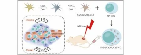
Figure 1 Schematic illustration of the preparation of the nanoplatform GNS@CaCO3/Ce6-NK and applications in bimodal imaging directed photothermal therapy (PTT)/photodynamic therapy (PDT) and immunotherapy (IT).
Synthesis of GNS@CaCO3/Ce6 nanoparticles
GNSs were synthesized according to the protocol described elsewhere31. At first, to obtain the seeds solution, when 100 mL of HAuCl4solution (1.0 mM) with severe stirring was heated to boiling, 15 mL of trisodium citrate solution (1%) was added. After the solution reached to room temperature,nitrocellulose membrane (0.22 μm) was used to filter the solution. 1 mL of gold seeds solution and 1 mL HCl solution(1 M) was added to 100 mL of HAuCl4(1.6 mM) successively with moderate stirring. After that, 1 mL of AgNO3(3 M) and 0.5 mL of L-ascorbic acid (1 M) were injected into the above mixture under the same rate of stirring. After the reaction was finished, 10 μL of HS-PEG-COOH (0.1 M) was added directly into the above solution. The reaction solution was carried out with suitable stirring speed in room temperature for overnight and GNSs can be obtained from the solution.
Next, the process of GNSs coating with calcium carbonate was conducted following previous literature with minor modifications26. In brief, 1 mL of CaCl2solution (10 mg mL-1)and 20 μL of Ce6 (0.5 mg mL-1) were added into 50 mL of the as-prepared GNSs with moderate stirring. After 24 h, the mixture was subjected to centrifugation at 10,000 rpm for 10 min. The deposits were redispersed in 20 mL of deionized water and 1 mL of Na2CO3solution (10 mg mL-1) and 20 μL of Ce6 (0.5 mg mL-1) were added. The reaction solution was carried out with suitable stirring speed in room temperature for 24 h. Then the precipitates were gathered and kept at 4-8°C for next use.
Isolation and culture of NK cells
On day 1, 2-3 × 10⁷PBMCs were acquired in a volume of 20 mL by density gradient centrifugation. The PBMCs were resuspended at the concentration of 1-2 × 10⁶mL-1in XVIVOTM15 medium supplement with 5% autologous plasma and IL-2 (400 U mL-1) and co-cultured with the same number of irradiated K562 feeder cells which can secret IL-21 and 4-1BB at 37°C and with 5% CO2. Fresh X-VIVOTM15 medium with autologous plasma and IL-2 (with the same concentration) were supplemented to make sure the cell densities were between 1-2 × 10⁶mL-1. On day 7, the same number of irradiated K562 feeder cells were added again to stimulate the NK cells. On day 14, NK cells were collected and washed with PBS, then the cells were stained with FITCCD3, APC-CD56, PE-NKG2D, PE-2B4, PE-NKp30, PENKp44, PE-NKp46 and analyzed by FACS Calibur system(BD Bioscience, USA).
Cancer cell culture and tumor model
The lung cancer cell lines A549 and NCI-H889 were chosen for in vitro or in vivo study. The A549 and NCI-H889 cells were cultured in high-glucose DMEM containing 10% FBS and were kept at 37°C and with 5% CO2.
Female BALB/c nude mice (4-5 weeks old) and male C57BL/6 mice (4-6 weeks old) were ordered from SLAC Laboratory Animal Co., Ltd. (Shanghai, China) and housed in a pathogen-free facility. All the animal experiments were conducted with the permission of Shanghai Jiao Tong University’s institutional Animal Use and Care Committee(Approval No. YXK2007-0025).
Cellular uptake assay
Flow cytometry (FCM) and Confocal laser scanning microscopic (CLSM) were applied to determine cellular uptake efficiency. In brief, 2 × 10⁵cells mL-1NK cells were cultured on 24-well plates with the addition of free Ce6,GNSs, GNS@CaCO3/Ce6 (equivalent Ce6 5 μg mL-1),respectively. After 4 h or 12 h incubation, one part of cells was collected for FCM and the fluorescence data were acquired via FACS Calibur (BD Bioscience, USA). The other part of cells was further fixed with 4% paraformaldehyde for 30 min, then stained with Hoechst 33342 and rinsed with PBS twice. The distribution of free Ce6 or GNS@CaCO3/Ce6 in the inner cell were examined by a confocal laser scanning microscopic (TCS SP8, Leica, German) at the wavelength of 405 nm and 633 nm.
Cell viability assay
CFSE+7AAD+ assay was used to detect the cytotoxicity of NK cells to the lung cancer A549 cells. In brief, 5-10 ×106mL-1single-cell suspension of A549 cells were prepared after rinsed with PBS twice. Then, the cells were treated with 1-5 μM CFSE and were incubated for 10 min at room temperature and kept from light. Cold complete media(containing 10% serum) was used to stop the CFSE labeling process. After washed with PBS for three times, the CFSElabeled A549 cells were prepared. To proceed the CFSE+7AAD+ assay, 5 × 10⁴CFSE-labeled A549 cells (as target cells) per well were seeded on the 24-well plate, then different number of NK cells (as effector cells) were added at different E (as effector cells): T (as target cells) ratios from 40 : 1, 30 : 1, 20 : 1 to 10 : 1. After 4 h incubation, the cells were collected and washed with PBS for twice and stained with 7AAD. The fluorescence data of CFSE+7AAD+ were acquired via FACS Calibur (BD Bioscience, USA).
To investigate the cytotoxicity of NK cells carried with free Ce6 (at the concentration of 0.1-10 μg mL-1) or GNS@CaCO3/Ce6 (at the equivalent Ce6 concentration of 0.1-10 μg mL-1) on A549 cells with or without laser irradiation, CCK-8 assay was conducted to examine the cell viability according to manufacturer’s protocol. The cell viability was calculated according to this formula:

Effects of GNS@CaCO3/Ce6 nanoparticles on human NK Cells
NK cells and NK cells loaded with GNS@CaCO3/Ce6 were respectively added into A549 cells at the E: T ratios from 1 : 1,10 : 1, 20 : 1, 30 : 1 to 40 : 1. Then, CCK-8 assay was conducted to examine the cell viability according to manufacturer’s protocol.
ELISA was applied to detect the cytokines secreted by NK cells loading with GNS@CaCO3/Ce6 nanoparticles. In brief,1 × 106mL-1NK cells were cultured on 24-well plate and treated with PBS (as control group), free Ce6, GNSs or GNS@CaCO3/Ce6 (equivalent Ce6 5 μg mL-1). 72 h later, the supernatant was collected and used for detection of the secreted cytokines (Human IFN-γ, TNF-α, GM-CSF,Granzyme-A, TRAIL (tumor necrosis factor-related apoptosis-inducing ligand) and FASL) via ELISA.
The intracellular ROS detection of GNS@CaCO3/Ce6 nanoparticles on human NK Cells
The intracellular ROS generation was examined by staining the NK cells with DCFH-DA. When entered into cells, the DCFH-DA would be hydrolyzed into nonfluorescent 2,7-dichlorofluorescin (DCFH) and then oxidized to fluorescent 2,7-dichlorofluorescein (DCF) by intracellular ROS. After respectively incubated with PBS, GNSs, free Ce6 (5 μg mL-1)or GNS@CaCO3/Ce6 (equivalent Ce6 5 μg mL-1) for 24h, the NK cells were collected and incubated with 1.5 × 10⁻⁵M DCFH-DA for 20 min. One part of the cells was further irradiated with a 633 nm He-Ne laser at a power of 100 mW cm-2for 1 min, while the other part of cells was kept in dark.Afterwards, FCM was used to detect the fluorescence intensity of the NK cells.
The PTT and PDT effects of GNS@CaCO3/Ce6-NK cells in vitro
At first, the PTT effects of GNS@CaCO3/Ce6 was assessed in vitro via an infrared thermal imaging camera. In short, after irradiated with an 808 nm laser (0.8 W cm-2) for ten minutes, the temperature of the solutions of PBS, Ce6(5 μg mL-1), GNSs (57.5 μg mL-1), and GNS@CaCO3/Ce6(57.5 μg mL-1) were monitored every minute using an infrared thermal imaging camera. Then the PTT and PDT effects of GNS@CaCO3/Ce6-NK on A549 cells were examined via MTT and FCM assays. 5 × 104549 cells per well were seeded on the 24-well plate for overnight. Then the cells were incubated with or without free Ce6, GNSs or NK cells(with or without GNS@CaCO3/Ce6) at 37°C and with 5%CO2. After 12 h incubation, the A549 cells was irradiated with a 633 nm He-Ne laser at a power of 100 mW cm-2for 3 min.Then all the cells were collected and rinse with PBS for twice and stained with Annexin V-FITC and PI. The fluorescence data were acquired via FACS Calibur (BD Bioscience, USA).
In vivo photoacoustic imaging
To establish tumor models, 5 × 10⁶A549 cells were implanted into the nude mouse’s back. When the tumor size reached about 50 mm3, 100 μL of PBS (as control), GNSs(57.5 μg mL-1) or 1 × 10⁶GNS@CaCO3/Ce6-NK cells (the same elemental Au concentration) were injected into the mice after anesthetized via isoflurane. Then the tumor sites were carried out with 808 nm irradiation by NIR laser. After 24 h, PA imaging of the tumor sites were collected by Endra Nexus 128 PA scanner (Ann tbor, MI).
In vivo photothermal therapy of GNS@CaCO3/Ce6-NK
When the tumor size reached about 50 mm3, 100 μL of PBS(as control), GNSs (57.5 μg mL-1) or 1 × 10⁶GNS@CaCO3/Ce6-NK cells (the same elemental Au concentration) were respectively injected into the mice after anesthetized via isoflurane. After 72 h, the tumor sites were irradiated with an 808 nm laser instrument for 3 min. The temperature data of corresponding tumor sites were collected via an infrared thermal imaging camera. And the tumor sizes were recorded every 5 days. On 25thday after treatment, the tumor tissues from the nude mice were fixed with 4% paraformaldehyde for histological analysis on H&E (hematoxylin and eoxin)staining assay.
In vivo immunotherapy of nanoparticles loaded NK cells
After the mice were injected with PBS or 1 × 10⁶GNS@CaCO3/Ce6-NK cells via tail for 72 h, the corresponding mice sera were collected for the detection of secreted cytokines such as TNF-α, IFN-γ, Granzyme A, Perforin, FASL, TRAIL and IL-18.
Statistical analysis
All values were expressed as mean ± SD (standard error)unless otherwise declared. A two-tailed student′s t-test was used for statistical analysis. *P < 0.05 was represented as statistically significant.
Results and discussion
Synthesis and characterization of GNS@CaCO3/Ce6
According to reported method, the prepared GNSs were further shrouded by a shell of CaCO3, in which the photosensitizer Ce6 were simultaneously encapsulated in the shell31. As shown in Figure 2A, the transmission electron microscopy (TEM) images significantly indicated that the synthesized GNSs possessed a uniform dominant size with a width of 96.3 ± 7.5 nm, and with outstanding spikes. When the surfaces of GNSs were coated with CaCO3shell, the spikes became shorter and the whole of GNS@CaCO3/Ce6 exhibited opaquer (Figure 2B). The characteristics of CaCO3shell were further verified by Scanning electron microscope(SEM) image (Figure 2C) and EDS (Energy-dispersive X-ray spectroscopy) mapping images. As shown in Figure 2D,elemental Ca ions were evenly distributed on the surface of elemental Au ions, which was consistent with the characteristic of the TEM image of GNS@CaCO3/Ce6.
As shown in Figure 2E, the original GNSs have a localized surface plasmon resonance (LSPR) peak at around 850 nm,while the peak showed a tiny blue shift to 795 nm after CaCO3shell encapsulated with Ce6 attached on the surface.As illustrated in the UV-Vis spectrum, the absorption spectrum of free Ce6 or GNS@CaCO3/Ce6 both possessed a remarkable peak at 405 nm. However, when the Ce6 encapsulated in the CaCO3shell, compared with that of free Ce6, the absorption peak of GNS@CaCO3/Ce6 exhibited a red-shift of about 23 nm from 645 to 668 nm, corresponding to the variation from the loaded-Ce6 environment. In addition, the NIR fluorescence intensity of Ce6 became weak in GNS@CaCO3/Ce6 compared to that of free Ce6(Figure 2F). The extinction spectrum clearly illustrated the efficient loading of the photosensitizer into the nanoplatform. Furthermore, by means of the UV-Vis spectrum, the loading of Ce6 in GNS@CaCO3/Ce6 was measured as about 8.7%. The release of Ce6 from GNS@CaCO3/Ce6 was detected at different pH values. As shown in Figure 2G, when the environment pH value was at 6.4, a significant release of the entrapped Ce6 from GNS@CaCO3/Ce6 was found compared to that of pH value was at 7.4 after GNS@CaCO3/Ce6 was exposed to an acidic environment for 12 h or longer. The results clearly demonstrated that the loaded Ce6 can be released from the GNS@CaCO3/Ce6 in the acidic environment (pH at 6.4).
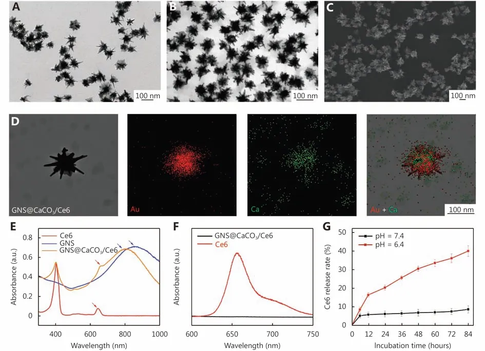
Figure 2 Characterization of GNS@CaCO3/Ce6. TEM images of GNSs (A) and GNS@CaCO3/Ce6 (B). SEM image of GNS@CaCO3/Ce6 (C).STEM and EDS elemental mapping images of GNS@CaCO3/Ce6 (D). Normalized UV-Vis spectra of free Ce6, GNSs and GNS@CaCO3/Ce6 (E).Fluorescence spectra of free Ce6 and GNS@CaCO3/Ce6 (F). Time course of Ce6 release from GNS@CaCO3/Ce6 at pH value of 7.4 and 6.4 (G).
Due to the nanoplatform would be used for PTT with NIR laser irradiation, the PTT property of GNS@CaCO3/Ce6 was assessed by an infrared thermal imaging camera. As shown in Supplementary Figure S1, during the laser irradiation period, the temperature changes of GNSs and GNS@CaCO3/Ce6 solution had no significant difference. The results suggested that the GNS@CaCO3/Ce6 nanoplatform had a similar ability to that of GNSs in inducing hyperthermia for PTT.
Phenotypes of NK cells incubated with or without GNS@CaCO3/Ce6
Human NK cells are important lymphocytes for anti-viral infection and anti-tumor immune response, accounting for 5% to 15% of the total number of peripheral blood lymphocytes32. NK cells have many characteristics, such as independent of antigen stimulation, targeting tumor sites,directly killing tumor cells, secreting cytokines or recruiting more lymphocytes to tumor sites33. By utilizing NK cells’strong killing ability and their excellent targeting ability, NK cells-based tumor immunotherapy is getting more and more attention. Furthermore, NK cells can be designed and used as a carrier to transport anticancer drugs or nanoparticles. Due to the high expression of numerous cell surface markers related to killing ability, such as CD56, CD314 (NKG2D),CD244 (2B4), CD337 (NKp30), CD336 (NKp44) and CD335(NKp46), NK cells own excellent natural killing ability34. The percentage of these markers after 12 days culturing could reach 92.7% (CD3- CD56+), 98.9% (CD56+ NKG2D+),99.2% (CD56+ 2B4+), 96.3% (CD56+ NKp30+), 91.6%(CD56+ NKp44+) and 86.1% (CD56+ NKp46+)respectively, which implying the successful expansion of NK cells (Figure 3A and Supplementary Figure S2).

Figure 3 (A) FCM results of several cell surface markers of NK cells on Day 14. APC-CD56, FITC-CD3, PE-NKG2D, PE-2B4, PE-NKp30, PENKp44 and PE-NKp46. (B) Viabilities of NK cells treated with different concentrations of free Ce6 and GNS@CaCO3/Ce6 for 12 h. (C) FCM results of cell surface markers of NK cells after incubated with GNS@CaCO3/Ce6 for 12 h. (D) ELISA results of the secreted cytokines from NK cells respectively treated with PBS, GNSs, free Ce6 or GNS@CaCO3/Ce6 for 72 h.
To obtain an optimal concentration of GNS@CaCO3/Ce6 for NK cells incubation, CCK-8 assay was carried out to assess the cytotoxicity caused by GNS@CaCO3/Ce6 nanoparticles. As shown in Figure 3B, after NK cells incubated with different concentrations of GNS@CaCO3/Ce6 for 12 h, we observed that cellular activity was not affected with the concentration increasing, whereas, in the free Ce6 group and GNSs group, cellular activity was declined with the concentration increasing. Finally, an equivalent dose of 5 μg mL-1of Ce6 was chosen.
Afterward, we examined the cell surface markers of NK cells after treated with GNS@CaCO3/Ce6 (equivalent to 5 μg mL-1Ce6) for 24 h. As shown in Figure 3C, compared to the untreated group, the percentage of the cell surface markers of the NK cells treated with GNS@CaCO3/Ce6 such as CD3- CD56+ (93.4%), CD56+ NKG2D+ (99.4%), CD56+2B4+ (99.2%), CD56+ NKp30 (94.3%), CD56+ NKp44+(95.1%) and CD56+NKp46+ (83.5%) were not influenced,which further confirmed the good biocompatibility of GNS@CaCO3/Ce6. Then, ELISA was performed to further examine the cytokines secretion of NK cells after incubated with GNS@CaCO3/Ce6, free Ce6, GNSs or PBS respectively.As shown in Figure 3D, the secretion of IFN-γ, GM-CSF,Granzyme-A, TNF-α, TRAIL and FASL were not changed in the PBS, GNSs or free Ce6 groups, whereas in the GNS@CaCO3/Ce6 group, the secretion of these cytokines increased significantly, especially for the secretion of TRAIL which could kill cancer cells by inducing apoptotic pathway.These data together indicated that the recognition and secreting functions of NK cells were not affected after incubated with the GNS@CaCO3/Ce6.
In vitro cellular uptake
Due to NK cells’ characteristic of actively targeting to tumor cells and directly killing ability, CFSE+ 7AAD+ assay was performed to optimize the E (as effector cells): T (as target cells) ratio of NK cells to the lung cancer A549 cells. As shown in Supplementary Figure S3, NK cells showed excellent killing ability to A549 cells when the E : T ratio reached 40 : 1. Finally, the ratio of 40:1 was selected for this study.
Due to cytoplasm’s natural acidic environment, the loaded Ce6 molecules would progressively be released from GNS@CaCO3/Ce635,36. As illustrated in the CLASM images(Figure 4A), after incubated with GNS@CaCO3/Ce6(equivalent to 5 μg mL-1Ce6), free Ce6 (5 μg mL-1), GNSCe6 (equivalent to 5 μg mL-1Ce6), or PBS respectively for 4 h, the GNS@CaCO3/Ce6 group revealed similar fluorescence signals with the GNS-Ce6 group which was due to the CaCO3layer and pH-triggered Ce6 release, while after 12 h, the fluorescence signal intensity in the GNS@CaCO3/Ce6 group became strongest than the other groups,indicating efficient cellular uptake of GNS@CaCO3/Ce6. In addition, FCM data further demonstrated the efficient accumulation of GNS@CaCO3/Ce6 in NK cells after incubation for 4 h and 12 h (Figure 4B).
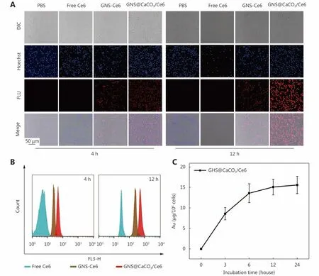
Figure 4 Cellular uptake of GNS@CaCO3/Ce6. (A) Confocal images of NK cells treated with PBS, free Ce6, GNS-Ce6 and GNS@CaCO3/Ce6 respectively for 4 h and 12 h. (B) Flow cytometry analysis of NK cellular uptake treated with free Ce6, GNS-Ce6 and GNS@CaCO3/Ce6 respectively for 4 h and 12 h. (C) Quantitative cellular uptake of GNS@CaCO3/Ce6 by NK cells, measured by ICP-MS.
To further assess the retention of GNS@CaCO3/Ce6 nanoparticles in NK cells, Inductively Coupled Plasma Mass Spectrometry (ICP-MS) was carried out to quantitatively measure the content of elemental gold at different time points. As shown in Figure 4C, in the first 4 hours, the uptake rate of GNS@CaCO3/Ce6 increased significantly,while, 12 hours later the uptake rate reached a plateau, which was consistent with the CLSM and FCM results of cellular uptake of GNS@CaCO3/Ce6 nanoparticles.DCFH-DA staining assay was used to examine the intracellular reactive oxygen species (ROS) production due to the interaction between photosensitizer and molecular oxygen15. As shown in Figure 5A, upon laser irradiation the NK cells treated with GNS@CaCO3/Ce6 displayed stronger fluorescence intensity than that of the groups treated with free Ce6 with or without laser irradiation, meanwhile, other groups treated with PBS or GNSs showed lower or negligible fluorescence intensity with or without laser irradiation. The results illustrated a prominent potential of GNS@CaCO3/Ce6-NK for enhanced PDT. Taken together, these results fully demonstrated the efficient uptake of GNS@CaCO3/Ce6 in NK cells and the Ce6 molecules can be released into the cytoplasm.
In vitro PDT and PTT
CCK-8 assay was performed to investigate the combination effects of gold nanoparticles, photosensitizer and NK cells on A549 cells with or without the NIR laser irradiation. As shown in Supplementary Figure S4, after NK cells loaded with free Ce6 or GNS@CaCO3/Ce6 were added into the A549 cells to co-culture in a cell-incubator at 37°C for 24 h, the cell viability of A549 cells dropped dramatically compared to that of the groups treated with PBS or free Ce6 regardless of whether or not been subjected to laser irradiation. This indicated that the NK cells’ killing ability was not influenced by the laser irradiation. Especially, after NIR laser irradiation,the cell viability of A549 cells co-cultured with GNS@CaCO3/Ce6-NK cells were declined to 50.1%, which was lower than that of the group without laser irradiation(61.3%). This indicated that our new designed nanoplatform would have a superimposed photodynamic therapy effect on tumor cells.

Figure 5 (A) FCM results of ROS generation of NK cells after treated with PBS, free Ce6, GNSs and GNS@CaCO3/Ce6 for 12 h respectively,and then exposed to a 633 nm laser for 1 min. (B) FCM results of cell viabilities of A549 cells treated with PBS, free Ce6, NK cells, GNSs and GNS@CaCO3/Ce6 for 12 h respectively and then with or without laser irradiation.
Furthermore, Annexin-V FITC/PI assay was used to assess the apoptosis and necrosis of the A549 cells after incubation with free Ce6, NK cells, GNSs, or GNS@CaCO3/Ce6-NK cells with or without laser irradiation. As shown in the Figure 5B,the percentage of apoptotic and necrotic A549 cells increased significantly after treated with NK cells with or without laser,which demonstrated the killing ability of NK cells to A549 cells compared with the other groups. Especially, the percentage of viable A549 cells treated with GNS@CaCO3/Ce6 decreased to 30.55% after laser irradiation, which was not only due to the killing effect of NK cells, but also caused by the superposition of PTT and PDT from GNS@CaCO3/Ce6 nanoparticles. The result was consistent with CCK-8 assay. Taken together, these results fully demonstrated that the nanoplatform of GNS@CaCO3/Ce6-NK have a good antitumor effect on the lung cancer A549 cells through the synergism of immunotherapy, PTT and PDT.
In vivo fluorescence imaging and PA imaging
Encouraged by the prominent results of GNS@CaCO3/Ce6-NK in vitro, multimodal imaging and treatment were conducted on A549 cells tumor model. At first, to study the actively targeting ability of GNS@CaCO3/Ce6-NK nanoplatform in vivo, the metastatic lung tumor NCI-H889 bearing mice were used to investigate the distribution at the lung sites. As shown in Figure 6A, 24 h post-injection with GNS@CaCO3/Ce6-NK cells, strong fluorescence signal can be found in the lung sites, which illustrated that the NK cells had a good active targeting performance of identifying and migrating to the tumor sites. In addition, the fluorescence image of the excised major organs further demonstrated the GNS@CaCO3/Ce6-NK nanoplatform could actively target to the lung sites.
Then, the distribution of GNS@CaCO3/Ce6-NK in vivo was then investigated in A549-bearing mice using the in vivo imaging system. As shown in Figure 6B, stronger fluorescence signals appeared in the tumor site of mice at 24 h post intravenous injection of GNS@CaCO3/Ce6-NK compared with the control group (treated with saline), which further indicating the good targeting ability of GNS@CaCO3/Ce6-NK at the tumor sites. In addition, the fluorescence image of the excised major organs further demonstrated the GNS@CaCO3/Ce6-NK nanoplatform could actively target to the tumor sites and the Ce6 molecules were released from the GNS@CaCO3/Ce6 in NK cells in vivo.
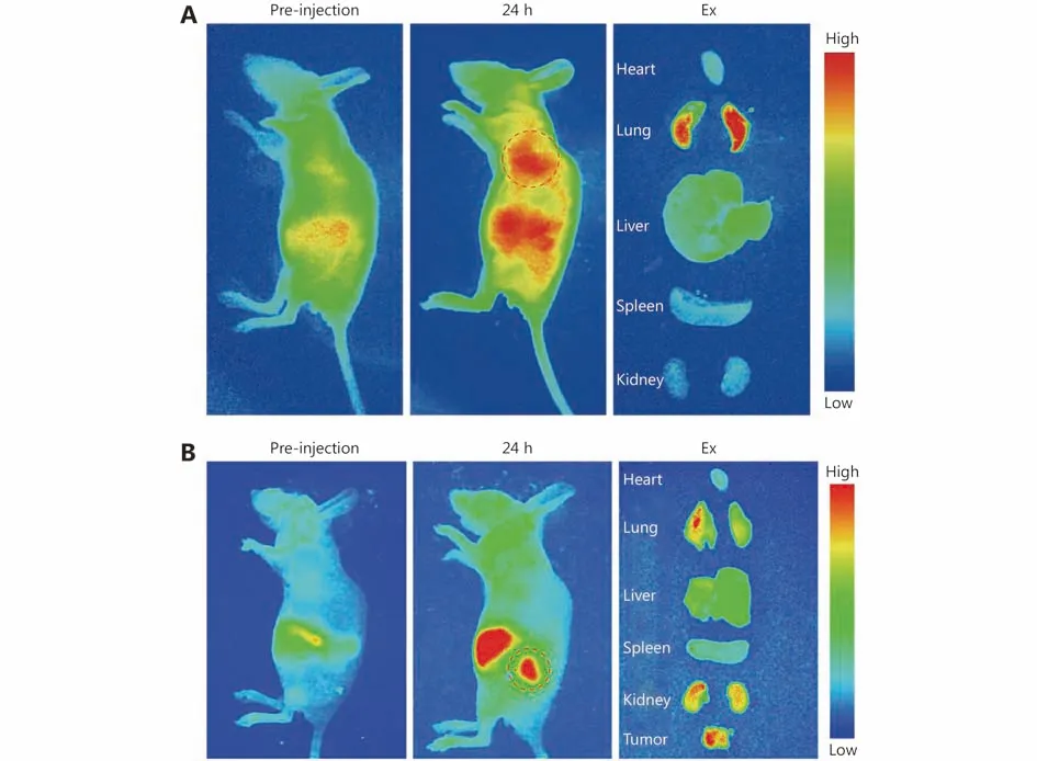

Figure 6 (A) Fluorescence images of nude mice bearing NCI-H889 tumors before and after tail vein injection of GNS@CaCO3/Ce6-NK cells and the fluorescence images of excised major organs at 24 h after injection of GNS@CaCO3/Ce6-NK cells. The red circle points the NCIH889 tumors located in the lung site. (B) Fluorescence images of nude mice bearing A549 tumors before and after tail vein injection of GNS@CaCO3/Ce6-NK cells and the fluorescence images of excised major organs at 24 h after injection of GNS@CaCO3/Ce6-NK cells. The red circle points the tumor site. (C) PA images of tumors acquired before and at 24 h after injection of saline or GNS@CaCO3/Ce6-NK(excitation: 810 nm). (D). Infrared microscopic imaging: the tumor model mice upon NIR laser irradiation (808 nm, 0.8 W cm-2, 3 min) at 72 h injected with PBS, GNS@CaCO3/Ce6 and GNS@CaCO3/Ce6-NK for 72 h.
Due to the correlation between the signal intensity of PA images and the concentration of gold nanoparticles, PA imaging is considered to be an outstanding technique for monitoring the accumulation and distribution of gold nanoparticles in tumors37,38. A photoacoustic imaging scanner was used to investigate whether NK cells-mediated nanoparticles delivery can accumulate in the tumor site and get well distributed. As shown in Figure 6C, almost no changes of the PA signal can be found in the blood vessels at the tumor sites from the picture of the control group (PBS)at 24 h post-injection. However, stronger PA signal can be found at the tumor sites at 24 h after treated with GNS@CaCO3/Ce6-NK. The results suggested that the NK cells could efficiently deliver the GNSs to the tumor sites and with a good distribution at the tumor sites.
In vivo PTT and immunotherapy
To investigate the antitumor effect of PTT brought by GNS@CaCO3/Ce6-NK based nanoplatform in vivo, infrared imaging was taken to illustrate the photothermal effect of GNSs. As previous report, gold nanoparticles have an outstanding photothermal conversion rate after irradiation with laser, the produced high temperature could bring damage to normal tissues39,40. To ensure the temperature of the thermotherapy does not harm healthy tissue, the temperature in the tumor sites with laser irradiation would be lower than 45°C41,42. As shown in Figure 6D, upon laser irradiation, the temperature of the different tumor groups injected with saline, GNS, or GNS@CaCO3/Ce6-NK reached to 36.6°C, 43.4°C and 44.2°C respectively. The results demonstrated that the new designed GNS@CaCO3/Ce6-NK nanoplatform could target to the tumor sites and induce an increased temperature for PTT and finally obtain a better therapeutic effect in vivo.
To assess the immunotherapy effect of immune cells, lung cancer A549 cells were inoculated to the abdomen of C57BL/6 mice to establish tumor models. As shown in Supplementary Figure S5, the tumor volumes of the groups treated with NK cells and GNS@CaCO3/Ce6-NK were both significantly smaller compared with the control group(saline). The results illustrated that NK cells (treated with or without GNS@CaCO3/Ce6) could significantly suppress the tumor growing, which owning to NK cells’ natural characteristics, i.e. directly killing tumor cells, secreting cytokines and recruiting more immune cells to the tumor sites.
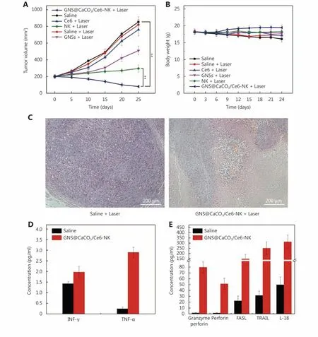
Figure 7 In vivo combination therapy of GNS@CaCO3/Ce6-NK. (A) Tumor growth in the various groups after irradiation treatment.(B) Body weight statistic data of mice after irradiation treatment. **P < 0.01 (n = 5). (C) Representative H&E sections of the tumor after laser irradiation therapy for 25 days. (D) and (E) Cytokines expression of mice for 72 h after injected with PBS or GNS@CaCO3/Ce6-NK.
To further research the consequence of the synergism therapy, nude mice (BLB/c) inoculated with lung cancer A549 cells were used for the implement of immunotherapy,PTT and PDT. As shown in Figure 7A, upon with laser irradiation, the tumor volumes of the groups treated with NK cells or GNS@CaCO3/Ce6-NK showed significant decrease compared with that of the groups respectively treated with saline, free Ce6 or GNSs. Particularly, the group of GNS@CaCO3/Ce6-NK showed the best inhibition efficacy on tumor growth than other treated groups. The results demonstrated that NK cells or GNS@CaCO3/Ce6-NK both exhibited an effective immunotherapeutic effect and the GNSs or GNS@CaCO3/Ce6-NK both exhibited an effective thermal therapy. NK cells performed excellent active targeting efficiency and immunotherapy effect, while the GNSs exhibited PTT effect, and Ce6 exhibited PDT therapy by generating1O2with NIR light irradiation43-45. Taken together, the results illustrated the successful and effective combination of immunotherapy, PTT and PDT based on our new designed nanoplatform, which achieving a triple therapeutic effect.
Furthermore, as shown in Figure 7B, the body weight of mice in the GNS@CaCO3/Ce6-NK group (with laser irradiation) showed higher than that of other groups. Also, as shown in Figure S6, no significant organ damage was observed in the main organs (heart, lung, liver, spleen and kidney) of the mice treated with GNS@CaCO3/Ce6-NK,which means that our new designed nanoplatform would hardly cause toxicity to the normal organs. As shown in Figure 7C, upon with laser irradiation, some damaged cells could be found in the H&E staining images of the GNS@CaCO3/Ce6-NK group. In contrast, nearly no damaged cells could be found in the control group. These results were consistent with the previous results of tumor volume reduction, which both illustrated that the new nanoplatform GNS@CaCO3/Ce6-NK performed excellent efficacy in inhibiting the tumor growth in vivo.
Cytokines variation of immunotherapy in vivo
According to previous report, NK cells’ excellent immunotherapy effects have strong relation with the secreted cytokines such as TNF-α, IFN-γ, Granzyme A, perforin,FASL, TRAIL and IL-1846-49. Under the above results of actively targeting to tumor sites and shrinking tumor volumes after treated with GNS@CaCO3/Ce6-NK, related cytokines were investigated to study the potential mechanism of immunotherapy via new nanoplatform GNS@CaCO3/Ce6-NK. As shown in Figure 7D and 7E, after treated with GNS@CaCO3/Ce6-NK, the secreted cytokines levels of TNFα, IFN-γ, Granzyme A, perforin, FASL, TRAIL and IL-18 were all increased obviously compared with that of the groups treated with PBS. IFN-γ, also regarded as type class II interferon, mainly secreted by NK cells, not only does it play a critical role in innate and adaptive immunity, but also it becomes a key to activating the macrophages or other immune cells. Likewise, TNF-α is essential to regulate immune cells to exert effects. Both Granzyme A and perforin are very important to inflammatory response. The increased level of IFN-γ and TNF-α were complied with verification of the effectiveness of immunotherapy of GNS@CaCO3/Ce6-NK treatment in vivo. Fas ligand (FASL or CD95L) and TNFrelated apoptosis-inducing ligand (TRAIL) both belong to the TNF family and can induce tumor cells apoptosis, taking a crucial part in the regulation of human body’s immune system and the progression of tumors. As expected, the levels of TRAIL and FASL have been increased due to the cancer cells’ death in vivo. IL-18, belonging to the IL-1 superfamily,can stimulate NK cells and some T lymphocytes to produce IFN-γ. As expected, after treatment with GNS@CaCO3/Ce6-NK, the increased level of IFN-γ and IL-18 mutually verified that the nanoplatform could still get alive in vivo and apply its function into anticancer. Taken together, these results clearly demonstrated that the nanoplatform could exert a prominent potency in the treatment of cancer.
Conclusions
In this study, GNS@CaCO3/Ce6 were successfully synthesized and incubated with NK cells to fabricate a new bioactive nanoplatform GNS@CaCO3/Ce6-NK. NK cells could transport GNS@CaCO3/Ce6 to A549 lung cancer sites in mice for enhanced photothermal/ photodynamic therapy, during which, the GNS@CaCO3/Ce6 displayed much higher loading capacity and protection of Ce6 owing to the CaCO3layer. The in vitro assays showed good biocompatibility and no cytotoxicity after the GNS@CaCO3/Ce6 were internalized by NK cells and GNS@CaCO3/Ce6-NK possessed high anticancer effect upon laser irradiation.Moreover, GNS@CaCO3/Ce6-NK still can actively target to the tumor sites, secret cytokines and kill the lung cancer cells in vivo. Most importantly, based on the GNS@CaCO3/Ce6-NK, the anticancer effects can be augmented with the combination of PTT, PDT and IT treatments. The strategy provides a particular direction for multimodal diagnosis and treatment of cancer by the combination of nanoparticles and NK cells.
Acknowledgements
This work was supported from 973 Project (Grant No.2015CB931802 and 2017YFA0205301), Chinese National Natural Scientific Fund (Grant No.81327002 and 81803094)and China Postdoctoral Science Foundation (Grant No.2017M621486). Funding from Shanghai Engineering Research Center for Intelligent diagnosis and treatment instrument (Grant No.15DZ2252000) is acknowledged.
Conflict of interest statement
No potential conflicts of interest are disclosed.
 Cancer Biology & Medicine2019年4期
- Cancer Biology & Medicine的其它文章
- Interpretation of breast cancer screening guideline for Chinese women
- Breast cancer screening guideline for Chinese women
- Erratum to Simultaneous inhibition of PI3Kα and CDK4/6 synergistically suppresses KRAS-mutated non-small cell lung cancer
- The correlation and overlaps between PD-L1 expression and classical genomic aberrations in Chinese lung adenocarcinoma patients: a single center case series
- Nomogram based on albumin-bilirubin grade to predict outcome of the patients with hepatitis C virus-related hepatocellular carcinoma after microwave ablation
- Omics-based integrated analysis identified ATRX as a biomarker associated with glioma diagnosis and prognosis
