First report of leaf-spot disease caused by Sphaeropsis visci on Asian mistletoe[Viscum coloratum(Kom.)Nakai]in China
Jie Chen•Xuefeng Liu•Hanqi Jia•Wenbo Zhu
Abstract In January 2017,symptoms of a leaf-spot disease were observed on Viscum coloratum plants in Yichun,China.The infected leaves were chlorotic,while sunken lesions formed on diseased branches,which were initially light brown and later turned dark brown.Moreover,diseased branches and leaves formed semi-buried,small,and spherical pycnidia.In total,20 leaves and 19 branches diseased samples from Yichun,China were collected and examined.The fungus was isolated and identified using Koch’s postulates,morphological characteristics and DNA sequence data.The data presented herein con firmed that the pathogen responsible for the disease was Sphaeropsis visci.To the best of our knowledge,this is the first report of it in China.
Keywords Botryosphaeriales·Viscum coloratum ·Morphology·Phylogeny·Sphaeropsis visci·New pathogen
Introduction
Asian mistletoe[Viscum coloratum(Kom.)Nakai]is a perennial,evergreen,hemiparasitic shrub that infects Populus spp.,Salix babylonica,and other woody species(Zuber 2004).This plant species adversely affects wood quality and quantity,inhibits fruiting,and increases the susceptibility of its hosts to other pathogens,including insects and wood-decay fungi(Qiu 1988).Asian mistletoe exhibits medicinal properties.For example,dried leaf petioles have been used to treat rheumatoid arthritis,lethargy,weakness,and other conditions.
The classification of the pathogen recently underwent a considerable change.Sphaeropsis visci(Alb&Schwein)Sacc,Michelia 2(no.6):105.1880.and Phaeobotryosphaeria visci(Kalchbr.)A.J.L.Phillips and Crous,Persoonia 21:47.2008.were determined to be the same species(Phillips et al.2008).The connection between the asexual and sexual morph of the fungus was established by Phillips et al.(2008)by the discovery that the ascomycete P.visci occurring on Viscum album produces conidia typical of S.visci.
Phillips et al.(2008)applying a one fungus,one name concept chose Phaeobotryosphaeria in favor of Sphaeropsis.But the 18th Botanical Congress adopted the Melbourne Code(McNeill 2012)rati-fying that priority of names will no longer be based on the life stage of fungi.Thus,the older name Sphaeropsis(1880)took priority over Phaeobotryosphaeria(1908).The changes in the name of this pathogen are summarized by Phillips et al.(2013).
Sphaeropsis visci can be found in Austria,Czechoslovakia,Egypt(Phillips et al.2013),Romania(Sutton 1980)and Ukraine(Phillips et al.2008),Hungary(Poczai et al.2015),Serbia,and Luxemburg(Zlatkovic et al.2016).It is a hyperparasitic fungal plant pathogen[S.visci(Alb.&Schwein.)Sacc],that causes leaf-spot disease in European mistletoe(V.album L.).And as a tool for biological control,S.visci can potential of European mistletoe(V.album L.)(Varga et al.2012;Stojanović1989).Three to six weeks after inoculation,the fungal infection spreads all over the leaves,branches,and berries;a few months later the whole shrub becomes dark yellow and necrotic(Sutton 1980).
We found and isolated another fungus that can infect the entire hemiparasitic plant,including berries,leaves,and branches.The leaves and branches of V.coloratum(Kom.)Nakai(Asian mistletoe)also exhibited disease symptoms.The leaf-infection sites were initially yellow and chlorotic,with buried pycnidia that appeared as small black dots.Dark brown lesions developed on inoculated leaves,and the resulting dead leaves were yellow–brown,with semiburied spherical pycnidia.Infected branches were light brown to brown,and eventually turned taupe.The infection sites of branches were yellow–brown,with swollen lesions.These eventually became sunken,with semi-buried,small sphaerical pycnidia.
The objective of this study was to identify a new disease affecting Asian mistletoe in China,using morphological characteristics and DNA sequence data for the internal transcribed spacer(ITS)rDNA.
Materials and methods
Sample collection and Sphaeropsis visci isolation
Diseased Asian mistletoe leaves and branches were collected in Yichun City,Heilongjiang province,China in January 2017.In the diseased tissue,if you found 1–2 spores,you can directly obtain a single strain.And it can have less contamination of bacteria.So it is conducive to the separation of pathogens.So we use a single-spore separation method(Chai and Jin 1991).The pycnidia was extracted from the conidia under the anatomical lens and microscope in a laminar- flow bench.The surface of the branches and leaves were sterilized with 70%alcohol and then blotted with absorbent paper.Pycnidia were collected from diseased leaves and branches,using a scalpel,and then put on an agar block,under a microscope,which was shifted to find a single spore.Then,an agar block with a single spore was cut with the side of a specially inoculated needle and put it onto potato dextrose agar plates.(200 g potato,20 g glucose,and 17 g agar per liter)(Beijing AOBOXING BIO-TECH CO.,LTD).The colonies were moved into the slant medium to be preserved.The inoculated media were incubated at 25°C.
Morphological identification
The size and morphological characteristics of conidia(n=20),conidiophores(n=20),and pycnidia(n=20)were determined using an optical light microscope and images from a digital camera.
DNA extraction,polymerase chain reaction,and sequencing
Mycelial plugs were incubated on a 5-day culture-medium,using a 0.75 cm stopper borer.Three mycelial plugs were used to inoculate 100 mL potato dextrose broth in conical flasks,which were incubated at 25°C with shaking(120 rpm)for about 10 days.The resulting mycelium(approximately 100 mg)was frozen with liquid nitrogen and then ground to a powder for a subsequent DNA extraction,using the Plant Genomic DNA Kit(Tiangen).
The internal transcribed spacer(ITS)region of the nuclear rDNA was amplified by a polymerase chain reaction(PCR)using universal fungal primers(ITS1 and ITS4)and previously described PCR conditions(White et al.1990).The 50 ul PCR reaction mixture consisted 34.5 μL double-distilled H2O,3 μL DNA template,1 μL each primer,5 μL dNTP,5 μL 10 × PCR buffer,and 0.5 μL Taq polymerase.The PCR conditions were as follows:94°C for 5 min;35 cycles of 94 °C for 30 s,56 °C for 30 s,and 72 °C for 1 min;and 72 °C for 7 min.The amplified products were analyzed by agarose gel electrophoresis and then sequenced in both directions using primers ITS1 and ITS4.
The DNA sequences were compared to other sequences of S.visci deposited in GenBank using BLAST.Sequence alignments were done using MEGA(version 5.0)program.Phylogenetic relationships were inferred using the maximum-likelihood methods with a heuristic search(Poczai et al.2015).Bootstrap support values based on 1,000 replications were calculated for tree branches in both phylogenetic analyses.Similarities(%)were calculated using the MEGALIGN program(DNASTAR Inc.).The pathogen DNA sequence was submitted to the NCBI database.
Pathogenicity test using Asian mistletoe branches
Conidia of S.visci were suspended in sterile double-distilled water.Control branches were wrapped with gauze soaked in sterile water.Twenty branches were inoculated respectively.Inoculated branches were incubated at 20°C for 25 days in a sterilized glass container filled with watersoaked cotton wool to maintain relative humidity at 75%(Grover and Nicole 1940).The incubation conditions simulated the spring-like continental climatic conditions of Heilongjiang province to induce the infection.Then,using a single-spore separation method,the suspected causal agent was subsequently re-isolated from symptomatic branches on potato dextrose agar.
Results
Morphological characteristics
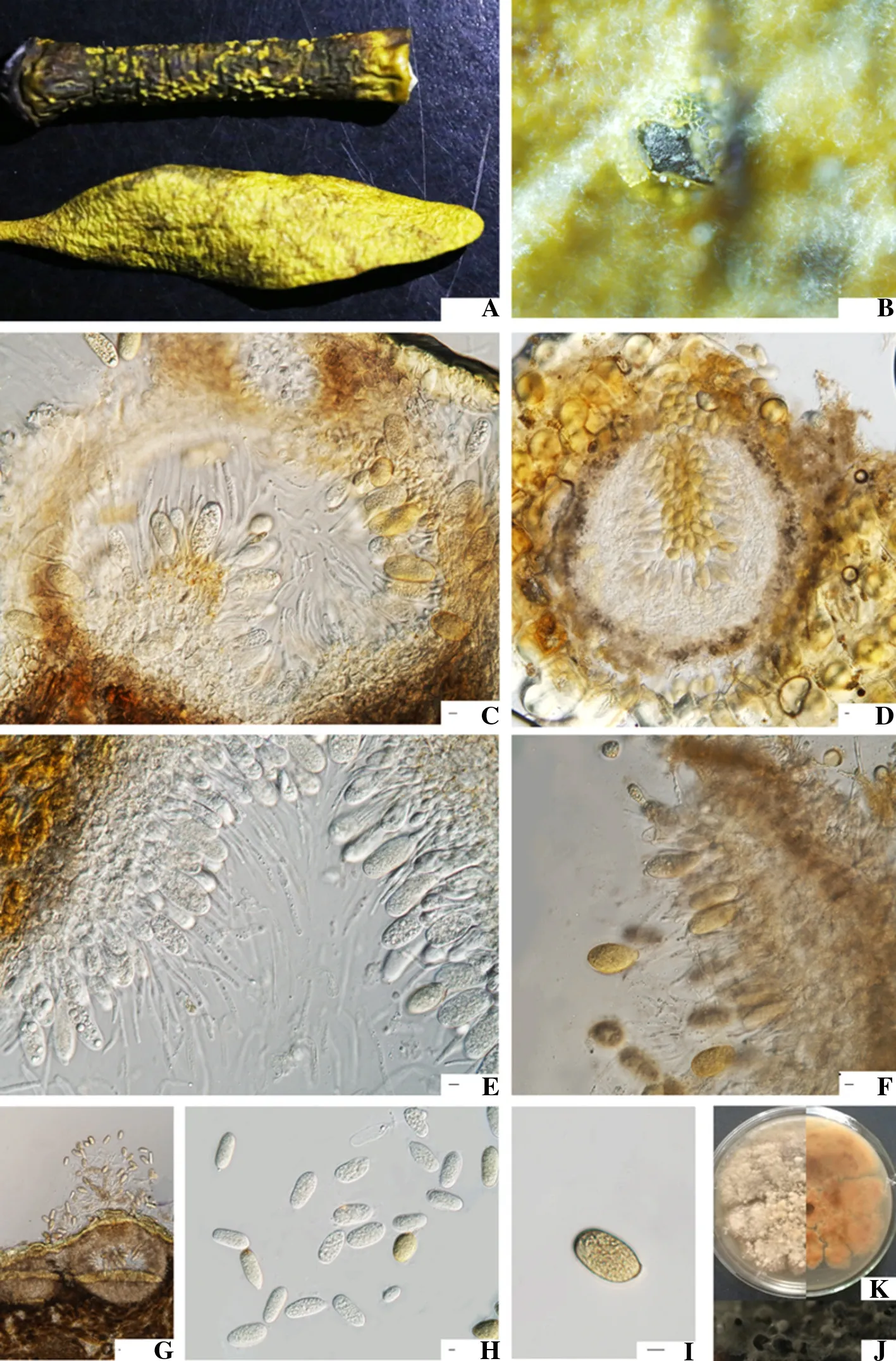
Colonies of S.visci were white and irregularly shaped.After eight days,the surface of the radially growing colony appeared dark gray,yellow,and white(from the center outward),while the underside was black.The observedunilocular and spherical pycnidia(442–490 × 400–420 μm)were initially buried and then exposed or semi-buried in the epidermis.The ostiole initially appeared as a diamond-shaped crack before being exposed.The ostioles from pycnidia growing on branches and leaves were 140–220 × 30–38 μm and 160–220 × 40–120 μm(n=20).

Fig.1 Morphological characteristics of Sphaeropsis visci.a Pycnidia formed on infected leaves and branches of mistletoe;b Pycnidium erupting from Asian mistletoe leaf;c Cross section of pycnidium;d longitudinal section of pycnidium;e developing conidia;f Conidiogenous cells and conidia of Sphaeropsis visci;g Cross section of pycnidium;h Immature conidia;i Mature conidia;j Culture characteristics(for 30 days);k Culture characteristics(for 6 days);Scale bars=10 μm
The pycnidial wall was composed of a dark-brown textura angularis,while the inner wall was hyaline.The inner wall underwent whole wall budding to produce conidiogenous cells that generated a single conidium.The conidia were oval or ovate[37.5–50.0 × 17.5–24.5 μm(n=20)],with an obtuse or truncate base.They were initially hyaline,but eventually turned brown(Fig.1).
Phylogenetic analyses
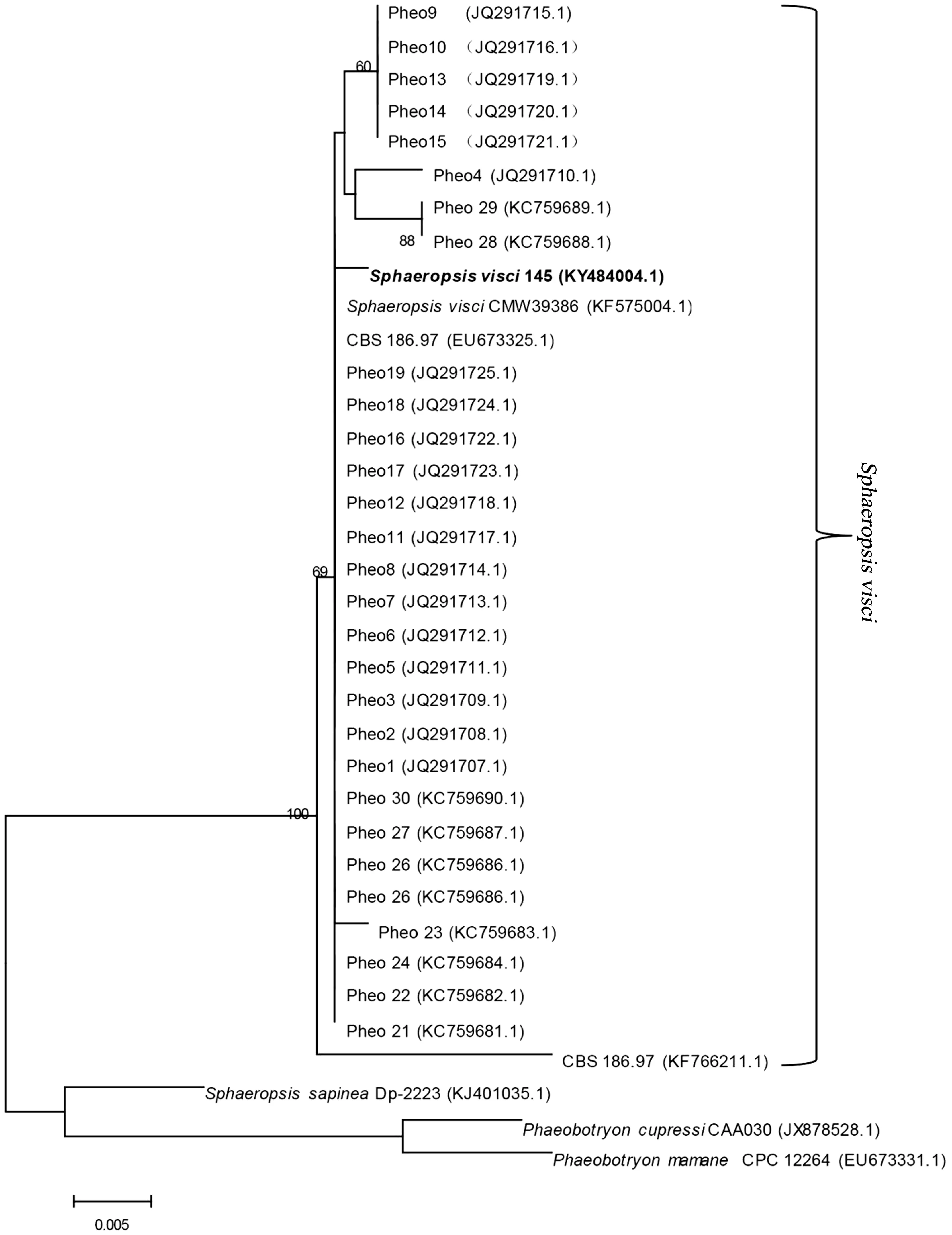
A BLAST search of the NCBI database revealed that the sequenced DNA was 99 percent similar to the S.visci(registration number:KF575004.1)sequence.A phylogenetic tree was constructed using the Maxium Likelihood tree method.The evolutionary distances were computed using the Kimura two-parameter method,and are presented as the number of base substitutions per site(scale).The analysis involved 36 nucleotide sequences.All positionswith gaps or missing data were eliminated,and 510 positions were ultimately analyzed in the final dataset.All gene sequences were derived from the NCBI database(Fig.2).

Fig.2 Phylogram inferred from ML analysis of ITS sequence data.The percentage of replicate trees in which the associated taxa clustered together in the bootstrap test(1000 replicates)is indicated next to the branches.The values above/below the branches indicate ML bootstrap support values
Pathogenicity test using Asian mistletoe branches
Branches shifted to fade green 7–10 days after inoculation.The semi-buried spherical pycnidia appeared approximately 20 days after inoculation.Twenty branches of inoculated pathogens,a total of 14 branches,found pycnidia.The pathogen infection rate was up of 70%.A total of 18 single colonies were re-isolated from different branches,of which two were Penicillium spp.and the rest were S.visci.The success of re-isolating the pathogen was closed to 88%.The S.visci was also re-isolated and its identity was con firmed,according to Koch’s postulates.
Discussions
Our study identified S.visci in Asian mistletoe and compared it to previous studies.The size of the conidia(37.5–50.0 × 17.5–24.5 μm)examined in this study was consistent with those reported by Phillips et al.(2008).The conidia observed in the earlierstudy were (27-)29–33(-50)× (14.5-)15.5–19(-22)μm,oval,initially hyaline but then brown,and externally smooth.They had an obtuse apex,obtuse or truncate base,and a moderately thick wall(Phillips et al.2008).At the molecular level,it has a common ITS sequence matching with sequences known from other countries in Europe(Phillips et al.2008),and virtually identical with KF575004,up to 99%in the Western Balkans(Zlatkovic et al.2016).
We conducted a scale survey in Changchun(Liaoning Province),Harbin,and Yichun(Heilongjiang Province)in China.We found that S.visci was widely infected with mistletoe.However,we did not find S.visci teleomorphs,only anamorphs on V.coloratum hosts.This finding parallels Varga et al.(2014),who were unable to find teleomorphs on V.album hosts in Hungary during 2010.Teleomorphs are rarely found and the connection between its anamorph was only recently shown by Phillips et al.(2008).The presence of different ITS types in the population may be attributed to sexual recombination as shown by Ahvenniemi et al.(2009)for Rhizoctonia solani.
The known distribution of S.visci is in Austria,Czechoslovakia,Egypt(Phillips et al.2013),Romania(Sutton 1980)and Ukraine(Phillips et al.2008),Hungary(Poczai et al.2015),Serbia,Luxemburg(Zlatkovic et al.2016).In our study,we found the first samples of S.visci in China,which causes leaf spot disease in Asian mistletoe(V.coloratum(Kom.)Nakai).Thus,two species of mistletoe are clearly susceptible to the pathogenic fungus responsible for causing leaf-spot disease(i.e.,S.visci),which con firms that S.visci can cause diverse mistletoe leaf-spot diseases.
In summary,our research proves that the disease capable of to completely destroying Asian mistletoe is caused by S.visci.Calder and Reid con firmed that mistletoe-infected trees are associated with signi ficantly higher mortality rates than trees lacking European mistletoe V.album L.(Calder and Bernhardt 1983;Reid et al.1994).Our finding that S.visci is clearly a potential biocontrol agent to manage diseases caused by mistletoe is important.Our future studies will focus on the pathogen infection cycle,which may be relevant for inhibiting the growth of Asian mistletoe.
AcknowledgementsWe thank all my colleagues who helped in writing this thesis.
 Journal of Forestry Research2018年6期
- Journal of Forestry Research的其它文章
- Black locust(Robinia pseudoacacia L.)as a multi-purpose tree species in Hungary and Romania:a review
- The impact of the environmental factors on the photosynthetic activity of common pine(Pinus sylvestris)in spring and in autumn in the region of Eastern Siberia
- Osmoregulators in Hymenaea courbaril and Hymenaea stigonocarpa under water stress and rehydration
- Effect of nitrogen levels on photosynthetic parameters,morphological and chemical characters of saplings and trees in a temperate forest
- Free amino acid content in trunk,branches and branchlets of Araucaria angustifolia(Araucariaceae)
- Exogenous application of succinic acid enhances tolerance of Larix olgensis seedling to lead stress
