
Growth and photosynthesis of Chlorella strains from polar,temperate and tropical freshwater environments under temperature stress*
2018-08-02 02:51:00KokKeongLEEPhaikEemLIMSzeWanPOONGChiewYenWONGSiewMoiPHANGJohnBEARDALL
Kok-Keong LEE , Phaik-Eem LIM , Sze-Wan POONG Chiew-Yen WONG ,Siew-Moi PHANG , John BEARDALL
1 Institute of Ocean and Earth Sciences, University of Malaya, 50603 Kuala Lumpur, Malaysia
2 Institute of Graduate Studies, University of Malaya, 50603 Kuala Lumpur, Malaysia
3 School of Health Sciences, International Medical University, 57000 Kuala Lumpur, Malaysia
4 National Antarctic Research Centre, University of Malaya, 50603 Kuala Lumpur, Malaysia
5 Institute of Biological Sciences, University of Malaya, 50603 Kuala Lumpur, Malaysia
6 School of Biological Sciences, Monash University, Clayton, Victoria, 3800, Australia
Abstract Elevated temperatures as a consequence of global warming have signi ficant impacts on the adaptation and survival of microalgae which are important primary producers in many ecosystems. The impact of temperature on the photosynthesis of microalgae is of great interest as the primary production of algal biomass is strongly dependent on the photosynthetic rates in a dynamic environment. Here, we examine the effects of elevated temperature on Chlorella strains originating from different latitudes,namely Antarctic, Arctic, temperate and tropical regions. Chlorophyll fluorescence was used to assess the photosynthetic responses of the microalgae. Rapid light curves (RLCs) and maximum quantum yield ( F v/ F m)were recorded. The results showed that Chlorella originating from different latitudes portrayed different growth trends and photosynthetic performance. The Chlorella genus is eurythermal, with a broad temperature tolerance range, but with strain-speci fic characteristics. However, there was a large overlap between the tolerance range of the four strains due to their “eurythermal adaptivity”. Changes in the photosynthetic parameters indicated temperature stress. The ability of the four strains to reactivate photosynthesis after inhibition of photosynthesis under high temperatures was also studied. The Chlorella strains were shown to recover in terms of photosynthesis and growth (measured as Chl a) when they were returned to their ambient temperatures. Polar strains showed faster recovery in their optimal temperature compared to that under the ambient temperature from which they were isolated.
Keyword: Antarctic; Arctic; F v/ F m; microalgae; pigments; recovery
1 INTRODUCTION
Anthropogenic in fluences have led to signi ficant changes in climate. The average global temperature has risen by about 0.8°C since 1880 and the average global surface temperature is expected to increase by 4–5°C over the next century (IPCC, 2007). In addition to higher average temperatures, global climate change is also resulting in higher temperature variability, thus increasing the risks to species’ tolerance limits.Temperature is one of the major factors affecting the distribution and productivity of microalgae.
Understanding the growth and photosynthetic response of microalgae to the changes in their thermal environment is crucial to the characterization of their ecophysiology in nature. Temperature effects on the growth rate and photosynthesis of microalgae is of great interest as the primary production of algal biomass is strongly dependent on the prevailing photosynthetic rates in a dynamic environment.Photosynthesis is known to be a heat-sensitive process, and can be inhibited by high temperature before other symptoms of stress are detected (Camejo et al., 2005). Photosystem II (PS II) is reported to be the most thermosensitive component of the photosynthetic apparatus compared with the electron transport chain, stromal enzymes, PSI activity and the chloroplasts envelope (Georgieva and Yordanov,1994; Ralph, 1998). The capacity for photochemical work is in fluenced by the stress levels of cells and any damage to the photochemical apparatus caused by photoinhibition (Eggert et al., 2007).
Elevated temperature has a signi ficant effect on most metabolic processes. Photoinhibition occurs before other cell functions are damaged (Harrison and Platt, 1986; Davison, 1991). Extreme temperatures induce damage to PSII by limiting electron transport and carbon fixation of the algae (Levasseur et al.,1990; Anning et al., 2001). Davison (1991) suggested that elevated temperatures were able to modulate the cellular concentrations of RUBISCO and other Calvin cycle enzymes that in turn decreased the operating quantum efficiency of PSII, Δ F/ Fm. Elevated temperatures will thus affect the photosynthesis rate or may induce phenotypic and genotypic changes.Moreover, high temperature stress caused inactivation of PSII reaction centres (Bukhov and Carpentier,2000; Yamamoto, 2016), causes a shift of the redox equilibrium between the primary acceptor plastoquinone ( QA) and the secondary acceptor plastoquinone ( QB) (Pospíši and Tyystjärvi, 1999;Tóth et al., 2007), and induces dissociation of the peripheral antenna complex of PSII from its core complex (Armond et al., 1980; Wen et al., 2005).When exposed to different temperatures above and below their ambient temperature, microalgae show different photosynthetic responses (Salleh and McMinn, 2011). High temperature may cause thylakoid membrane instability, speci fically affecting the membrane lipid composition while extreme low temperature causes reduced flexibility of membranes which then become crystalline or freeze (Falkowski and Raven, 2013).
Chlorella species (Chlorophyta), are small,ubiquitous, coccoid phototrophic eukaryotes found in diverse habitats including hot springs and other extreme environments (Phang and Chu, 2004). In nature, these microalgae play a role as primary producers in aquatic ecosystems, but can also serve as a source of nutraceuticals, feedstocks for biofuel, and play a role in wastewater treatment (Chu et al., 2009;Lim et al., 2010). They also show potential for electricity generation (Ng et al., 2014). The biochemical responses of Chlorella to temperature stress have been studied (Teoh et al., 2004, 2013). The protein contents of polar Chlorella were markedly affected by temperature while no clear trends were observed in their lipid and carbohydrate contents(Teoh et al., 2004).
Numerous studies have been done on the various stress factors on algal cells which involve morphological and biochemical changes that are apoptosis-like (Moharikar et al., 2006; Lee and Hsu,2013). In some cases, cells are not killed even though their photosynthetic activity may have been completely inhibited in the face of moderate to extreme temperature fluctuations. Their performance may decline to a certain extent but when the conditions are favourable, microalgal cells have the ability to fully recover (Lee and Hsu, 2013). Polar Chlorella species have shown eurythermal adaptivity and higher growth rates at temperatures higher than ambient(Teoh et al., 2004; Cao et al., 2016). Therefore,slightly warmer temperatures are predicted to accelerate the recovery of the heat-stressed Chlorella cells, within physiological limits.
In the present study, four Chlorella strains originating from different latitudes were exposed to a range of temperatures to investigate their responses based on growth and photosynthetic performance.Their ability to recover from stress caused by exposure to unfavourable temperature was also investigated.
2 MATERIAL AND METHOD
2.1 Algae culture
Four freshwater Chlorella strains were obtained from the University of Malaya Algae Culture Collection (UMACC), namely UMACC237( Chlorella-Ant), UMACC263 ( Chlorella-Arc),UMACC248 ( Chlorella-Temp) and UMACC001( Chlorella-Trop). The identi fication of the Chlorella spp. was done based on morphological and molecular studies (results not shown). Chlorella-Ant was isolated from a soil sample collected near a wastewater pond at Casey Station, Antarctica; Chlorella-Arc was isolated near the Arctic Research Station at Ny-Ålesund, Norway; and Chlorella-Temp (original code: LB259) was originally isolated from a freshwater lake in the Netherlands. Chlorella-Trop was isolated from a fish pond at University of Malaya(Phang, 2004). The strains were maintained at temperatures close to those of their original habitats to avoid long-term cultivation effects (Lakeman et al.,2009), although some microalgal strains may show high genetic stability despite long term cultivation(Marija and Dieter, 2014). The cultures were maintained in a controlled-environment incubator at 4°C for the Arctic and Antarctic strains, and at 18°C and 28°C for the temperate and tropical strains respectively. All cultures were maintained in Bold’s Basal Medium (BBM) (Nichols and Bold, 1965) and illuminated with TLD 18W/54-765 cool white fluorescent lamps (Philips, Amsterdam, the Netherlands) providing ~42 μmol photons/(m2·s)photosynthetic active radiation (PAR) on 12 h light:12 h dark photoperiod.
2.2 Experimental design
Batch mode culturing was conducted at 4 to 33°C for the polar strains ( Chlorella-Ant and Chlorella-Arc), 18 to 38°C for Chlorella-Temp, and 18 to 43°C for Chlorella-Trop. The cultures incubated at 4°C for the Chlorella-Ant and Chlorella-Arc, and at 18°C and 28°C for the Chlorella-Temp and Chlorella-Trop strains respectively, were set as control in this experiment. The inoculum was 10% (v/v) from the exponential phase cultures standardized at an optical density of 0.2 at 620 nm (OD620). Triplicate cultures of each strain were grown in 1 L Erlenmeyer flasks with 500 mL BBM and maintained in controlled environment shaking incubators at 80 r/min (Hotech Model 718, Taiwan, China) at the range of temperatures mentioned earlier. Irradiance of~42 μmol photons/(m2·s) PAR was provided directly from above the flasks with a 12 h light:12 h dark cycle and was measured with a Li-Cor Light Meter (Model L1-250A). Cultures were allowed to grow until stationary phase (day 10).
Growth was monitored by measuring absorbance at OD620using a UV-1800 UV-VIS spectrophotometer(Shimadzu, Japan). Cell density was estimated by cell counting using a haemocytometer (improved Double-Neubaur, Assistent, Germany). Speci fic growth rate( μ, /d) was calculated using the following formula:

where N1and N2represent the cell concentrations at times t1and t2, respectively, within the whole logarithmic phase.
Chlorophyll- a (Chl a) content was analyzed spectrophotometrically after extraction of the filtered samples (Whatman GF/C, 0.45 μm) in acetone(Strickland and Parsons, 1972). The absorbance of the pigment extract was measured at 665 nm (OD665),645 nm (OD645) and 630 nm (OD630). The concentration of Chl a was calculated using the following formula:

where, Ca=11.6OD665–1.31OD645–0.14OD630; Va=volume of the acetone (mL) used for extraction; Vc= volume of culture (L); Chl a (mg/L)=Chl a (mg/m3)/1 000.
The extraction and quanti fication of total carotenoids followed the method described by Vonshak and Borowitzka (1991). The absorbance of the pigment extract was measured at 452 nm (OD452).The total carotenoid content (μg/mL) was calculated using the following formula:

where, Ve=volume of the acetone (mL) used for extraction; Vc=volume of culture (mL).
2.3 Measurement of photosynthesis performance
Non-invasive fluorescence measurements were obtained using a high-resolution Water-Pulse Amplitude Modulated (PAM) fluorometer (Walz GmbH, Effeltrich, Germany). The cultures were dark adapted for a period of 15 min before the generation of rapid light curves (RLCs). A weak measuring light(0.15 μmol photons/(m2·s)) from the PAM fluorometer was used to measure the fluorescence yield without inducing photosynthesis, termed as minimum fluorescence ( Fo, open PSII reaction centers). A saturating pulse (>3000 μmol photons/(m2·s) for 0.8 s) was used to determine maximum fluorescence( Fmin the dark) when all PSII reaction centres were closed. PSII quantum yield, also known as the maximal efficiency of PSII, was determined as followed according to Schreiber et al. (1995):
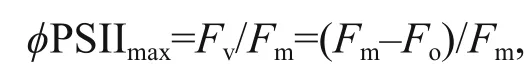
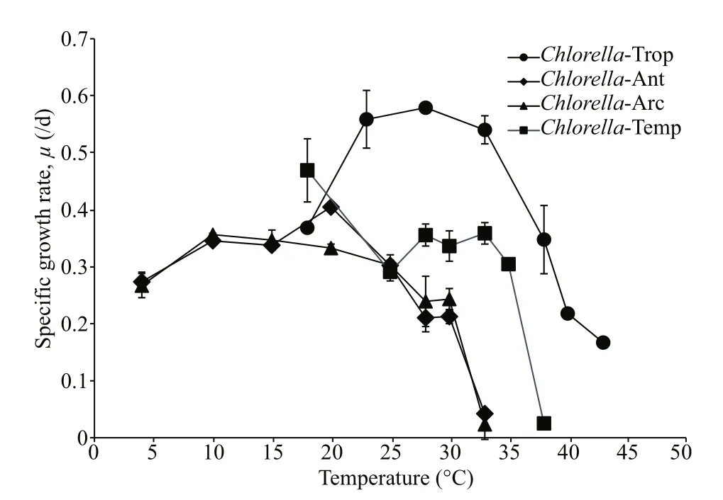
Fig.1 Speci fic growth rates, μ (/d) of the four strains at various temperatures
where Fmand Foare the maximum and the minimum fluorescence yields in the dark-adapted state,respectively. RLCs were generated for all samples using the Water PAM software (WinControl, Walz).Light-emitting diodes (LEDs) provided the eightstepwise increment of actinic light used in the generation of RLCs. The actinic light levels used were 0, 48, 105, 158, 233, 358, 530, 812 and 1 216 μmol photons/(m2·s), each lasting for 10 s.While we recognise that a short exposure time,especially in dark-adapted cultures, will not provide accurate measures of steady state electron transport,all samples were processed in the same way so the changes between treatments provide a reliable measure of the relative alterations to cell performance.Photosynthetic parameters such as photosynthetic efficiency ( α), maximum rate of relative electron transport (rETRmax) and photoadaptive index ( Ek)were determined to compare the RLCs quantitatively(Ralph and Gademann, 2005). The relative electron transport rate (rETR) at a given irradiance was calculated by multiplying the effective photochemical efficiency and the irradiance (Genty et al., 1989).RLCs were constructed by plotting rETR against PAR data and subsequently fitted mathematically to a single exponential function (Platt et al., 1980), using a Marquardt-Levenberg regression algorithm:
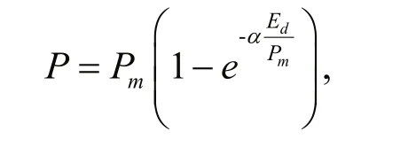
where P is the rETR at a given irradiance, Pmis the maximum potential rETR (rETRmax), α is the initial slope of the fitted RLC before the onset of saturation(light-limiting condition efficiency) and Edis the incident irradiance (400–700 nm). The function becomes an asymptotic maximum rETR value by assuming that there is no photoinhibition (Jassby and Platt, 1976). The intercept of the α value with rETRmaxgave the saturation irradiance for electron transport( Ek) which is de fined as:

Non-Photochemical Quenching (NPQ) is a measurement of the photoprotective xanthophyll cycle whereby excessive light is dissipated as heat to avoid negative impacts to the photosynthetic electron transport chain. NPQ was calculated using the formula of Schreiber (2004):

where Fmis the maximal fluorescence after a dark adaptation period and Fm' is the maximum fluorescence in the steady state light-adapted state (attained after 5–10 min). NPQ was calculated and quanti fied by measuring the change in Fmto the final value Fm(Δ F%). The relative inhibition of α and rETRmaxin relation to the control was calculated as:

where X are the values of α or rETRmax.
2.4 Stress and recovery
The stress-inducing temperatures for each strain were selected based on the initial experiments with a range of increasing temperatures. Temperatures of 34°C were used for both Chlorella-Ant and Chlorella-Arc, 40°C for Chlorella-Temp and 44°C for Chlorella-Trop. These temperatures represented the condition when the Fv/ Fmdecreased sharply during the growth cycle (see results), indicating a highly-stressed condition of the culture. These selected temperatures may lead to culture death with prolong exposure. The Chlorella strains were allowed to recover by returning them to their ambient temperatures after the high temperature treatments. Two threshold levels were selected; Fv/ Fm≈0.4 (assigned as point ‘A’) and Fv/ Fm≈0.2 (assigned as point ‘B’). These points were selected on the basis that at Fv/ Fm≈0.4, most of the culture was still able to grow while at Fv/ Fm≈0.2, no net increase of biomass was observed. Biomass (Chl a) was estimated at Fv/ Fm≈0.4 and Fv/ Fm≈0.2. The relative rate of recovery is de fined as the time taken for the culture to recover from Fv/ Fm≈0.4 and Fv/ Fm≈0.2, to its original Fv/ Fm.
2.5 Statistical analysis
Data were analyzed by one-way analysis of variance (ANOVA) followed by a post-hoc test using Tukey’s HSD test. Statistical analyses were carried out using Statistica 10 (StatSoft Inc., USA).
3 RESULT
3.1 Microalgal growth and pigment contents
The Chlorella strains used in this study tolerated a wide range of temperatures. Chlorella-Trop displayed a higher speci fic growth rate ( μ, /d) than the rest of the strains under various temperature treatments (Fig.1)with the highest μ (0.579/d) attained at 28°C, but μdecreased markedly with increasing temperature above the optimum. Chlorella-Ant and Chlorella-Arc showed similar growth trends across the temperature treatments. Both polar strains could grow from 4°C to the upper temperature limit of 33°C. The upper temperature limit is de fined as the highest temperature that a culture can tolerate. The μ of polar strains increased with increasing temperatures, whereby the highest μ values for Chlorella-Ant and Chlorella-Arc were recorded as 0.405/d at 20°C and 0.356/d at 10°C,respectively. However, there was no consistent growth trend observed for Chlorella-Temp but its highest μ was recorded as 0.469/d at 18°C. The lethal temperature (temperature at which the culture could not survive) was the lowest for Chlorella-Ant and Chlorella-Arc. Temperatures at and above 33°C,38°C and 43°C were fatal to the polar, temperate and tropical Chlorella respectively.
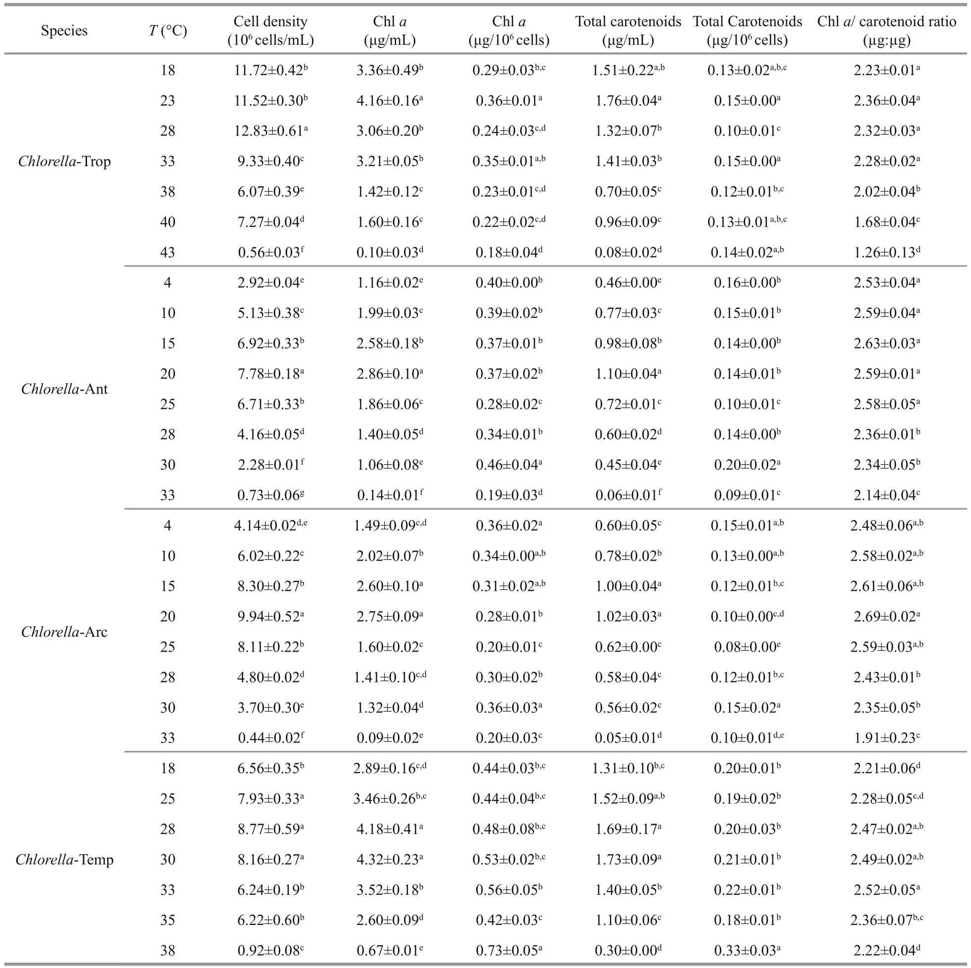
Table 1 Comparison of growth parameters and pigment contents of Chlorella-Trop, Chlorella-Ant, Chlorella-Arc and Chlorella-Temp cultivated at a range of temperatures

Fig.2 Maximum quantum yield ( F v/ F m) of (a) Chlorella-Trop, (b) Chlorella-Ant, (c) Chlorella-Arc and (d) Chlorella-Temp grown in different temperatures Data are mean±SD of triplicate samples.
The Chl a:carotenoids ratio (μg:μg) was also used as an indicator of the stress condition of the culture(Table 1). The Chl a:car ratio (μg:μg) were signi ficantly lower in Chlorella-Ant, Chlorella-Arc and Chlorella-Trop at temperatures above 25, 30, and 33°C, respectively. Chlorella-Temp grown at 18°C showed the lowest Chl a:car ratio amongst the strain grown at different temperatures. The highest Chl a:car ratio in Chlorella-Ant and Chlorella-Arc were observed at 15°C and 20°C, respectively. It was found that under the in fluence of cultivated temperatures,Chlorella-Ant, Chlorella-Arc and Chlorella-Trop showed a strong correlation between their speci fic growth rate and Chl a:car ratio (Table S1) with R2=0.933, 0.864 and 0.900 respectively (Pearson correlation, P <0.01).
3.2 Photosynthetic performance
Effects of temperature on photosynthetic parameters varied amongst strains with regards to their latitudinal origin. During cultivation, Chlorella-Ant, Chlorella-Arc and Chlorella-Trop were able to maintain a relatively stable Fv/ Fm(= ϕ PSIImax) at their ambient temperatures and at temperatures near Topt(Fig.2).However, ϕ PSIImaxof Chlorella-Trop presented a downward trend from day 6 at 38°C and 40°C (Fig.2a). For both Chlorella-Ant and Chlorella-Arc, ϕ PSIImaxvalues were at maximum values during incubation at temperatures near 10°C, but declined as temperature rose to above 20°C. ϕ PSIImaxin both polar Chlorella were somewhat stable, albeit with comparatively lower values after day 6 at 28°C and 30°C (Fig.2b, c). ϕ PSIImaxof Chlorella-Ant could still be detected on day 8 while ϕ PSIImaxof Chlorella-Arc was already zero on day 6 at 33°C. A sharp decrease in ϕ PSIImaxwas observed at 33°C for both polar Chlorella and at 43°C for Chlorella-Trop. The variation in ϕ PSIImaxfor Chlorella-Temp was temperature-independent, whereby ϕ PSIImaxfluctuated around 0.5 from 18°C to 38°C (Fig.2d).

Fig.3 Percentage inhibition of light harvesting efficiency (α) and maximal relative electron transport rate (rETRmax) of (a)Chlorella-Trop, (b) Chlorella-Ant, (c) Chlorella-Arc and (d) Chlorella-Temp grown at different temperatures
The four Chlorella strains grown at various temperatures showed different trends in the inhibition of both rETRmaxand light harvesting efficiency ( α)(Fig.3). Generally, the inhibition of rETRmaxand α was increased with increasing temperature. For Chlorella-Trop, increasing temperature up to 38°C did not inhibit rETRmax, however, temperatures beyond 38°C lowered the rETRmax(Fig.3a).Chlorella-Ant did not show inhibition of rETRmaxfrom 10 to 25°C, but the percentage of inhibition increased signi ficantly ( P <0.05) with further increase in temperature (Fig.3b). Although the inhibition of rETRmaxin Chlorella-Arc was relatively higher than Chlorella-Ant, the Antarctic strain ( R2=0.916,P <0.01) displayed better correlation (Table S2)between temperature and inhibition of rETRmaxthan Chlorella-Arc ( R2=0.775, P <0.01) and Chlorella-Trop ( R2=0.604, P <0.01). Chlorella-Arc showed 26.8%, 7.1%, 58.7%, 82.7% and complete inhibition of rETRmaxat 20°C, 25°C, 28°C, 30°C and 33°C,respectively (Fig.3c). Inhibition of α was essentially temperature dependent especially at temperatures beyond the Toptfor all strains studied.The values of α consistently declined in both polar Chlorella and Chlorella-Trop with increasing temperature. The effect of temperature on α was the most strongly correlated for Chlorella-Trop ( R2=0.669, P <0.01)followed by Chlorella-Arc ( R2=0.636, P <0.05) and Chlorella-Ant ( R2=0.598, P <0.01). Signi ficant inhibition ( P <0.05) of rETRmaxwas only observed in Chlorella-Temp at 38°C with 30.8% inhibition but α was generally suppressed across 25°C to 38°C(Fig.3d).
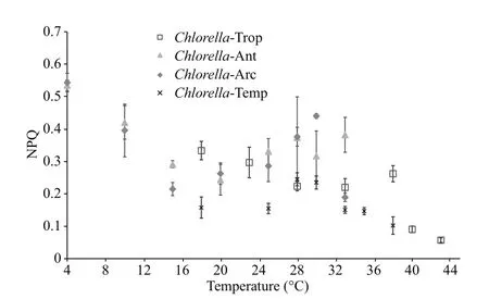
Fig.4 The NPQ of Chlorella-Trop, Chlorella-Ant, Chlorella-Arc and Chlorella-Temp grown at different temperatures
Generally, at 1 216 μmol photons/(m2·s) Chlorella-Temp presented the lowest NPQ values ranging from 0.147 to 0.245 across the experimented temperatures(Fig.4). NPQ in both polar Chlorella strains was activated in response to temperature stress. NPQ at 1 216 μmol photons/(m2·s) peaked at 4°C with 0.535 followed by 0.382 at 33°C for Chlorella-Ant. For Chlorella-Arc, NPQ at 1 216 μmol photons/(m2·s)peaked at 4°C with 0.543 followed by 0.440 at 30°C.Chlorella-Trop recorded a largely downward trend in NPQ with increasing temperature.
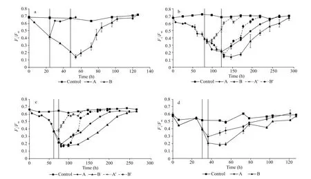
Fig.5 (a) Chlorella-Trop, (b) Chlorella-Ant, (c) Chlorella-Arc and (d) Chlorella-Temp grown at 44°C, 34°C, 34°C and 38°C respectively until the F v/ F m decreased reaching the thresholds of A≈0.4 and B≈0.2
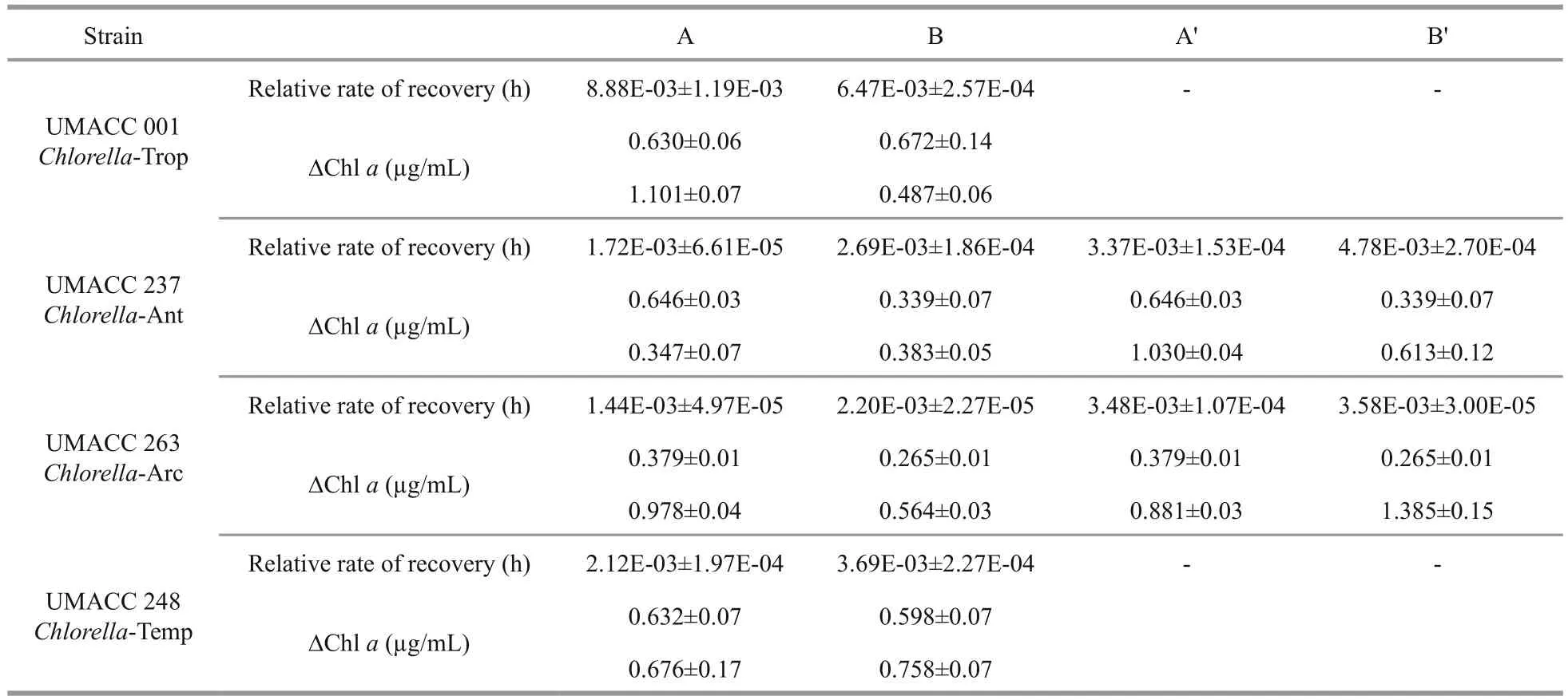
Table 2 Relative recovery rate and changes in biomass before and after recovery (Chl a)
All the Chlorella strains were able to recover from exposure to high temperature after being returned to their ambient or optimal temperature (Fig.5, Table 2).The recovery rate of ϕ PSIImaxdepended on the degree of damage. Recovery from Fv/ Fm≈0.2 was generally faster than from Fv/ Fm≈0.4 (Table 2). Only Chlorella-Trop showed the inverse trend. Comparatively,Chlorella-Trop had the highest relative rate of Fv/ Fmrecovery, followed by Chlorella-Temp. Meanwhile,Chlorella-Ant and Chlorella-Arc showed faster relative recovery rates at their optimal temperature of 20°C (denoted by A' and B') than in their ambient condition (4°C) (Fig.5b, c).
4 DISCUSSION
Despite the use of a low number of strains from each latitude, which hampers our ability to draw hard conclusions about a general relationship between temperature response and latitude, the results of this study are in broad agreement with other work on various Chlorella strains from different latitudes (Teoh et al., 2013; Cao et al.,2016) and the responses of growth and photosynthesis were similar to those found in psychrophilic and temperate species of other green algae such as Chlamydomonas (Lukeš et al., 2014). The four Chlorella strains were found to be able to grow and photosynthesize at temperatures one and a half to six times higher than their ambient growth temperatures.Although the exposure time in the present study was short, at a maximum of ten days, even at the extremes of growth-permissive temperatures, the cultures were able to undergo at least one cell division over this time. For example, the μ of Chlorella-Trop under stress conditions was 0.167/d which implies that there was a turnover of at least one generation within the ten days.
4.1 The eurythermal adaptivity of polar Chlorell a strains
This study extrapolated the possible interactions between the elevated temperatures associated with global climate change and the sensitivity of Chlorella from different latitudes, in terms of their growth and photosynthetic responses. Microalgae are able to acclimatize to changes in temperature within a short time span. The ranges of temperature used in this study varied according to the latitudinal origin of the strains and were designed to exceed their respective tolerated upper temperature limit. When exposed to a new temperature, microalgae would respond and adapt by altering their biochemical composition,pigment contents, and metabolites via regulation of gene expression (Morgan-Kiss et al., 2006; Song et al., 2014), to maintain their normal functions and prevent mortality from extreme stress. Beyond the optimal temperature range, algae would function less efficiently, and may die if the stress level is too extreme. The results showed that the polar Chlorella were able to grow at much higher temperatures, with faster growth rates than that achieved at the ambient temperature. According to Vincent (2007), the growth rates of polar species at low temperatures are less impressive compared to growth at higher temperatures. Generally, the growth rates of Chlorella increased until the respective optimum temperatures ( Topt) were reached. Incubation of the Chlorella strains at supra-optimal conditions signi ficantly affected their growth. Chlorella-Ant and Chlorella-Arc survived temperatures up to 34°C, although their optimum growth temperatures were around 20°C. Cold-adapted microorganisms include both psychrotolerant species and psychrophiles (Morgan-Kiss et al., 2006). Both polar Chlorella strains in this study are psychrotolerant as shown by the wide range of temperatures that they tolerated. As for the tropical Chlorella, an increase in temperature resulted in decreased μ and photosynthetic performance, indicating that the strain was already living at its upper limit of growth temperature. High temperature-induced stress was clearly evident when the strains were incubated at temperatures above their respective optimal temperature.
Elevated temperature had an adverse effect on the Chl a content per unit biomass, thus the photosynthetic rate per cell may decrease. Generally, Chl a decreased under temperature stress but carotenoids often remain constant or high. A reduction in Chl a might imply degradation of PSII and PSI reaction centres (Luder et al., 2002). Carotenoids not only serve as accessory light-harvesting pigments but also protect photosynthetic systems as non-enzymatic antioxidant compounds against reactive oxygen species (ROS)generated during stressful conditions (Phillips et al.,1995). Photosynthetic pigments are susceptible to attack by ROS and this could lead to a decrease in the growth rate of Chlorella spp. (Takahashi et al., 2004).Chlorella cells in this study appeared to have adjusted their photoprotective carotenoids to attenuate high temperature-induced stress. The strong correlation between Chl a:car ratio and speci fic growth rate suggested that this ratio can be used as an indicator of the physiological status of the Chlorella species.
4.2 Photosynthetic response to temperature variations
PAM- fluorometry offers a rapid and non-invasive way to measure high temperature induced photophysiological stress in the four Chlorella strains,with respect to geographical habitat differences. The present study showed signi ficant differences ( P <0.05)in the parameters of photosynthetic performance following exposure to various temperatures. The maximum quantum yield ( Fv/ Fm) of PSII was found to vary signi ficantly under incubation at different temperatures. The values of Fv/ Fmobserved under the ambient condition were 0.64–0.71, 0.65–0.71, 0.67–0.71, and 0.50–0.63 for Chlorella-Ant, Chlorella-Arc, Chlorella-Trop and Chlorella-Temp respectively,indicating healthily photosynthesizing cells. However,these values were signi ficantly decreased at higher temperatures and with longer exposure periods,indicating the increase of physiological stress. The decrease in Fv/ Fmsuggested functional disorder of the photosynthetic apparatus and damage to PSII, and cells subsequently increased their NPQ (Ralph and Gademann, 2005; Campbell et al., 2006).
Apart from regulating light utilization for photosynthesis, Chlorella cells also control the amount of light absorbed. The value of α quanti fies the light harvesting efficiency (Ralph and Gademann,2005). Values of α in all strains showed different degree of inhibition under the ranges of temperature tested. In contrast to the observation by Cao (2016)and co-workers, the current finding indicated downregulation of light capture efficiency under high temperature stress. Changes in α can be attributed to the variation in pigment composition of light harvesting complexes (LHCs) and the efficiency of energy transfer from the light-harvesting antenna to PSII reactions centres (Ralph and Gademann, 2005;Serôdio et al., 2006). Damage to the photosynthetic apparatus (as re flected in the decline in Fv/ Fm) arising from excessively high temperature may also contribute to the drop in α.
Excessively high temperature causes a decrease in the electron transport rate, as was observed in this study. The rETRmaxprovides an approximation of the rate of electron flow via PSII into the photosynthetic electron transport chain and is related to the overall photosynthetic capacity of microalgae (Juneau et al.,2005). The percentage inhibition of rETRmaxwas higher for the cultures of Chlorella-Ant, Chlorella-Arc and Chlorella-Trop, but not for Chlorella-Temp,exposed to higher temperature, indicating stress to photosynthesis which intensi fied with increasing temperature. The rETRmaxvalues of Chlorella-Ant and Chlorella-Arc were decreased above 25°C and 20°C,respectively. This implied that the electron transport rates of the microalgae were higher at temperatures below their respective optimum temperatures. It has been suggested that the enzymes of the dark reaction system might operate more rapidly and thus be able to consume NADPH2and ATP at a faster rate (Kirk,1994) as temperatures increase until it reaches the optimum temperature. Defew et al. (2004) observed a similar pattern in a temperate microalgal community,whereby rETRmaxincreased with temperature until the optimal temperature (20°C) was reached and then declined as the enzymes that control the RUBISCO activity, for example RUBISCO activase and others involved in carbon fixation, were deactivated(MacIntyre et al., 1997).
It has been demonstrated that the effect of high temperature and high irradiance can be minimized if NPQ is activated (Salleh et al., 2010). NPQ helps to regulate and protect PSII against photoinhibition.Normally, NPQ is associated with the xanthophyll cycle. The xanthophyll cycle in Chlorophyta consists of the pH-dependent conversion from violaxanthin to an intermediate, antheraxanthin, and subsequently to zeaxanthin (Müller et al., 2001). When the absorption of light energy exceeds the capacity for light utilization, NPQ is activated to dissipate light energy in the form of heat (Müller et al., 2001). Enhanced NPQ activities were observed in Chlorella-Ant and Chlorella-Arc at 4°C and at a higher temperature of 33°C for Chlorella-Ant and 30°C for Chlorella-Arc.The marked decline in NPQ observed in Chlorella-Trop as temperature increased above 28°C suggest the diminishing ability of this strain to dissipate excess irradiance as heat. Adverse temperatures are believed to disrupt the NPQ process in cells. With regards to this, Chlorella strains are likely to show species-speci fic xanthophyll cycle parameters such as rate constants and the extent and kinetics of depoxidation (Lavaud et al., 2004).
4.3 Recovery from high temperature stress
The inhibition of Fv/ Fmof the Chlorella species was temperature and latitude dependent. Here, Fv/ Fmwas used as an indicator of the physiological health of the microalgae. As the selected strains in normal conditions showed variation in Fv/ Fmwith values fluctuating between 0.5 and 0.7, thus this range was selected to represent a “healthy” photosynthetic state of these strains. In addition, it was observed that by exposing different stresses the Fv/ Fmvalue began to decline below this range. Therefore, the value of 0.5 was chosen as a threshold de fining the onset of stress along with the values 0.4 and 0.2 considered different levels of stress. Also as noted by Reeves et al., (2011),Fv/ Fmvalues of ~0.1 are usually considered representative of dead cells. We noticed that at Fv/ Fm0.4, all of the strains were still able to increase biomass(Chl a), whereas most of the strains (excluding Chlorella-Trop) showed no net increase of biomass at Fv/ Fm0.2. Although the recovery process took several days, the Chlorella strains in this study were able to restore their photosynthetic fitness from two different levels of stress ( Fv/ Fm≈0.4 and Fv/ Fm≈0.2) even at high temperatures of 34°C for polar Chlorella, 38°C for Chlorella-Temp and 44°C for Chlorella-Trop.Chlorella-Trop had the highest relative rate of recovery amongst the four strains when returned to its optimum temperature. Polar strains showed higher relative rate of recovery at 20°C than 4°C, which suggested that the protein and enzymes for repair processes are at their optimal condition at 20°C. A number of studies (Rautenberger et al., 2015; Wong et al., 2015; Rivas et al., 2016) stated that repair processes such as the turnover and re-synthesis of D1 protein are enzymatically driven, thus the repair processes are strongly dependent on temperature(within physiological limits).
Long term exposure to high temperatures above the ambient condition may result in conformation changes and denaturation of proteins (Jensen and Knutsen, 1993). Extreme temperatures decrease the repair rate of D1 protein and thus can have damaging effects on the operation of the photosynthetic apparatus. Long et al. (1994) demonstrated that photoinhibition is associated with the loss of D1 protein, and this blocks the recovery of the PSII. It was predicted that longer exposure time to excessively high temperature will cause irreversible damage to the photosynthesis apparatus of the cells and may eventually be fatal if no net recovery process takes place.
5 CONCLUSION
The growth and photosynthetic performance of Chlorella spp. incubated at different temperatures were characterized. The findings showed how Chlorella from different geographical regions responded differently to temperature stress with changes in the photosynthetic parameters and pigment contents. Different Chlorella species are adapted to grow under different thermal habitats. Chlorella-Ant and Chlorella-Arc were found to exhibit eurythermal adaptability with a wide temperature tolerance. They are presumed to have evolved from the temperate regions and possessed different mechanisms to cope with abiotic stresses. While Chlorella-Temp was rather insensitive to the range of tested temperatures,Chlorella-Trop was already living at its upper growth temperature limit. Hence further increase in temperature negatively affected the growth of this strain. The stable temperature in the tropical freshwater environment may be the reason for Chlorella-Trop’s lack of ability to tolerate large temperature fluctuations. Although variations in temperature may alter the primary productivity of Chlorella spp. to a certain extent, the rise in temperature per se is unlikely to cause negative impacts on the polar Chlorella as they were able to survive at rather high temperatures. Future work should include identifying the genes and biological pathways involved in the response to heat stress. A fundamental understanding of the thermal tolerance of Chlorella from different latitudes is crucial to predict how these microalgae will fare in the future climate scenario of rising global temperatures.However, work on additional strains from each latitudinal region is required to make de finitive conclusions about the relationship between latitude and temperature response.
6 DATA AVAILABILITY STATEMENT
All data generated or analysed during this study are included in this published article and its supplementary information files.
 Journal of Oceanology and Limnology2018年4期
- Journal of Oceanology and Limnology的其它文章
- Editorial Statement
- Effects of seawater acidi fication on the early development of sea urchin Glyptocidaris crenularis*
- Dietary effects of A zolla pinnata combined with exogenous digestive enzyme (Digestin™) on growth and nutrients utilization of freshwater prawn, Macrobrachium rosenbergii(de Man 1879)
- Preliminarily study on the maximum handling size, prey size and species selectivity of growth hormone transgenic and non-transgenic common carp Cyprinus carpio when foraging on gastropods*
- Hydrodynamic characteristics of the double-winged otter board in the deep waters of the Mauritanian Sea*
- De novo transcriptome sequencing reveals candidate genes involved in orange shell coloration of bay scallop Argopecten irradians*