腰椎间盘退行性病变的MRI-DTI定量分析
赵爽,彭如臣,沈秀芝,张双,钟佳利,宋海龙
腰椎间盘退行性病变的MRI-DTI定量分析
赵爽,彭如臣*,沈秀芝,张双,钟佳利,宋海龙

作者单位:
首都医科大学附属北京潞河医院放射科,北京 101149
目的探讨磁共振扩散张量成像(diffusion tensor imaging,DTI)相关测量指标作为定量分析方法在腰椎间盘病变的应用,并对扩散梯度方向对测量结果的影响进行研究。材料与方法选取来我院的腰腿疼患者50例,其中男32例,女18例,年龄24~62岁,平均年龄(46.9±16.2)岁,全部患者进行常规MRI及矢状位DTI腰椎间盘扫描,DTI扩散梯度方向分别为6、12、20。按扩散梯度方向将腰椎间盘分为DD6、DD12、DD20组;按Pfirrmann (Pm)分级将椎间盘分为Ⅱ、Ⅲ、Ⅳ及Ⅴ组,测量并比较不同扩散梯度方向组间、不同Pm分级组间椎间盘髓核区的表观扩散系数(apparent diffusion coefficient,ADC)、各项异向性分数(fractional anisotropy,FA)、纵向弥散系数(λ1)。结果不同扩散梯度方向组间ADC值差异无统计学意义(P>0.05),而FA值、λ1差异有统计学意义(P<0.05);不同Pm分级组间,扩散梯度方向为6时,FA值在Ⅳ与Ⅴ组间差异无统计学意义(P>0.05),其余分组间差异均有统计学意义(P<0.05);扩散梯度方向为12和20时,FA值在Ⅱ与其他组间差异有统计学意义(P<0.05),而Ⅲ、Ⅳ、Ⅴ组间差异无统计学意义(P>0.05)。扩散梯度方向为6、12、20时,ADC值在Ⅱ、Ⅲ组间差异无统计学意义(P>0.05),其余组间差异有统计学意义(P<0.05)。在扩散梯度方向为6、12、20时,各组λ1差异均无统计学意义(P>0.05)。结论DTI可应用于腰椎间盘病变的定量分析,扩散梯度方向为6时,可在较短的扫描时间内获得较高质量的图像及测量数据,并可应用FA值对不同Pm分级椎间盘进行定量分析。
磁共振成像;扩散张量成像;椎间盘退化;表观扩散系数
腰椎间盘突出症现已成为临床上常见的一种疾病,MRI可作为首选的、无创的腰椎间盘检查方法,传统的MRI通过信号强度变化及形态改变对椎间盘进行诊断分析。近年来,MRI已发展到功能成像阶段,其中磁共振扩散张量成像(diffusion tensor imaging,DTI)技术可以反映细胞细微结构和功能的改变,主要用于中枢神经系统,但在椎间盘退变诊断中应用尚报道较少,本文将对DTI在腰椎间盘退行性病变中的应用及诊断价值进行探讨。
1 材料与方法
1.1 病例资料
志愿者纳入标准:无腰椎外伤史、存在或无腰椎间盘突出者;无幽闭恐惧症且能配合静躺者;无腰椎手术、炎症、肿瘤或其他严重全身性疾病者;知情同意参加本研究者。按以上纳入标准,收集2016年11月至2017年1月在我院接受腰椎MRI检查者作为样本,共50例,其中男32例,女18例,平均年龄(46.9±16.2)岁。
1.2 MRI仪器、序列及参数
MRI检查采用西门子公司Skyra超导型3.0 T磁共振机扫描,应用32通道脊柱线圈。扫描参数:矢状面T2WI:TR=2900 ms,TE=98 ms,矩阵320×240,NEX=2;矢状面T1WI:TR=650 ms,TE=8.1 ms,矩阵320×240,NEX=2;横断面T2WI:TR=3000 ms,TE=101 ms,层厚4 mm,层间距0.8 mm,矩阵218×256,NEX=2。矢状面DTI:采用RESOLVE弥散序列,TR=2000 ms,TE=87 ms,层厚5 mm,层间距0 mm,FOV 240 mm×240 mm,采集矩阵为174×192;共扫11层,NEX=1。弥散加权系数(b)分别取0和400 s/mm2,扩散梯度方向分别取6、12、20,扫描时间分别为:2 min 4 s、3 min 44 s、5 min 58 s。
1.3 椎间盘分组
根据不同扩散梯度方向,将所有椎间盘分为DD6、DD12、DD20组。根据矢状面图像上椎间盘髓核信号强度、髓核与纤维环分界及椎间隙高度,参照国际公认的评价椎间盘退变程度的Pfirrmann (Pm)分级标准[1],收集退变程度为Ⅱ~Ⅴ级的腰椎间盘。以上图像分析由两位有丰富MRI诊断经验的高年资医师完成,对有分歧者经两者商议后达成一致。
1.4 图像后处理及数据测量
全部数据使用西门子Syngo.via后处理工作站进行采集。由两名高年资影像科诊断医师对所有椎间盘分别进行分级,对分级一致的椎间盘进行相关测量。使用神经3D进行数据收集,包括表观扩散系数(apparent diffusion coefficient,ADC)、各向异性分数(fractional anisotropy,FA)及纵向弥散系数(λ1)。在神经3D矢状T2加权图像中手动勾画椎间盘中间约1/2区域作为感兴趣区(region of interst,ROI),各椎间盘重复测量3次,以平均值作为各项指标测量结果。整个测量过程由一位有5年以上工作经验的医师完成。
1.5 统计学分析
应用SPSS 19.0统计学软件进行统计学分析,结果采用均数±标准差表示。(1)按不同扩散梯度方向(6、12、20)对椎间盘进行分组,采集ADC值、FA值、λ1分别进行单因素方差分析;(2)按相同扩散梯度方向-不同Pm分级对椎间盘进行分组,测量数据并行单因素方差分析;方差不齐时采用Tamhane's T2进行两两比较,以P<0.05为差异有统计学意义。
2 结果
2.1 腰椎间盘Pm分级情况
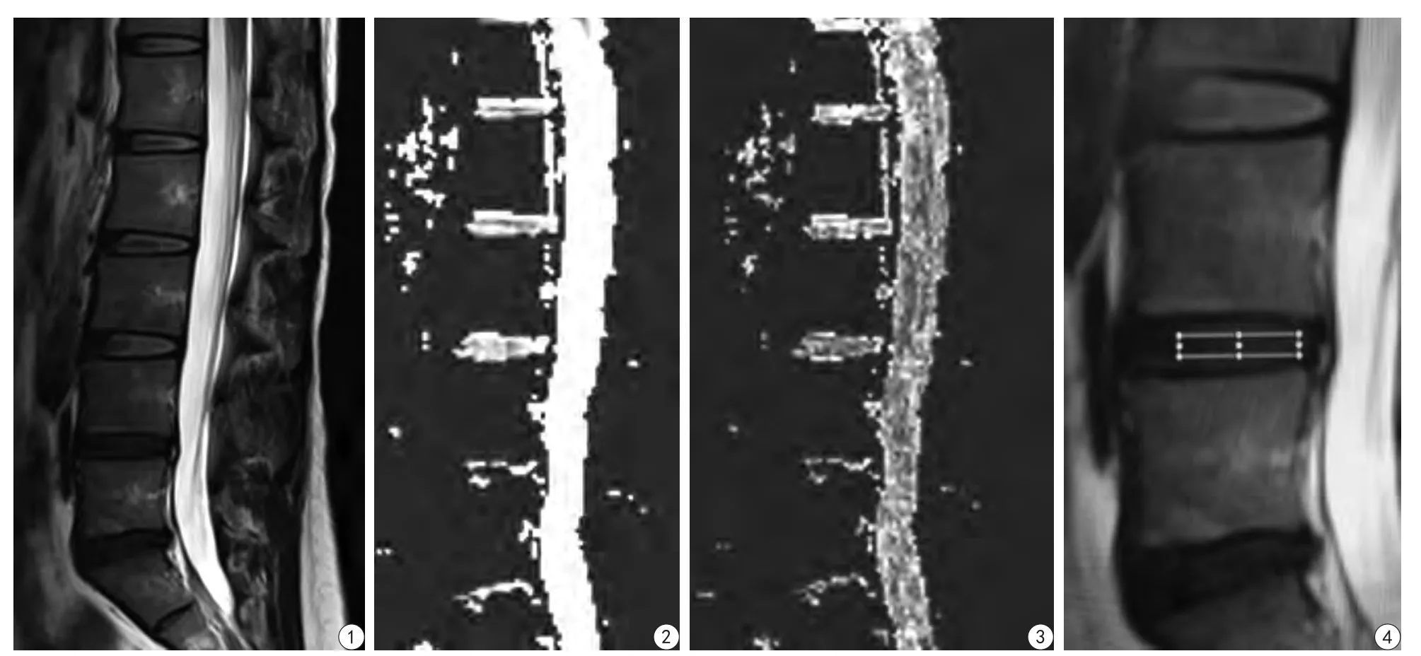
采用西门子RESOLVE序列,所有患者均得到较理想的图像(见图1~4),腰椎间盘Pm分级中,Ⅱ、Ⅲ、Ⅳ、Ⅴ分别对应的椎间盘数为33、98、63、56。
2.2 按不同扩散梯度方向分组椎间盘ADC值、FA值、λ1比较

图1 女,40岁,该患者矢状位T2加权像,L1/2~L3/4腰椎间盘为Pm Ⅲ级,L4/5、L5/S1为Pm Ⅳ级 图2~3 同一患者,扩散梯度方向为6时与矢状位T2加权像同一层面ADC图及FA图。L4/5、L5/S1信号明显减低 图4 椎间盘ROI的选取Fig. 1 Female, forty–year-old, sagittal T2WI image, L1/2-L3/4 for Pm Ⅲ, L4/5 and L5/S1 for Pm Ⅳ. Fig. 2—3 DGD for 6, ADC image and FA image respectively of the same patient, in same layer with T2WI. The signals of L4/5, L5/S1 were decreased. Fig.4 Shows the selection of ROI.
以扩散梯度方向进行分组,分为DD6、DD12、DD20组,对测量的所有椎间盘FA值、λ1进行单因素方差分析并两两比较,其中ADC值在不同弥散梯度组间差异无统计学意义(P>0.05),其余各组数据差异均有统计学意义(P<0.05,表1)。
2.3 相同扩散梯度方向-不同分级椎间盘FA值、λ1比较
以椎间盘分级分为Ⅱ、Ⅲ、Ⅳ及Ⅴ组,并对相同扩散梯度方向时ADC值、FA值、λ1进行比较(表2)。

表1 不同扩散梯度方向组间FA、ADC、λ1比较Tab.1 Comparison of the ADC value and FA value and λ1of different DGD groups
3 讨论
鉴于多方位成像、高组织分辨率及对组织水含量的增减有很强的敏感性等优势,MRI是目前椎间盘病变最好的检查手段,并可根据国际公认的评价椎间盘退变程度的Pfirrmann (Pm)分级标准进行分级[1]。但随着MRI技术的发展,传统的形态学变化已不能满足目前影像学的发展趋势,越来越多的MRI新技术将椎间盘病变的诊断由形态学带到定量分析的新阶段[2-4]。
DTI是基于扩散加权成像(diffusion weighted imaging,DWI)基础发展起来的一项磁共振新技术,利用组织中水分子扩散运动存在各向异性的原理,探测组织微观结构变化,并能够准确地评价多种组织纤维的损伤[5-6]。既往研究认为DTI可无创地反映椎间盘纤维环的形态及完整性,同时对椎间盘损伤、退变及负荷运动下椎间盘的变化情况方面也有重要价值[7],但对DTI检查时扩散梯度方向这一扫描参数对测量结果所产生的影响研究尚不足。本研究对所有椎间盘进行6、12、20 3个扩散梯度方向的DTI检查,按扩散梯度方向将椎间盘进行分组研究,发现不同扩散梯度方向组间,ADC值差异无统计学意义,其余各项指标差异均存在统计学意义(P<0.05),这一结果说明扩散梯度方向对椎间盘DTI测量结果存在影响,而选择适合的扩散梯度方向则对椎间盘DTI的定量分析具有重要意义。

表2 相同扩散梯度方向不同分级椎间盘FA值、ADC、λ1比较Tab.2 Comparison of the ADC value and FA value and λ1of different Pm grades groups
目前常用的DTI量化参数有很多,主要包括ADC值、FA、λ1,而应用最多的是FA值及ADC值[8]。ADC值与FA值与椎间盘变性时髓核储水能力及组织结构变化相关[9-11]。本研究按Pm分级对椎间盘进行分组,并对相同扩散梯度方向下的腰椎间盘DTI各指标进行比较,在扩散梯度方向为6时,FA值在Ⅳ与Ⅴ组间差异无统计学意义(P>0.05),在其余分组间差异均存在统计学意义(P<0.05),本研究结果显示随Pm分级增加,FA值呈升高趋势,这与以往报道相一致[12]。由于椎间盘髓核中基质退变,蛋白多糖合成数量及质量下降、胶原纤维不规则网状结构破坏等原因,其内水分子扩散的各向同性受限,相对各向异性扩散增加,因此导致FA值升高[13-14]。本研究结果Ⅳ与Ⅴ组间FA值差异无统计学意义,可能与Ⅳ与Ⅴ椎间盘主要区别在于形态、结构改变,而含水量差异不大有关[15]。扩散梯度方向为12和20时,FA值在Ⅱ与其他组间差异存在统计学意义(P<0.05),而Ⅲ、Ⅳ、Ⅴ组间差异无统计学意义(P>0.05),可能与扫描时间偏长、产热量较多,患者扫描过程中不耐受引起运动导致图像及测量质量下降有关。
扩散梯度方向为6、12、20时,ADC值在Ⅱ、Ⅲ组间差异无统计学意义(P>0.05),与其余组间差异有统计学意义(P<0.05),ADC值随Pm分级增高而减低的趋势与以往研究结果相符[15-17]。有研究表明[18],椎间盘变性髓核中胶原纤维会发生不同程度的扭曲,胶原纤维束间裂隙增大。Ⅱ级、Ⅲ级椎间盘的主要区别是髓核区的T2信号减低而无明显形态上的改变,本研究结果Ⅱ级、Ⅲ级椎间盘ADC值差异无统计学意义,笔者认为产生这一结果可能的原因是两级椎间盘间含水量确实存在差异,但微观结构上无显著破坏,水分子弥散受限程度变化不大。本研究显示Ⅳ级与Ⅴ级间ADC值差异存在统计学意义,与以往研究不符,这可能与Ⅴ级椎间盘形态改变较大,ADC图信号丢失较多有关。
纵向弥散系数λ1代表水分子在纵向的扩散能力[19]。本研究另一结果显示,扩散梯度方向为6、12、20时,各组λ1差异均无统计学意义。陈德胜等[20]研究认为,正常椎间盘内胶原纤维排列整齐、规则,纵横交错,形态及走行一致,而退变的胶原纤维主要表现为扭结成团。笔者认为可能由于正常椎间盘胶原纤维走行交错,而椎间盘变性时胶原纤维的扭曲不会影响水分子的纵向扩散能力。本研究也存在一些不足,如样本量较小,患者年龄跨度较大,因此这一结论尚需进一步验证。
DTI扫描中,扩散梯度方向的增加会延长DTI的扫描时间从而引起患者的不适,导致患者运动而影响测量结果,而扩散梯度方向的增加并不会引起FA值及信噪比(signal to noise ratio,SNR)的明显差异[21]。本研究中采用西门子RESOLVE弥散序列,并选取6、12、20 3个扩散梯度方向进行研究,扫描时间分别为2 min 4 s、3 min 44 s、5 min 58 s。结果显示扩散梯度方向为6时,扫描时间较短,且可得到较高质量的DTI后处理图像,测得的FA值和ADC值可对椎间盘变性进行定量分析,更能为临床所接受。
综上所述,DTI作为一种无创性的检查方法已广泛应用于多个系统的疾病诊断,腰椎间盘DTI检查可得到腰椎间盘多项定量指标,并能反映组织的含水量及细微结构变化,可为椎间盘变性的分级及治疗提供重要依据。
[References]
[1] Pfirrmann CW, Metzdorf A, Zanetti M, et al. Magentic resonance classification of lumbar intervertebral disc degeneration. Spine, 2001,26(17): 1873-1878.
[2] Fang Y, Liu LX, Li JL, et al. Prelimary study of lumbar intervertebral disc degeneration with magnetic resonance spectroscopy, T2 relaxation times and apparent diffusion coefficient. Chin J Magn Reson Imaging, 2011, 2(4): 278-282.方元, 刘兰祥, 李京龙, 等. 磁共振波谱成像、T2弛豫时间、扩散加权成像对腰椎间盘退变的初步研究. 磁共振成像, 2011, 2(4):278-282.
[3] Jiang XL, Zha YF, Wang J, et al. Study of correlation between cartilaginous endplate defects and intervertebral disc degeneration of lumbar spine with MR 3d-UTE technique. Radiology Practice, 2015,30(9): 952-955.蒋小莉, 查云飞, 王娇, 等. 3D-UTE评价腰椎间盘软骨终板缺损与椎间盘退变的相关性. 放射学实践, 2015, 30(9): 952-955.
[4] Zhu JY, Wang XM. The study of correlation between age and anatomical segment in healthy lumbar intervertebral discs by diffusion tensor imaging. Chin J Magn Reson Imaging, 2015, 6(7):518-523.祝静雅, 王晓明. 扩散张量成像对健康成人腰椎间盘与年龄及解剖部位的相关性研究. 磁共振成像, 2015, 6(7): 518-523.
[5] Zijta FM, Lakeman MM, Froeling M, et al. Evaluation of the female pelvic floor in pelvic organ prolapse using 3.0 Tesla diffusion tensor imaging and fibre tractography. Eur Radiol, 2012, 22(12): 2806-2813.
[6] Rousset P, Delmas V, Buy JN, et al. In vivo visualization of the levator ani muscle subdivisions using MR fiber tractography with diffusion tensor imaging. J Anat, 2012, 221(3): 221-228.
[7] Zu JY, Wang CG, Jia NY, et al. Initial study of the degeneration of lumbar intervertebral disc by magnetic resonance diffusion tensor imaging. Chin J Radiol, 2012, 46(11): 1002-1005.俎金燕, 王晨光, 贾宁阳, 等. 腰椎间盘退行性变的MR扩散张量成像初步研究. 中华放射学杂志, 2012, 46(11): 1002-1005.
[8] Yang LJ, Meng ZH, Cheng Y. Study on DTI of brainstem wallerian degeneration. J Shanxi Med Univ, 2014, 45(3): 213-216.杨丽娟, 孟志华, 程英. 脑干华勒氏变性的DTI研究. 山西医科大学学报, 2014, 45(3): 213-216.
[9] Budzik JF, Balbi V, Verclytte S, et al. Diffusion tensor imaging in musculoskeletal disorders. Radiographics, 2014, 34(3): 56-72.
[10] Gao SJ, Yuan X, Jiang XY, et al. Correlation study of 3.0 T MRDTI measurements and clinical symptoms of cervical spondylotic myelopathy. Eur J Radiol, 2013, 82(11): 1940-1945.
[11] Zhao DD, Li H, Qin H, et al. Initial application and clinical significance of DTI in normal adult patella cartilage. Chin J Magn Reson Imaging, 2016, 7(2): 131-135.赵丹丹, 李红, 秦灏, 等. DTI在正常人髌骨软骨的初步应用及临床意义. 磁共振成像, 2016, 7(2): 131-135.
[12] Niu G, Yang J, Wang R, et al. MR imaging assessment of lumbar intervertebral disk degeneration and age-relate changes: apparent diffusion coefficient versus T2 quantitation. AJNR Am J Neuroradiol,2011, 32(9): 1617-1623.
[13] Zhang Z, Chan Q, Anthony MP, et al. Age-related diffusion patterns in human lumbar intervertebral discs: a pilot study in asymptomatic subjects. Magn Reson Med, 2012, 30(2): 181-188.
[14] Siepe CJ, Heider F, Haas E, et al. Influence of lumbar intervertebral disc degeneration on the outcome of total lumbar disc replacement: a prospective clinical, histological, X-ray and MRI investigation. Eur Spine J, 2012, 21(11): 2287-2299.
[15] Chu XL, Ma JX, Qiu XL, et al. Application of diffusion tensor imaging in lumbar intervertebral disc degeneration: a preliminary study. Radiology Practice, 2016, 31(1): 81-85.褚相乐, 马景旭, 邱雪玲, 等. DTI在腰椎间盘退行性改变中的应用初探. 放射学实践, 2016, 31(1): 81-85.
[16] Wu N, Liu H, Chen J, et al. Comparison of apparent diffusion coefficient and T2 relaxation time variation patterns in assessment of age and disc level related interverte bral disc changes. PLoS One,2013, 8(7): e69052.
[17] Kealey SM, Aho T, Delong D, et al. Assessment of apparent diffusion coefficient in normal and degenerated intervertebral lumbar disks:initial experience. Radiology, 2005, 235(2): 569-574.
[18] He BR, Hao DJ, Xu ZW, et al. Correlation between clinical manifestations and ultrastructure of nucleus pulposus in lumbar dics herniation. Orthopaedics, 2010, 1(1): 33-36.贺宝荣, 郝定均, 许正伟, 等. 腰椎间盘突出症髓核的超微结构与临床对照研究. 骨科, 2010, 1(1): 33-36.
[19] Joong HK, David NL, Liang HF, et al. Noninvasive diffusion tensor imaging of evolving white matter pathology in a mouse model of acute spinal cord injury. Magn Reson Med, 2007, 58(2):254-260.
[20] Chen DS, Jin QH, Zhang Y. Observation of ultrapathology in the rat model study of experimental lumbar intervertebral disc degeneration.Orthopedic J Chin, 2007, 15(3): 223-225.陈德胜, 金群华, 张焱. 大鼠退变腰椎间盘组织的超微结构观察.中国矫形外科杂志, 2007, 15(3): 223-225.
[21] Fu ZH, Gong ZG, Zhou JH, et al. The effect of the number of diffusion gradient directions on the DTI parameters of SNR and FA.J Clin Radiol, 2014, 33(3): 437-440.付志辉, 贡志刚, 周建华, 等. 扩散梯度方向数目对DTI参数SNR和FA的影像. 临床放射学杂志, 2014, 33(3): 437-440.
Quantitative metabolites of lumbar intervertebral disc degeneration by diffusion tensor MR
ZHAO Shuang, PENG Ru-chen*, SHEN Xiu-zhi, ZHANG Shuang, ZHONG Jia-li,SONG Hai-long
Department of Radiology, Beijing Luhe Hospital, Capital Medical University, Beijing 101149, China
Objective:To investigate the MR diffusion tensor imaging (DTI) as an application of quantitative analysis methods in lumbar intervertebral disc degeneration and the effects of different diffusion gradient directions (DGD) of DTI.Materials and Methods:Selected 50 patients with pain of waist or legs, including 32 male and 18 female, from 24 to 62 years old with mean age 46.9±16.2 years old, all the patients underwent sagittal scans for regular lumbar MRI and DTI, and with 6, 12, 20 DGD of DTI. According to the DGD, lumbar intervertebral discs can be divided into DD6,DD12, DD20 groups and divided lumbar intervertebral discs into Ⅱ, Ⅲ, Ⅳ, and Ⅴgroups by Pfirrmann grading system. ADC and FA and λ1valuse were measured and analyzed among the different DGD groups and Pm groups.Results:There were no statistical difference of ADC values among the different DGD groups (P>0.05), while statistical difference of FA and λ1values were found (P<0.05); DGD for 6, FA values were statistical differences among the different Pm groups (P<0.05), except Ⅳ andⅤ(P>0.05); DGD for 12 and 20, there were no statistical difference of FA values in all PM groups (P>0.05), except Ⅱ. DGD for 6, 12, 20, ADC values were statistical difference in all Pm groups, except Ⅱ and Ⅲ. In DGD for 6, 12, 20, λ1values had no statistical difference in all Pm groups (P>0.05).Conclusion:DTI can be applied to the quantitative analysis of lumbar intervertebral disc, DGD for 6 can get high quality images and data in a relatively short time, and FA value can be used for quantitativeanalysis of different Pm grading lumbar intervertebral disc.
Magnetic resonance imaging; Diffusion tensor imaging; Intervertebral disc degeneration; Apparent diffusion coefficient
21 Jan 2017, Accepted 16 Mar 2017
彭如臣,E-mail:13501271260@163.com
2017-01-21
接受日期:2017-03-16
R445.2;R681.57
A
10.12015/issn.1674-8034.2017.06.011
赵爽, 彭如臣, 沈秀芝, 等. 腰椎间盘退行性病变的MRI-DTI定量分析. 磁共振成像, 2017, 8(6): 457-461.
*Correspondence to: Peng RC, E-mail:13501271260@163.com

